
Joint imaging
By
Dr. Firas Abdullah

Aims of our lecture:
Radiological signs of joint disease
Diagnosis of arthritis
Different types of arthritis
Other joint pathology
MRI of knee and shoulder
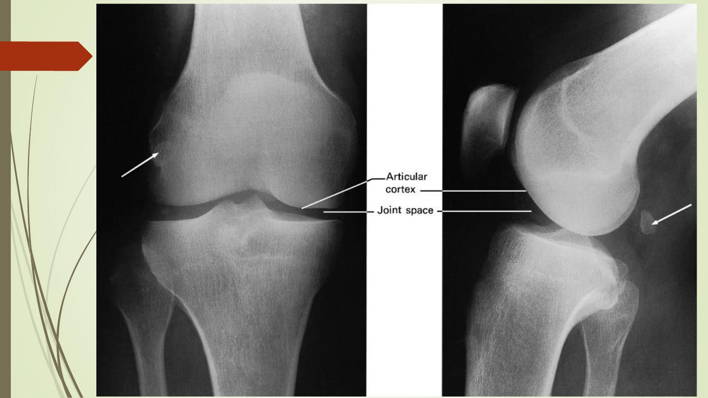
Plain film signs of joint disease
❖
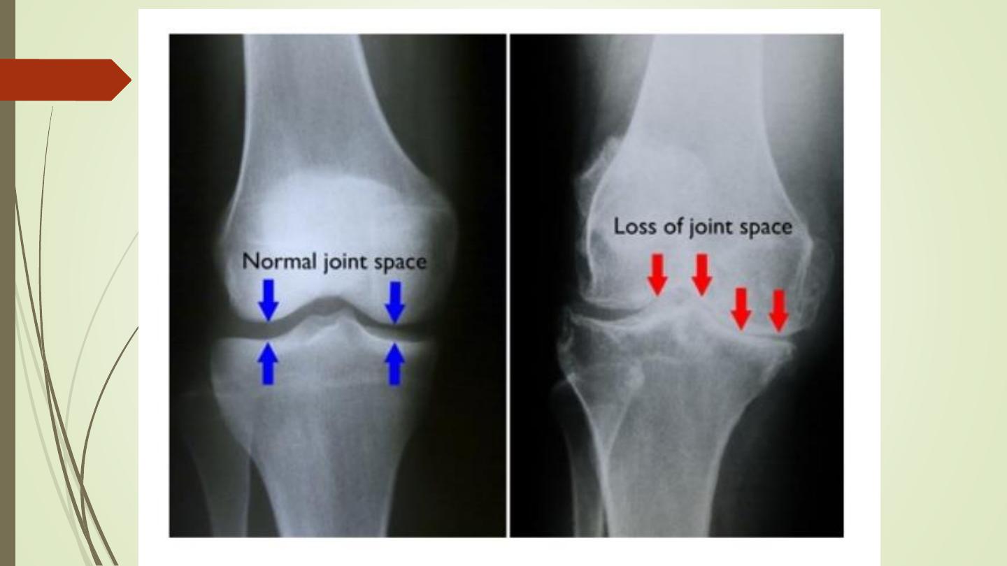
Joint space narrowing
: due to destruction of articular
cartilage. It occurs in practically all forms of joint
disease, except avascular necrosis.
❖
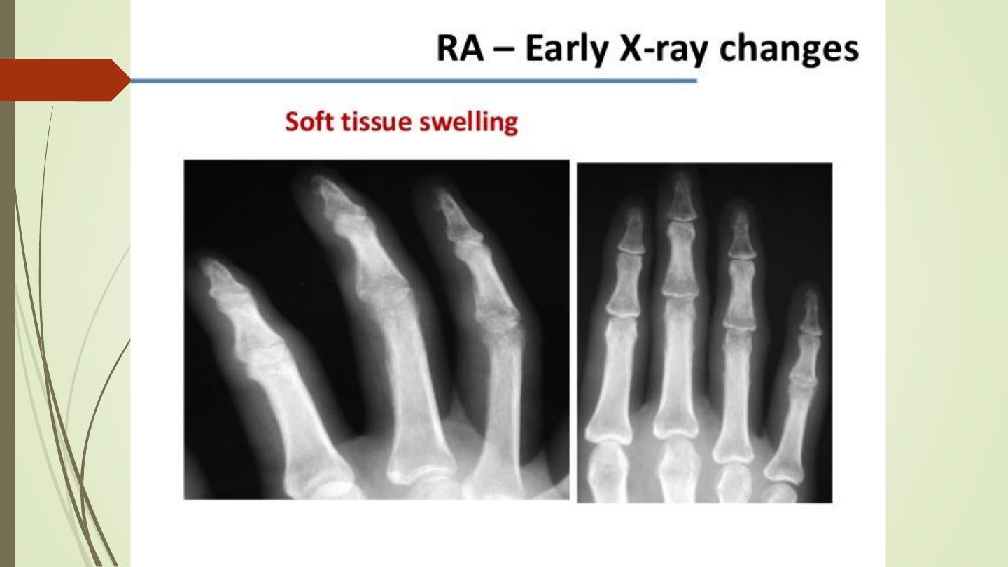
Soft tissue swelling
: a feature of inflammatory, and
particularly infective arthritis. Also can be seen in
gouty tophi.
❖
Osteoporosis
: painful conditions and underuse of the
bones. E.g. rheumatoid and tuberculous arthritis.
❖
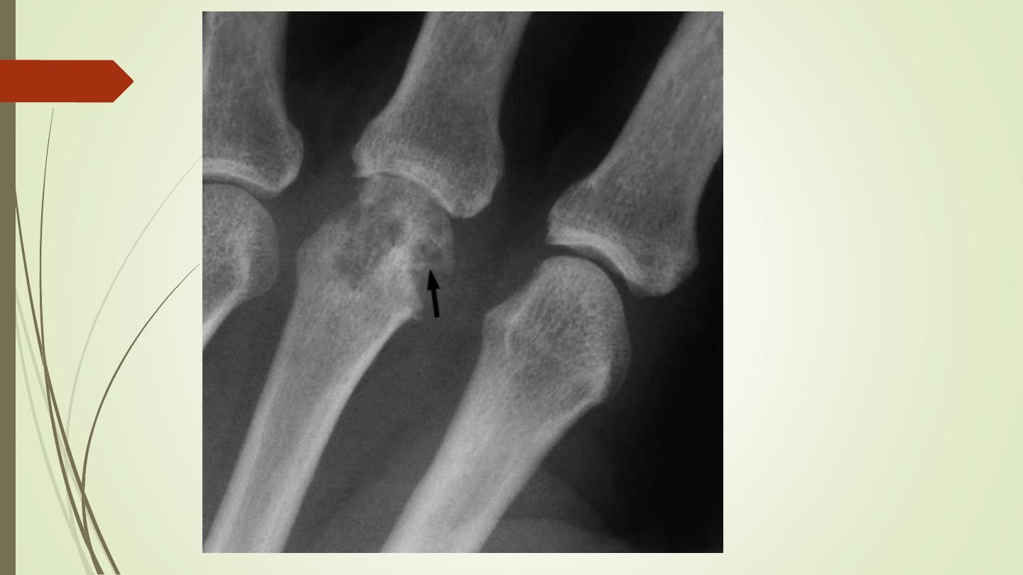
Articular erosions
: destruction of the articular cortex and
the adjacent trabecular bone
Causes
:
1- Inflammatory overgrowth of the synovium (pannus)
•
Rheumatoid arthritis , commonest
•
Juvenile rheumatoid arthritis (Still’s disease)
•
Psoriasis
•
Reiter’s disease
•
Ankylosing spondylitis
•
Tuberculosis.
2- Deposition of urate crystals in gout.
3- Infection: pyogenic arthritis and tuberculosis.
4- Repeated hemorrhage in hemophilia
5- Neoplastic, e.g. synovial sarcoma.
❖
Osteophytes, subchondral sclerosis and cysts
:
Features of osteoarthritis. A characteristic increase
in the density of subchondral bone is seen in
avascular necrosis
❖
Alteration in the shape of the joint
: slipped
epiphysis, developmental dysplasia of the hip,
osteochondritis dissecans and avascular necrosis in
its later stages.
Joint erosion

Diagnosis of arthritis
1. Whether one or more than one joint involved?
e.g.
rheumatoid arthritis, infections and synovial tumours.
2. Which joints are involved?
❑
Rheumatoid arthritis
❑
Psoriatic arthritis
❑
Gout characteristically
❑
Osteoarthritis
❑
Neuropathic arthritis
3. Is a known disease present?
e.g. haemophilia or
diabetes.

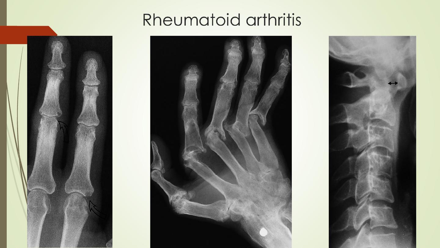
Rheumatoid arthritis
❖
A polyarthritis caused by inflammatory overgrowth of synovium
known as
pannus
.
❖
The earliest change is
periarticular soft tissue swelling
and
osteoporosis
.
❖
Joint space narrowing.
❖
Initially small bony
erosions
, at the joint margins. Later, extensive
erosions
❖
Metatarso- or metacarpophalangeal joints, proximal
interphalangeal joints and on the styloid process of the ulna.
❖
Advance changes:
Ulnar deviation
.
Arthritis mutilans
.

Rheumatoid arthritis
❖
With severe disease, there may be subluxation at the
atlantoaxial joint, possibility of neurological symptoms from
compression of the spinal cord by the odontoid process
❖
A widespread erosive arthropathy is almost diagnostic of
rheumatoid arthritis.

Osteoarthritis
❖
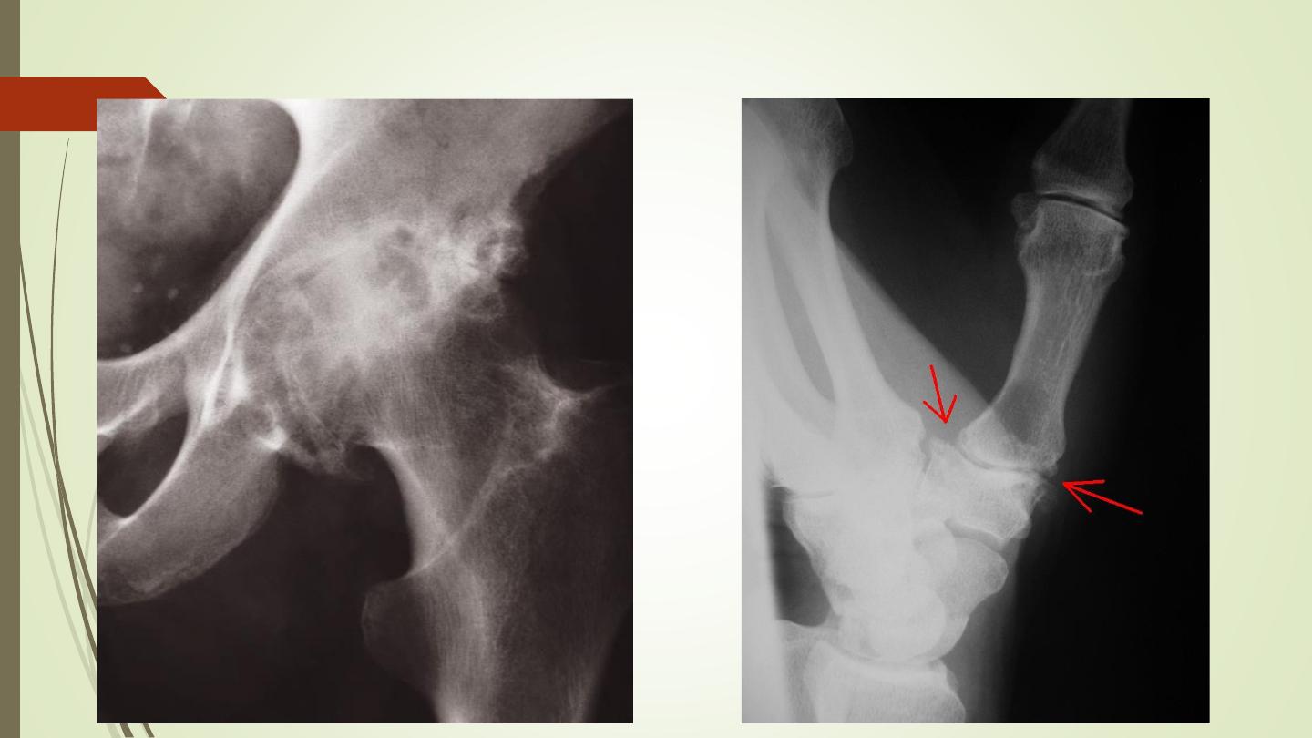
commonest form of arthritis.
❖
The hip and the knee are frequently involved, the ankle elbow
are infrequently affected.
❖
The wrist, joints of the hand and the metatarsophalangeal joint
of the big toe are also frequently involved.
❖
Radiological features:
❑
Joint space narrowing.
❑
Osteophytes
❑
Subchondral sclerosis
❑
Subchondral cysts
❑
Loose bodies
Osteoarthritis
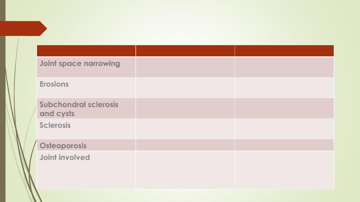
Comparison of osteoarthritis and rheumatoid arthritis
Radiological feature
Osteoarthritis
Rheumatoid arthritis
Joint space narrowing
Maximal at weight-
bearing site
Uniform
Erosions
Not occur
Is a characteristic feature
Subchondral sclerosis
and cysts
Seen
Not a feature
Sclerosis
Prominent feature
Not a feature
Osteoporosis
Not occur
Often present
Joint involved
Knee, hip
Metacarpophalangeal
Distal interphalangeal
Metacarpophalangeal
Proximal interphalangeal

Gouty Arthritis
❖
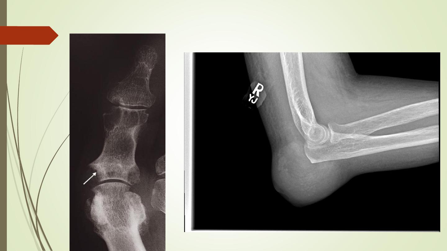
Most commonly affects the metatarsophalangeal joint of
the big toe.
❖
The earliest change is soft tissue swelling
❖
Erosions have a well-defined, often sclerotic overhanging
edge
❖
Usually no osteoporosis
❖
Localized soft tissue lumps, known as tophi, may occur in
the periarticular and occasionally show calcification.
Gouty Arthritis
Tophi

Joint Infection
❖
Pyogenic bacterial infection or tuberculosis
❖
In pyogenic arthritis, Staphylococcus aureus, there is rapid
destruction of the articular cartilage & subchondral bone
and a soft tissue swelling. A joint effusion is the earliest
finding
❖
TB arthritis, The hip and knee are the most commonly
affected. Joint space narrowing and erosions, articular
cortex destruction, and striking osteoporosis,
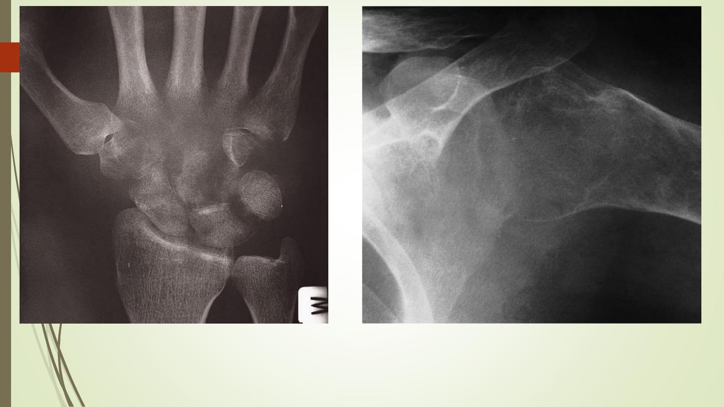
pyogenic arthritis
TB arthritis

Neuropathic joint (Charcot joint)
6 Ds of Charcot joint:
❑
Increased Density (subchondral sclerosis)
❑
Destruction
❑
Debris (intra-articular loose bodies)
❑
Dislocation
❑
Distention
❑
Disorganisation
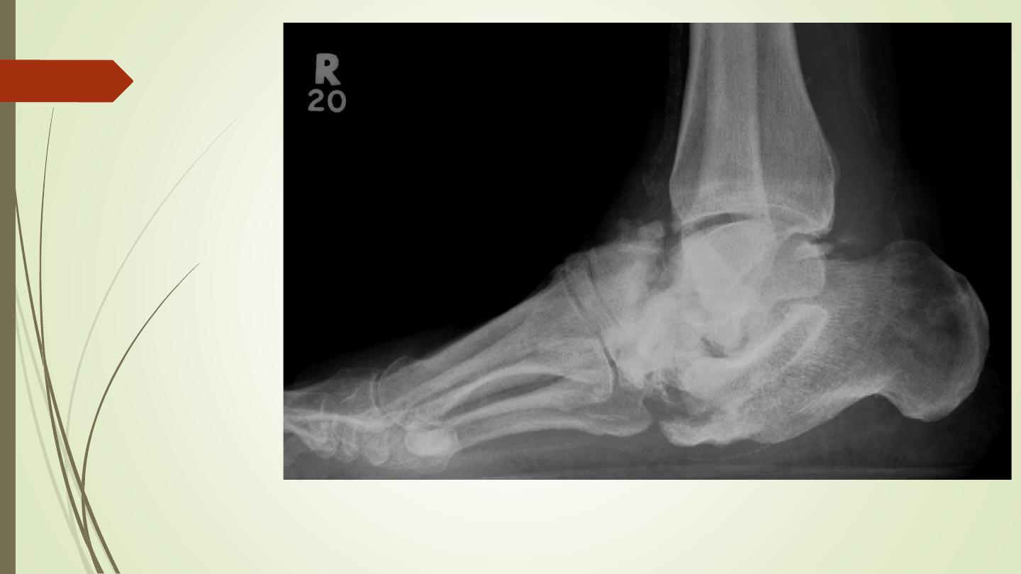
Charcot joint
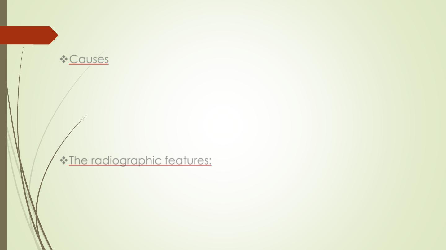
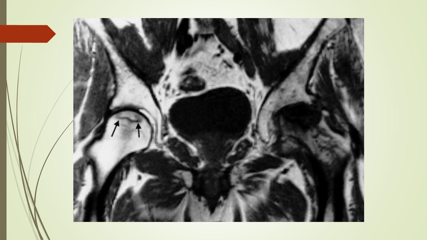
Avascular (aseptic) necrosis
❖
Causes
:
➢
steroid therapy
➢
Collagen vascular diseases
➢
Radiation therapy
➢
Sickle cell anemia
➢
Exposure to high pressure environments
➢
Fractures.
❖
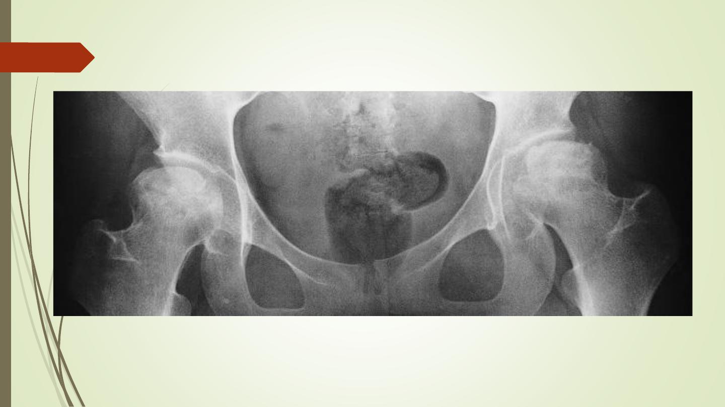
The radiographic features:
➢
Increased density of the subchondral bone with irregularity of the
articular contour or fragmentation of the bone
➢
A characteristic crescentic lucent line just beneath the articular
cortex.
➢
The cartilage space is preserved until secondary degenerative
changes supervene.

Osteochondrosis
❖
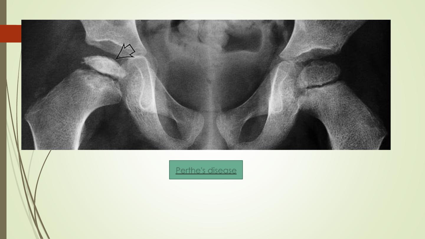
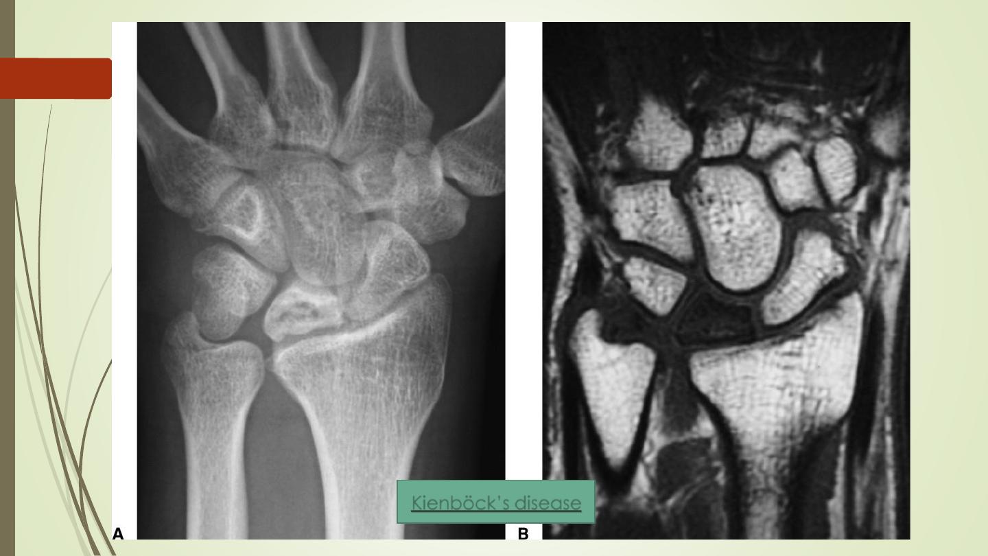
A group of disorders that affect the progress of bone
growth by bone necrosis. It is only seen in children and
adolescents who are still growing.
➢
Perthe's disease: femora head
➢
Freiberg’s disease: metatarsal heads
➢
Kohler’s disease: navicular bone of the foot
➢
Osgood–Schlatter’s disease: tibial tuberosity
➢
Kienböck’s disease: lunate bone in the wrist
Perthe's disease
Perthe's disease
Kienböck’s disease

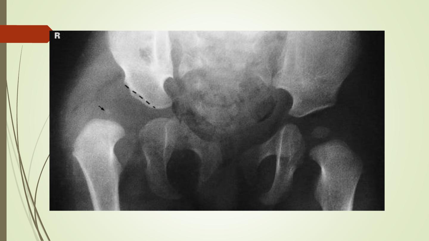
Developmental dysplasia of the hip (DDH)
Abnormal development of the hip joint resulting from an
abnormal relationship of the femoral head to the
acetabulum. There is a clear female predominance, and it
usually occurs from ligamentous laxity and abnormal position
in utero. Therefore, it is more common with oligohydramnios
pregnancies.
Risk factors include:
• female gender (M: F ratio ~1:8)
• firstborn baby
• family history
• breech presentation
• oligohydramnios

Investigation used for diagnosis:
❑
Ultrasound at early infancy
❑
X ray later in life
❑
The features: lateral and upper displacement
of the head of the femur. Increased slope to
the acetabular roof

Best Regards