Physiology of hearing
Anatomy &Introduction
The ears are paired sensory organs comprising the auditory system, involved in the detection of sound, and the vestibular system, involved with maintaining body balance/ equilibrium. The ear divides anatomically and functionally into three regions: the external ear, the middle ear, and the inner ear. All three regions are involved in hearing. Only the inner ear functions in the vestibular system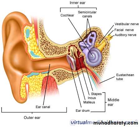
OUTER EAR AURICLE-/ framework of cartilaginous fibers except lobule -capture sound & funnel it
E.A.M.
2.5cm.
Direction
Histology : - cart.2/3 X bone 1/3
- hair /skin
- seb.&cerumucinous gl.
Isthmus
channel , tubal resonator amplifying sound pr.
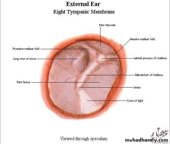
Tympanic membrane
Three layers : - ecto .- fibrous
- mucosal
• * cone-shaped /tension by T.T.M for
• better reception of vibrations of high frequency
•
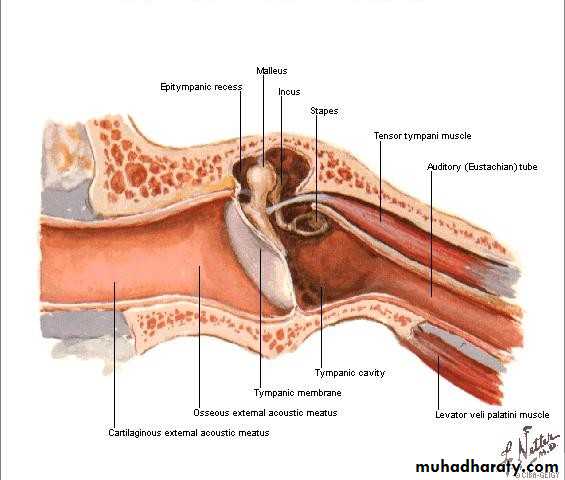
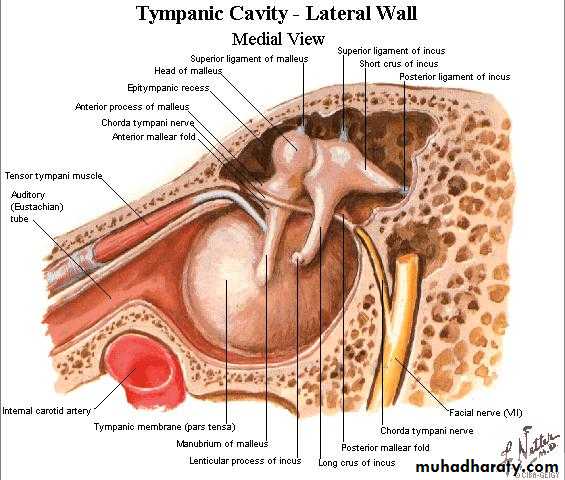
Middle ear
Walls15x13x2mm.
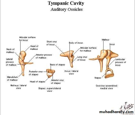
Ossicles : -malleus
-incus
-stapes
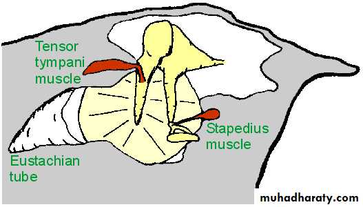
Muscles : - tensor tympani m.
- stapedius m.
Middle Ear Muscles
Tensor tympaniAttached to malleus
Innervated by V, trigeminal nerve
Stapedius
Attached to stapes
Innervated by VII, facial nerve
Middle Ear Muscle Function:
Help maintain ossicles in proper position
Protect inner ear from excessive sound levels
When ear exposed to sound levels above 70 dB, the muscles contract, decreasing amount of energy transferred to inner ear
This protective reflex termed "acoustic reflex"
• 1-ConductionConduct sound from the outer ear to the inner ea-2)ProtectionCreates a barrier that protects the middle and inner areas from foreign objectsMiddle ear muscles may provide protection from loud sounds3)TransducerConverts acoustic energy to mechanical energyConverts mechanical energy to hydraulic energy4)AmplifierTransformer action of the middle earonly about 1/1000 of the acoustic energy in air would be transmitted to the inner-ear fluids (about 30 dB hearing loss)
Function of Middle Ear
Eustachian Tube
The eustachian tube connects the front wall of the middle ear with the nasopharynxThe eustachian tube also operates like a valve, which opens during swallowing and yawning
This equalizes the pressure on either side of the eardrum, which is necessary for optimal hearing.
Without this function, a difference between the static pressure in the middle ear and the outside pressure may develop, causing the eardrum to displace inward or outward
This reduces the efficiency of the middle ear and less acoustic energy will be transmitted to the inner ear.
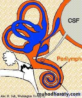
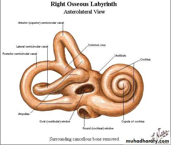
Inner ear
Bony labyrinth ___ perilymphMembranous labyrinth ___endolymph
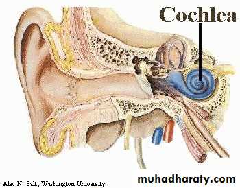
Cochlea
Vestibule(utricle ,saccule ,s.c.c.)
Function of Inner Ear
Convert mechanical sound waves to neural impulses that can be recognized by the brain for:Hearing
Balance
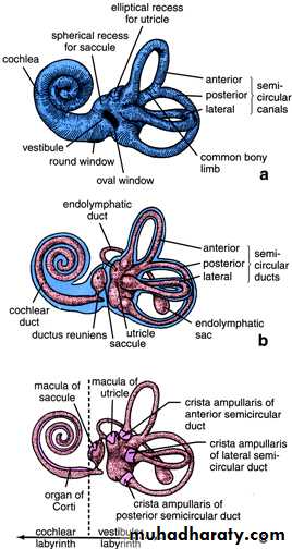
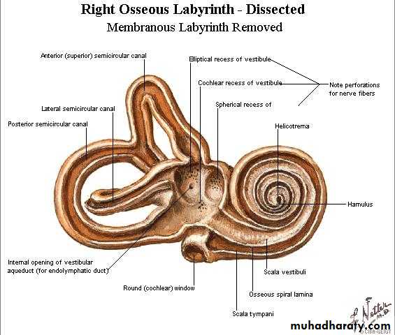
CochleaThe cochlea is a spiral structure, like a snail shell containing two and one half turns from its base at the oval window to its apex taken along the central pillar or modiolus,
*Small openingScala v.&scala t. communicate through, at apex called helicotema
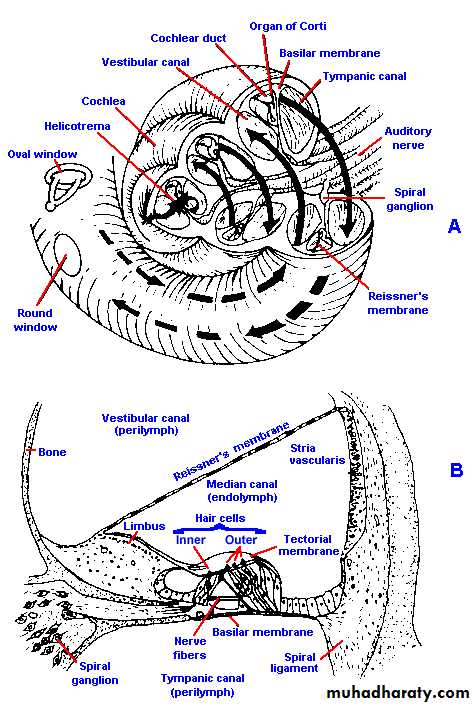
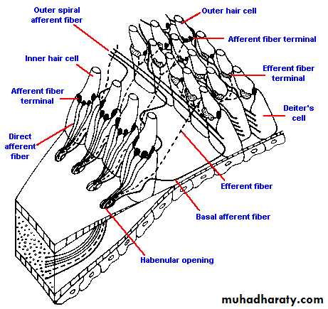
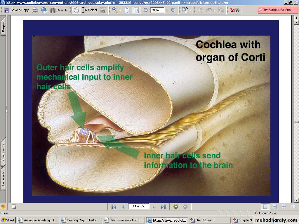
Organ of corti
Located on the basilar membraneContains the hair cells covered by tectorial membrane
-Outer h. c.
-Inner h. c.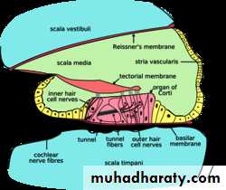
Hair cells
Outer hair cells*columnar in shape
* 12,000-20,000
*arranged in 3-4 rows lateral to rod of Corti
*carry 50-150 steriocilia
Inner hair cells
*bulbus in shape
*3,500
*single row medial to rod of corti
*120 steriocilia
OHC vs. IHC Function
Sound conduction pathways
• 1. Through Ossicular chain to oval window• 11. Directly cross middle ear to round window
• (large perforation)
• III. Bone conduction (vibration of skull bones)
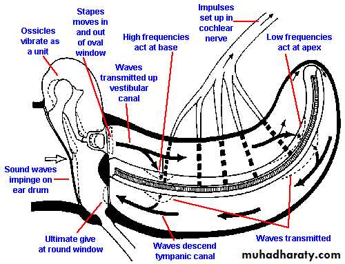
traveling waves
The impedance of the fluid in the cochlea is about 30 times greater than that of air, and if the sound were applied directly to the oval window, most of it (~97%) would be reflected, leaving only 3% transmission.Transformer/Amplifier
.
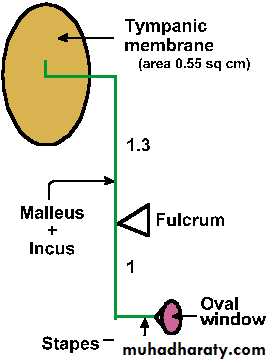
The middle ear enhances the transfer of acoustical energy in two ways:
The area of the eardrum is about 17 times larger than the oval windowThe effective pressure (force per unit area) is increased by this amount.
The ossicles produce a lever action that further amplifies the pressure
Without the transformer action of middle ear, about 1/1000 of acoustic energy in air transmitted to inner-ear fluids (about 30 dB loss).
Malleus and incus vibrate together, transmitting the sound waves from the eardrum to the footplate of the stapes (this pushes the oval window in and out)(mechanical energy)
Lever system
• Areal ratio =18:1
• Lever ratio=1.3:1• Transformer ratio=21:1
• Fig. 8-5. Schematic drawing of ossicle system to illustrate the lever arms and the position of the fulcrum. Relative areas of the tympanic membrane and the membrane of the oval window are shown.
When the stapes moves inward at the oval window pressure waves are transmitted to the perilymph of the scala vestibuli and thence through Reissner's membrane and the basilar membrane to the scala tympani. In the scala tympani, the vibrations pass again through perilymph to the round window at the base of the cochlea. The membranous covering of the round window bulges into the middle ear and forms the ultimate "give".The "give" at the round window is necessary to prevent pressure-wave reflections within the cochlea.
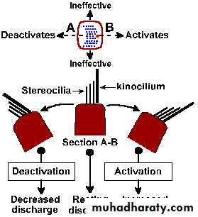
Movement of the cilia to kinocillium side results in a depolarization of the hair cell`s receptor that in turn releases a transmitter substance that finally depolarizes the afferent fibers that contact it.resulting in generation of action potential & transmission of the impulses
Central auditory pathways
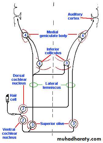
Sound localization
• Differences in the phase of the signals at each ear help in localization the source of low-frequency sound.• Differences in intensity are used to localize the source of high-frequency sound.
• The difference in time of arrival of a sound at the two ears
• Central system function .
Question 1
What is the purpose of the pinna?A. Cosmetics
B. Sound collector
C. Same side localization
D. A and B
E. A, B and C
Question :2 The pars tensa portion of the TM:
• Consists of 2 layers of tissue
• Consists of 4 layers of tissue
• Consists of 1 layer of tissue
• Consists of 3 layers of tissue
• Consists of 5 layers of tissue
Question :3
• The Eustachian tube:• Opens when one yawns
• Opens when one smiles
• Opens when one blinks
• It is always open
• Never opens
Question :4
• The middle ear:• Converts acoustic energy to hydraulic
• Converts hydraulic energy to mechanical
• Converts acoustic energy to mechanical
• Converts acoustic energy to electrical
• Converts mechanical to electrical
Question : 5
• The middle ear amplifies sound:
• About 15 dB
• About 25 dB
• About 35 dB
• About 20 dB
• About 30 dB
Question 6:
The function of the inner ear:• Balance
• Hearing
• Touch
• All the above
• A and B
QUESTION : 7
The channel that houses the organ of Corti:• Scala tympani
• Scala media
• Scala vestibuli
• Semicircular canals
• B and D
equilibrium
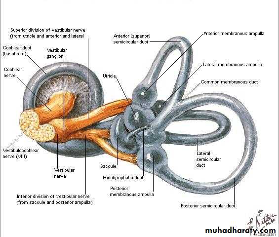
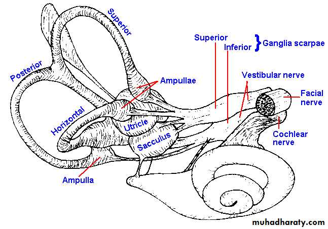
Vestibular anatomybony&membranous labyrinths
Semicircular canals
Sensory epithelial cells
1. The sensory epithelium of the utricle,the utricular macula,
2. The sensory epithelium of saccule ,the saccular macula
3. s.c.c crista ampullaris
Macula &crista
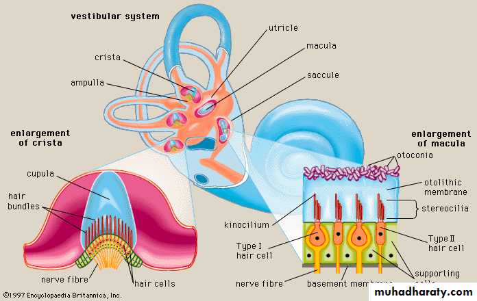
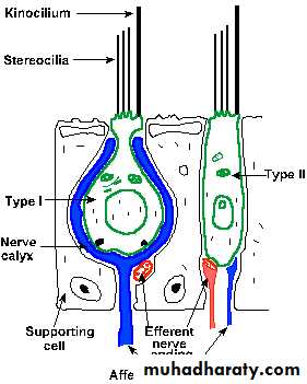
Vestibular sensory cells
Vestibular physiology
Utricular macula signals position of head& linear accelerationSaccule / exact function is not known
S.c.c. angular rotationMovement of the cilia to kinocillium side results in a de polarization of the hair cell, a receptor potential that in turn releases a transmitter substance that finally depolarizes (the generator potential) the afferent fibers that contact it.
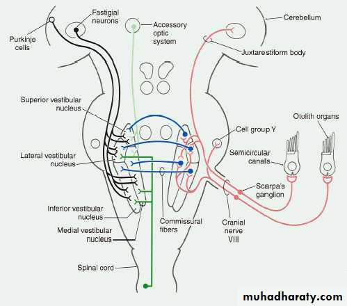
Vestibular pathways
The vestibular nerve fibers project to the vestibular nuclei and to the flocculonodular lobe of the cerebellum. The vestibular nuclei give rise both to the vestibulospinal tracts, which play a role in movement and posture,and to connections with cranial nerve nuclei III, IV, and VI and motoneurons of the neck by way of the medial longitudinal fasciculus
.
*The neural pathways of the reflex controlled by the horizontal semicircular canal
If the head is moved to the left, the eyes are moved* conjugately to the right, in order to maintain the gaze fixed on an object as the head moves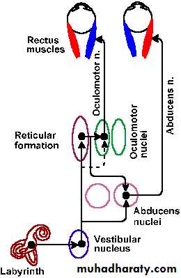
The vestibulo-ocular reflex has an important clinical use.
As a result of the reflex, when the head is moved from side-to-side, the eyes remain fixed on an object by moving from side-to-side but in the opposite direction.Vestibulo-occular reflux
Nystagmus and the caloric test
• The jerky movement of the eye observed at the start and end of a period of rotation is called nystagmus• *Types of nystagmus:
• Horizontal (frequently)
• Vertical (c.n.s. lesion)
• Rotatory (central)
Orientation in space
• Vestibular receptors• Vision
• Proprioceptors in joints capsule
• Cutaneous exteroceptors touch &pressure receptors
NICE TO MEET YOU