Anatomy & histology
Learning objectivesBy the end of this lecture; the student should be able to:
1-List the components of the integumentary system, including their physical relationships.
2-Specify the functions of the integumentary system.
3-Describe the main features and functions of the epidermis and dermis.
4-Explain the structure and function of the various skin appendages.
Largest organ in the body
Surafceabout 2 sq meters
Thickness varies according to area:
0.2-0.5 mm on eyelid & prepuce
3-5 mm on palm & sole
Weight
4-5 kg
20 kg with hypodermis
Functions of the skin
The most important function is protection:Serving as a barrier against infection, UV light & disease
Helping to regulate body temperature
Removing waste products from the body
Vitamin D3 synthesis
Sensory organ
Calorie reserve & heat insulation
Beauty organ


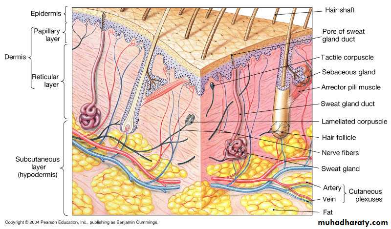
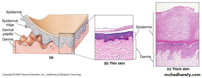
Layers of skin
EpidermisDermis
Subcutaneous fatty layer:
not part of skinThe Epidermis
The epidermis is the outer most layer of the skincomposed of 4-5 layers of keratinocytes
In thin skin = 4 layers (strata) as in hairy skin
In thick skin = 5 layers as in glabrous skin (palm & sole)
Histology of epidermis
Avascular stratified squamous epithelium, composed of:1- Keratinocytes: of ectodermal origin, constitute 90% of epidermal cells, arranged in 5 layers
Stratum germinativum (basal cell layer)
Stratum spinosum (prickle cell layer)
Stratum granulosum (granular cell layer)
Stratum lucidum: only in palm & sole
stratum corneum(horny layer)- non-viable epidermis
2-Dendritic cells: melanocytes, Langerhans's cells, merkel’s cells
Viable epidermis
The ultimate function of epidermis is to produce keratin by keratinocytes
• New cells are produced at the basal layer, they push older cells to the surface of the skin where they become flattened, lose their cellular content & start making keratin• It is a tough fibrous protein which forms the basic structure of hair, nail & skin
Eventually the keratinocytes die & form a tough, flexible, waterproof covering of the surface of the body
This is shed or washed away once every 14-28 days
Where do you expect this section of skin was taken from:
A- Sole. B- abdomen1-Basal cell layer
Single layer, tall columnar cells, have nuclei & all organellesSite of DNA synthesis & mitosis
Connected to each other by
desmosomes & to basement membrane
by hemidesmosome
2-Prickle cell layer
5-20 layers, polygonal, nucleated,cells, their cytoplasm becomes full
of keratin bundles that are attached
to desmosomes
Desmosomes are small interlocking cytoplasmic processes composed of thickenings on the cell membrane of
two opposing cells, allowing the sliding
of adjacent cells on each other without
separation upon trauma, links are
so strong that dead cells are shed in
sheets not individually.
Prickle cell layer
The upper part of this layer contain lamellar granules (Odland’s bodies, keratinosomes)which contain lipids &
polysaccharides & their
contents are discharged into
the intercellular space at the
interface with granular layer
Forming the hydrophobic
barrier
3-Granular cell layer
3-10 layers, flattened cells, cytoplasmfull of basophilic keratohyaline granules
Here there is dissolution of nucleus
& other cell organelles
Keratin filaments appear in large bundles
Keratinosomes migrate to the periphery of cells & discharge their lipid content
Horny cell layer
Flattened cells arranged in vertical
stacks that have lost nuclei &cellular organelles
Keratin filaments arranged into macro fibers under influence of fillagrin
Highly insoluble cornified envelope within plasma membrane
Desmosomes are lost
Epidermal cell cycle
After reaching the surface, corneocytes are shed continuously being replaced by newer cells from beneathThe whole cell cycle takes around 4 weeks normally from basal layer to be shed at the surface as a scale.
This rate is accelerated in certain
disease conditions such as
psoriasis to be less than 1 week
Dendritic cells: 1- melanocytes
Neuronal originlocalized between basal cells at a
rate of 1 in 10, fixed in all races
Contain melanosomes:
specialized organelles that synthetize
melanin from tyrosine under action
of tyrosinase enzyme then transfer
it to surrounding keratinocytes,
forming epidermal-melanin unit
Melanocytes
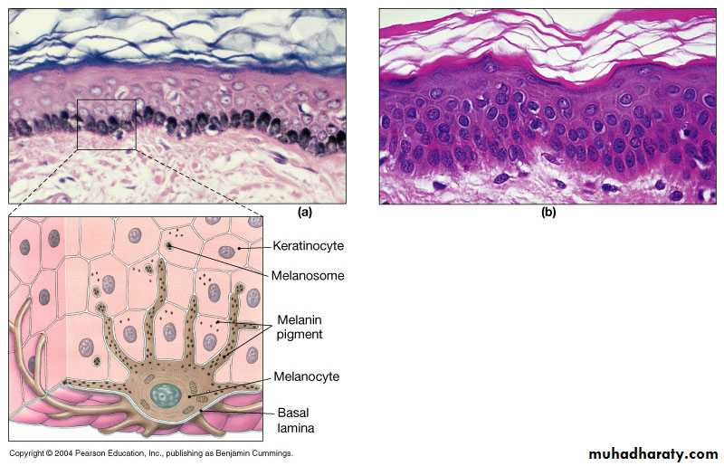
melanosomes are
responsible for thedifference in
normal skin color
between races;
being more in no.,
Larger & more
dispersed in darker
skin
Question What would happen to the skin if tyrosinase enzyme was deficient?
2- Langerhans's cellMesenchymal origin, localized in suprabasal layer
By electron microscope show Birbeck granules in cytoplasm
Antigen presenting cell in the skin: process antigens encountered on skin & present it to local
lymph nodes, thus have
a key role in adaptive
immune response.
3- Merkel’s cells:
Dendritic cells localized between basal cells directly above basement membrane
Associated with unmyelinated nerve endings & act as mechano-sensory receptors in response to touch
4- indeterminate cells:
they have the same ultrastructure of Langerhans’s cells but without Birbeck granulesDermo- epidermal junction
In light microscope is one layer, actually it is 3 layers:The upper part is formed by the
basement membrane of basal layer
with its attached hemidesmosomes
Lamina lucida
Lamina densa
Sub laminal fibrous band
The dermis
Dermis organizationPapillary layer
• Contains blood vessels,
• lymphatics, sensory nerves of
• epidermis
• Reticular layer
• Contains network of collagen and elastic fibers to resist tension
Dermis
The dermis forms the main bulk of the skin, lies under the epidermis & supports it both structurally and nutritionally, they interdigitate so that upward projections of the dermis (the dermal papillae) interlock with downward ridges of the epidermis (the rete-pegs), this increases the force of adhesion & the contact area.
Composition of dermis
Components of dermis:Cells: fibroblasts, mast cells, macrophages & all the cells in the blood
Fibers: 80-85% is collagen mainly type I & III, the remainder is composed of elastic & reticular fibers
Ground substance: composed of glycosaminoglycan/ proteoglycan macromolecules, they constitute 0.1-0.3% of the weight of dry dermis but are responsible for the hydration of the dermis due to the high water binding capacity of hyaluronic acid.
60% of the weight of the dermis is water
Skin appendages: Hair
Hair types1-Lanugo hair: intrauterine life, fine long hair
2-Vellus hair: peach fuzz; all over body, fine short hair
3-Terminal hair: coarse long
hair on scalp
the Anatomy of a Single Hair
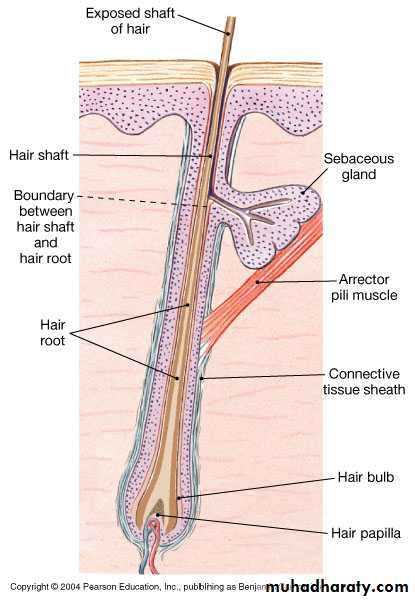
Composition of hair
Originate in hair follicleComposed of root and shaft
Root base (hair papilla) surrounded by hair bulb and root hair plexus
Hairs have soft medulla and hard cortex
Cuticle = superficial dead protective layer
Hair
A bundle of smooth muscle, the arrector pili,extends at an angle between
the surface of the dermis
and a point in the follicle wall.
supplied by adrenergic fibers
causing hair erection during
fear, anger, & cold.
Cycles of hair growth
Anagen: growth phase lasts 2-3 yearsCatagen: transition phase 2-3 weeks
Telogen: resting phase 2-3months, after which a club hair is shed
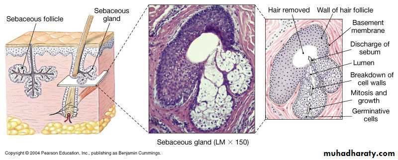
Sebaceous glands
Sebaceous glands
Present all over body but mostly in seborrheic areas:( scalp, face, upper part of: chest, shoulders & back)
Attached to hair follicles
Secrete sebum: a complex lipid which is bactericidal & fungistatic
Holocrine type of secretion: degeneration of the whole gland after it is filled & release of sebum
Pilosebaceous follicles
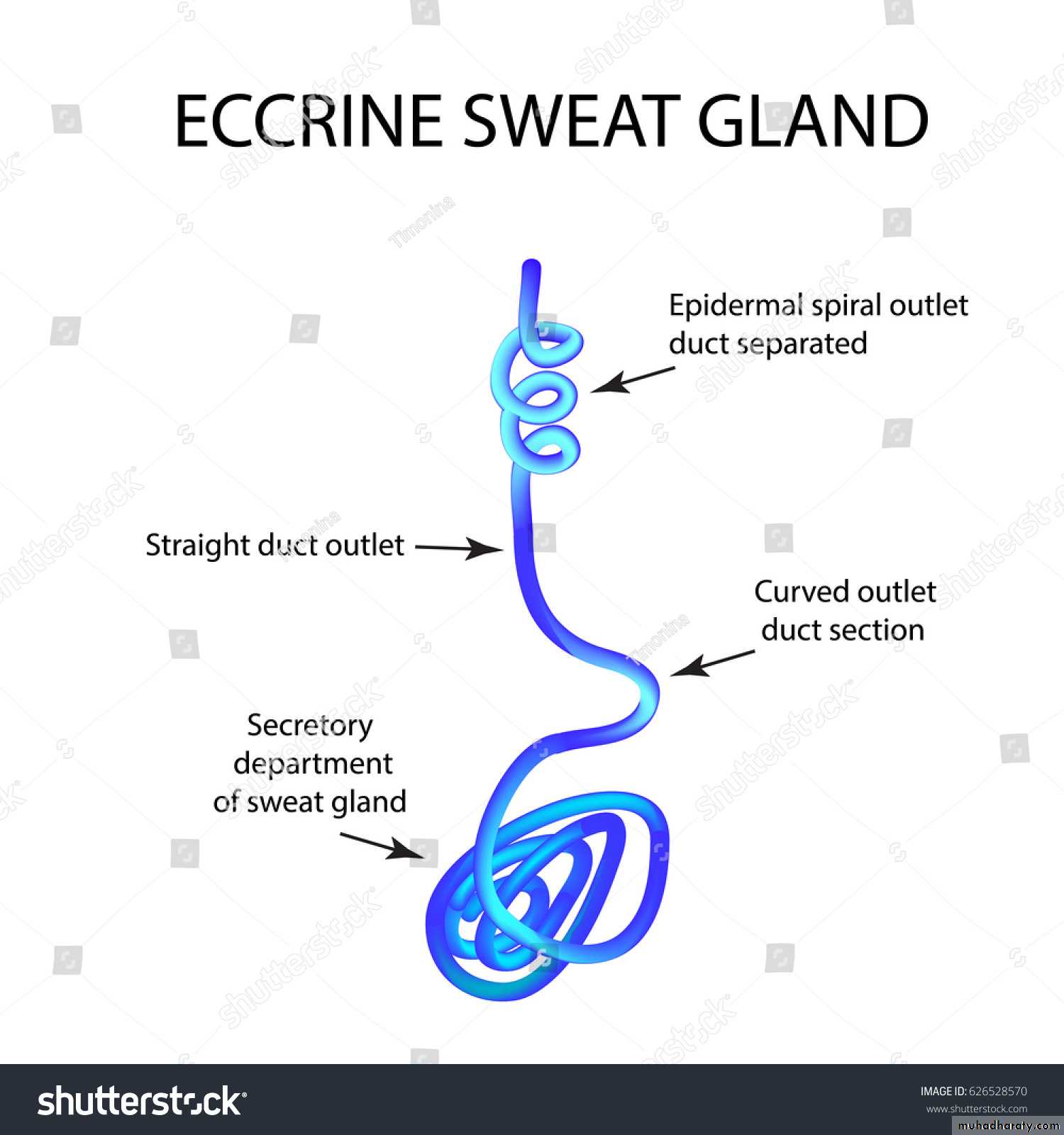
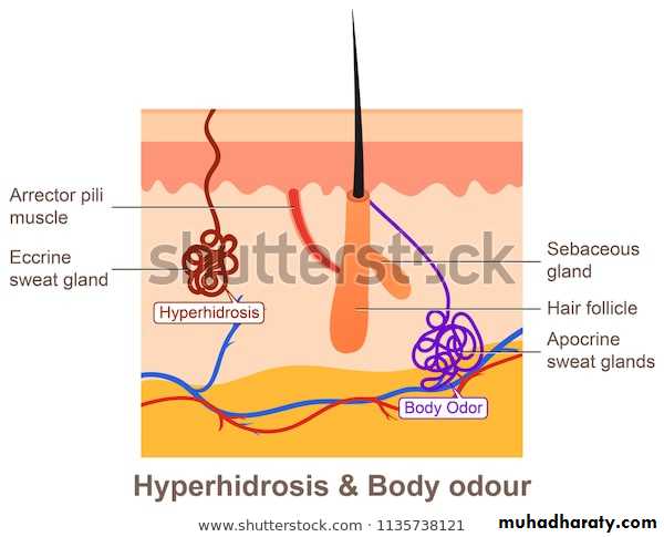
Eccrine sweat glands
2-4 millionsAll over body, mostly palms, soles & axillae
Two parts :
1- the secretory coil deep in the dermis
2- the duct: extends from the gland & opens directly onto skin surface independent of hair follicles
Sweat glands are innervated by cholinergic fibers of the sympathetic nervous system
Important in thermoregulation
Apocrine sweat glands
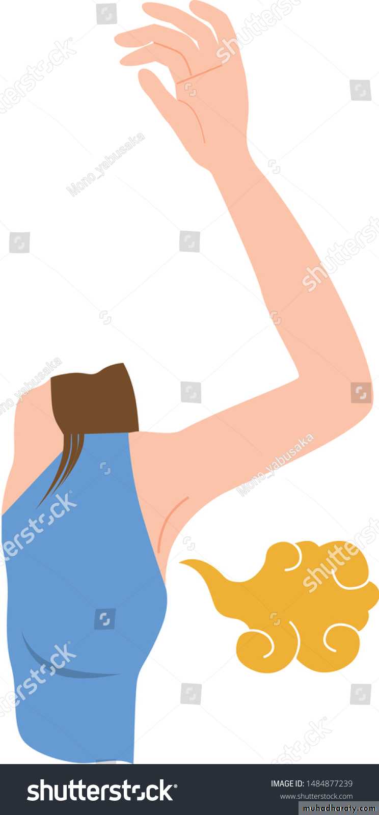
A modified sweat glands limited to the axillae,nipples, periumbilical area, perineum & genitalia
Opens directly into hair follicle
Secretion is by decapitation
Responsible for the odor of the body
Under action of androgen hormone
The nail
1) Nail plate2) Nail matrix
3) Nail bed
4) Nail folds
5)The cuticle
Blood supply of skin
The dermis is the source of nutrition of
the skin, the blood vessels lie in 2horizontal layers:
1- the deep plexus: just above the subcutaneous fat
2- a superficial plexus: in the papillary dermis
with interconnecting channels between the two.
summary
Now you should be familiar with:The components of the integumentary system, including their physical relationships.
The functions of the integumentary system.
The main features and functions of the epidermis and dermis.
The structure and function of the various accessory organs of the skin.
Thank you