1
بسم هللا الرحمن الرحيم
Lecture -1- Medical Physiology (GIT system)
2
nd
stage Dr. Noor Jawad(2020)
Gastrointestinal tract (Digestive system)
GENERAL PRINCIPLES OF GASTROINTESTINAL
Objectives of our lecture:
1. What is the physiological anatomy of the gastrointestinal wall?
2. What is the enteric nervous system?
3. Functional types of movements in GIT?
The digestive system is a tube running from mouth to anus.It
provides the body with a continual supply of water, electrolytes,
vitamins, and nutrients, which requires:
(1) movement of food through the alimentary tract;
(2) secretion of digestive juices and digestion of the food;
(3) absorption of water, various electrolytes, vitamins, and
digestive products;
(4) circulation of blood through the gastrointestinal organs to carry
away the absorbed substances; and
(5) control of all these functions by local, nervous, and hormonal
systems.
2
PHYSIOLOGICAL ANATOMY OF THE
GASTROINTESTINAL WALL
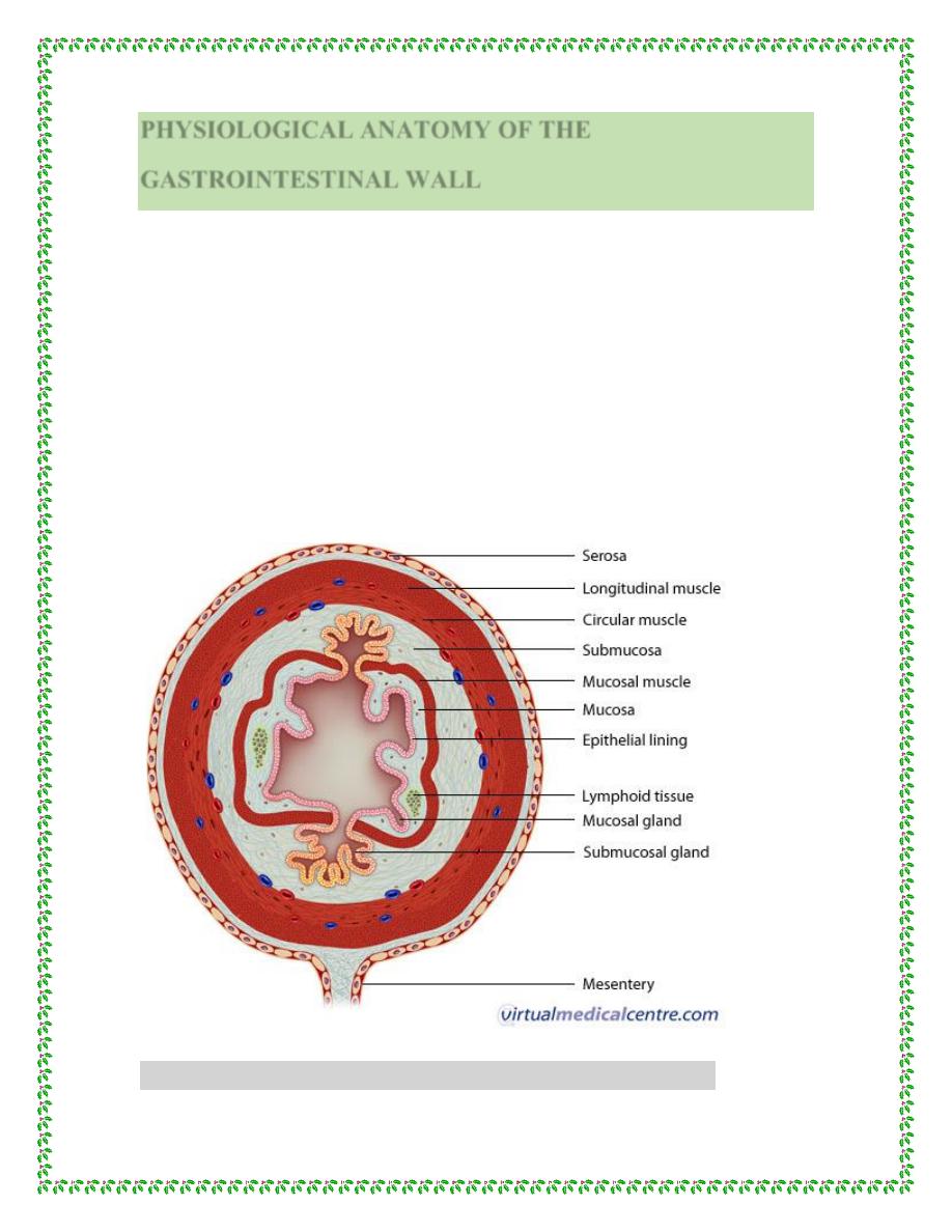
Figure below shows a typical cross section of the intestinal wall,
including the following layers from the outer surface inward: (1)
the serosa, (2) a longitudinal smooth muscle layer, (3) a circular
smooth muscle layer, (4) the submucosa, and (5) the mucosa. In
addition, sparse bundles of smooth muscle fibers, the mucosal
muscle, lie in the deeper layers of the mucosa. The motor functions
of the gut are performed by the different layers of smooth muscle.
oss section of the intestinal wall
Figure shows a typical cr

3
NEURAL CONTROL OF GASTROINTESTINAL
FUNCTION—ENTERIC NERVOUS SYSTEM
The gastrointestinal tract has a nervous system all its own called
the enteric nervous system. It lies entirely in the wall of the gut,
beginning in the esophagus and extending all the way to the anus.
The number of neurons in this enteric system is about 100 million,
nearly equal to the number in the entire spinal cord. This highly
developed enteric nervous system is especially important in
controlling gastrointestinal movements and secretion.
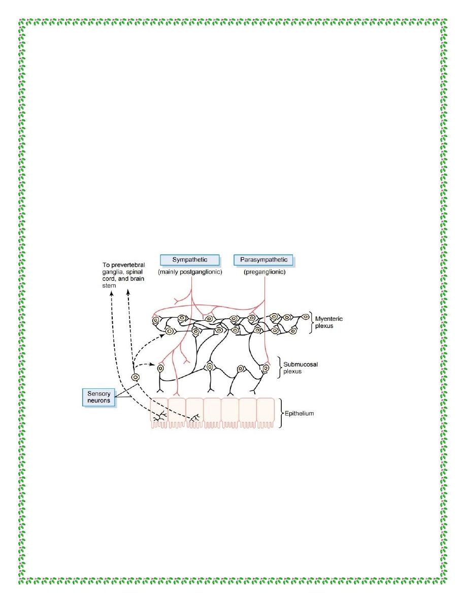
The enteric nervous system is composed mainly of two plexuses,
shown in Figure below: (1) an outer plexus lying between the
longitudinal and circular muscle layers, called the myenteric
plexus or Auerbach’s plexus, and (2) an inner plexus, called the
submucosal plexus or Meissner’s plexus, which lies in the
submucosa.
The myenteric plexus controls mainly the gastrointestinal
movements, and the submucosal plexus controls mainly
gastrointestinal secretion and local blood flow.. Although the
enteric nervous system can function independently of these
extrinsic nerves, stimulation by the parasympathetic and
sympathetic systems can greatly enhance or inhibit gastrointestinal
functions.
4
Also shown in Figure below are sensory nerve endings that
originate in the gastrointestinal epithelium or gut wall and send
afferent fibers to both plexuses of the enteric system, as well as (1)
to the prevertebral ganglia of the sympathetic nervous system, (2)
to the spinal cord, and (3) in the vagus nerves all the way to the
brain stem. These sensory nerves can elicit local reflexes within
the gut wall itself and still other reflexes that are relayed to the gut
from either the prevertebral ganglia or the basal regions of the
brain.
(Figure show (Neural control of the gut wall), showing (1) the myenteric and
submucosal plexuses (black fibers); (2) extrinsic control of these plexuses by the
sympathetic and parasympathetic nervous systems (red fibers); and (3) sensory
fibers passing from the luminal epithelium and gut wall to the enteric plexuses, then
to the prevertebral ganglia of the spinal cord and directly to the spinal cord and
brain stem)
5
DIFFERENCES BETWEEN THE MYENTERIC AND
SUBMUCOSAL PLEXUSES
The myenteric plexus consists mostly of a linear chain of many
interconnecting neurons that extends the entire length of the
gastrointestinal tract. Because the myenteric plexus extends all the
way along the intestinal wall and lies between the longitudinal and
circular layers of intestinal smooth muscle, it is concerned mainly
with controlling muscle activity along the length of the gut.
(1)
When this plexus is stimulated, its principal effects are
increased
(2)
increased tonic contraction, or “tone,” of the gut wall;
slightly increased rate
(3)
intensity of the rhythmical contractions;
increased velocity of
(4)
of the rhythm of contraction; and
conduction of excitatory waves along the gut wall, causing more
rapid movement of the gut peristaltic waves.
The myenteric plexus should not be considered entirely excitatory
because some of its neurons are inhibitory; their fiber endings
secrete an inhibitory transmitter, possibly vasoactive intestinal
polypeptide or some other inhibitory peptide. The resulting
inhibitory signals are especially useful for inhibiting some of the
intestinal sphincter muscles that impede movement of food along
successive segments of the gastrointestinal tract, such as the
pyloric sphincter, which controls emptying of the stomach into the
6
duodenum, and the sphincter of the ileocecal valve, which controls
emptying from the small intestine into the cecum.
The submucosal plexus, in contrast to the myenteric plexus, is
mainly concerned with controlling function within the inner wall
of each minute segment of the intestine. For instance, many
sensory signals originate from the gastrointestinal epithelium and
are then integrated in the submucosal plexus to help control local
intestinal secretion, local absorption, and local contraction of the
submucosal muscle that causes various degrees of infolding of the
gastrointestinal mucosa.
TYPES OF NEUROTRANSMITTERS SECRETED BY
ENTERIC NEURONS
Researchers have identified a dozen or more different
neurotransmitter substances that are released by the nerve endings
of different types of enteric neurons, including: (1) acetylcholine,
(2) norepinephrine, (3) adenosine triphosphate, (4) serotonin, (5)
dopamine, (6) cholecystokinin, (7) substance P, (8) vasoactive
intestinal polypeptide, (9) somatostatin, (10) leu-enkephalin, (11)
met-enkephalin, and (12) bombesin.
Acetylcholine most often excites gastrointestinal activity.
Norepinephrine almost always inhibits gastrointestinal activity, as
does epinephrine, which reaches the gastrointestinal tract mainly
7
by way of the blood after it is secreted by the adrenal medullae into
the circulation.
FUNCTIONAL TYPES OF MOVEMENTS IN THE
GASTROINTESTINAL TRACT
Two types of movements occur in the gastrointestinal tract: (1)
propulsive movements, which cause food to move forward along
the tract at an appropriate rate to accommodate digestion and
absorption, and (2) mixing movements, which keep the intestinal
contents thoroughly mixed at all times.
PROPULSIVE MOVEMENTS—PERISTALSIS
A contractile ring appears around the gut and then moves forward;
this mechanism is analogous to putting one’s fingers around a thin
distended tube, then constricting the fingers and sliding them
forward along the tube. Any material in front of the contractile ring
is moved forward. Peristalsis is an inherent property of many
syncytial smooth muscle tubes; stimulation at any point in the gut
can cause a contractile ring to appear in the circular muscle, and
this ring then spreads along the gut tube. (Peristalsis also occurs in
the bile ducts, glandular ducts, ureters, and many other smooth
muscle tubes of the body.)
The usual stimulus for intestinal peristalsis is distention of the gut.
That is, if a large amount of food collects at any point in the gut,
8
the stretching of the gut wall stimulates the enteric nervous system
to contract the gut wall 2 to 3 centimeters behind this point, and a
contractile ring appears that initiates a peristaltic movement. Other
stimuli that can initiate peristalsis include chemical or physical
irritation of the epithelial lining in the gut. Also, strong
parasympathetic nervous signals to the gut will elicit strong
peristalsis.
MIXING MOVEMENTS
Mixing movements differ in different parts of the alimentary tract.
In some areas, the peristaltic contractions cause most of the
mixing. This is especially true when forward progression of the
intestinal contents is blocked by a sphincter so that a peristaltic
wave can then only churn the intestinal contents, rather than
propelling them forward. At other times, local intermittent
constrictive contractions occur every few centimeters in the gut
wall. These constrictions usually last only 5 to 30 seconds; new
constrictions then occur at other points in the gut, thus “chopping”
and “shearing” the contents first here and then there.
Thank you
References : Guyton and Hall textbook of medical physiology,
thirteen edition.