
1
Muscles of lower limb
stage
st
1
Dr.Kalid Ali Zayer
Fascia lata
Contents
1. Anatomical Structure
2. Anatomical Relations
o 2.1 Iliotibial Tract (ITT)
o 2.2 Tensor Fascia Lata (TFL)
3. Attachments
o 3.1 Proximal
o 3.2 Lateral
o 3.3 Inferior
o 3.4 Central
Fascia
is a sheet or band of fibrous tissue lying deep to the skin. It lines, invests,
and separates structures within the body. There are three main types of fascia:
Superficial fascia
– blends with the reticular layer beneath the dermis.
Deep fascia
– envelopes muscles, bones, and neurovascular structures.
Visceral fascia
– provides membranous investments that suspend organs within
their cavities.
Anatomical Structure
The fascia lata is a deep fascial investment of the musculature of the thigh, and
is analogous to a strong, extensible, and elasticated stocking. It begins proximally
around the iliac crest and inguinal ligament, and ends distal to the bony
prominences of the tibia. It is continuous with what is renamed the deep fascia of
the leg (also known as the crural fascia).
The depth of the fascia lata varies considerably across the thigh. It is thickest
along the superolateral aspect of the thigh, where it arises from the fascial
condensations of gluteus maximus and medius. It is also thick around the knee
where the fascia receives reinforcing fibers from tendons of the quadriceps
muscles. The fascial investment is thinnest where it covers the adductor muscles
of the medial thigh.
2
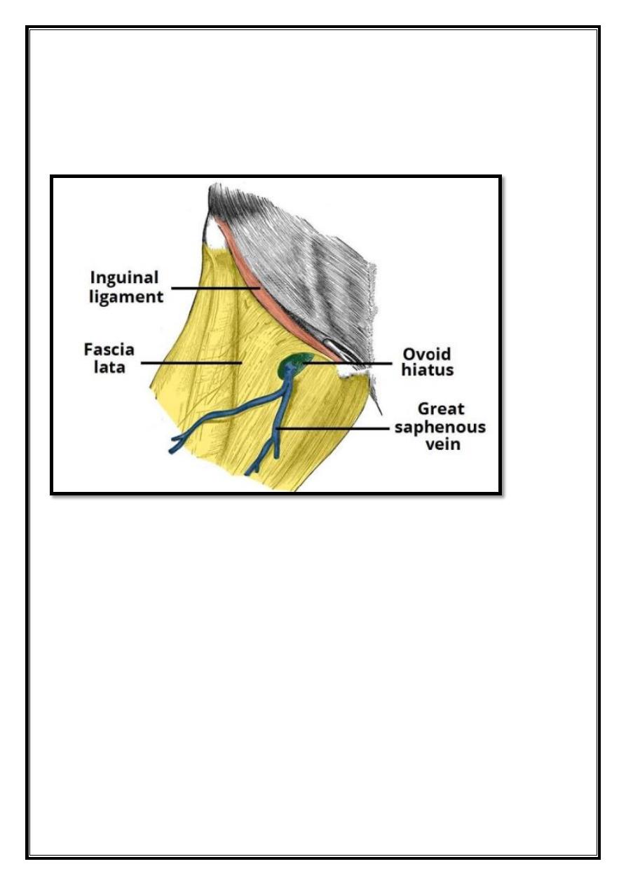
An ovoid hiatus known as the saphenous opening is present in the fascia lata
just inferior to the inguinal ligament. The opening serves as an entry point for
efferent lymphatic vessels and the great saphenous vein, draining into
superficialinguinal lymph nodes and the femoral vein respectively.
Fig 9 – The ovoid hiatus of the fascia lata.
Anatomical Relations
Iliotibial Tract (ITT)
The iliotibial tract (sometimes known as the iliotibial band or IT band) is a
longitudinal thickening of the fascia lata, which is strengthened superoposteriorly
by fibers from the gluteus maximus. It is located laterally in the thigh, extending
from the iliac tubercle to the lateral tibial condyle. The ITT has three main
functions:
Movement
– acts as an extensor, abductor and lateral rotator of the hip, with an
additional role in providing lateral stabilization to the knee joint.
Compartmentalization
– the deepest aspect of ITT extends centrally to form the
lateral intermuscular septum of the thigh and attaches to the femur.
Muscular sheath
– forms a sheath around the tensor fascia lata muscle.
3
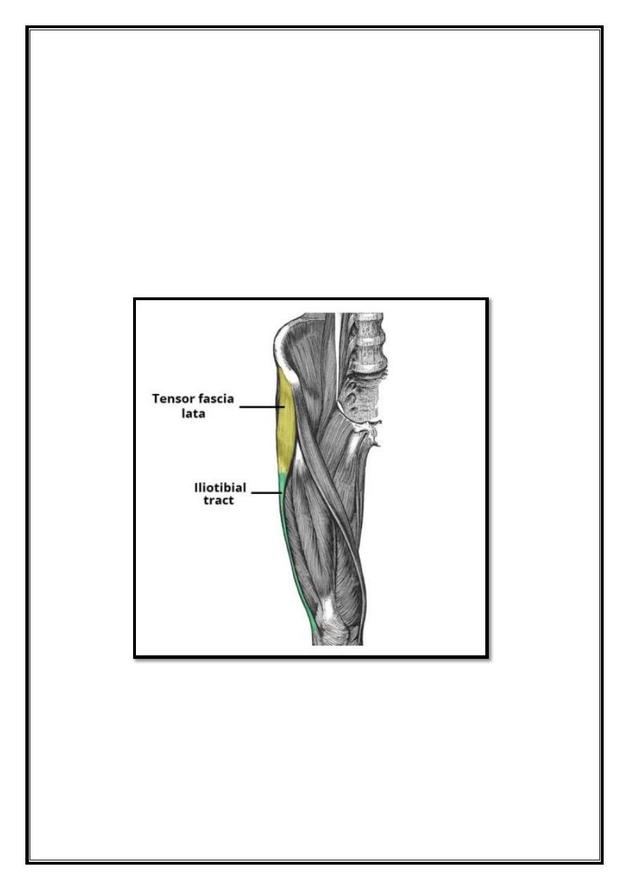
Tensor Fascia Lata (TFL)
The tensor fascia lata is a gluteal muscle that acts as a flexor, abductor, and
internal rotator of the hip. Its name, however, is derived from its additional role in
tensing the fascia lata. It is innervated by the superior gluteal nerve, like gluteus
medius and minimus, but is located more anterolaterally than the other gluteal
muscles. The muscle originates from the iliac crest, and descends inferiorly to the
superolateral thigh. At the junction of the middle and upper thirds of the thigh, it
inserts into the anterior aspect of the iliotibial tract. When stimulated, the tensor
fasciae lata tautens the iliotibial band and braces the knee, especially when the
opposite foot is lifted.
Fig 10 – The tensor fascia lata and iliotibial tract.

4
Gluteal region
Contents
1. The Superficial Muscles
2. The Deep Muscles
The gluteal region is an anatomical area located posteriorly to the pelvic girdle,
at the proximal end of the femur. The muscles in this region move the lower limb
at the hip joint. The muscles of the gluteal region can be broadly divided into two
groups:
Superficial abductors and extenders
– group of large muscles that abduct and
extend the femur. Includes the gluteus maximus, gluteus medius, gluteus
minimus and tensor fascia lata.
Deep lateral rotators
– group of smaller muscles that mainly act to laterally
rotate the femur. Includes the quadratus femoris, piriformis, gemellus superior,
gemellus inferior and obturator internus.
The arterial supply to these muscles is mostly via the superior and inferior gluteal
arteries – branches of the internal iliac artery. Venous drainage follows the
arterial supply.
The Superficial Muscles
The superficial muscles in the gluteal region consist of the three glutei and the
tensor fascia lata. They mainly act to abduct and extend the lower limb at the hip
joint.
5
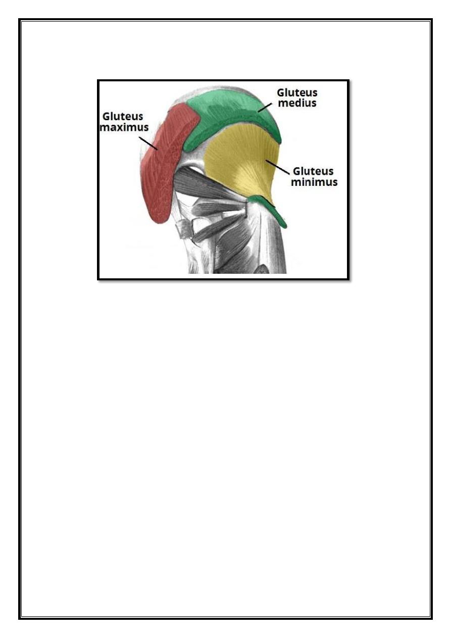
Gluteus Maximus
Fig 11 – The superficial muscles of the gluteal region. The gluteus
maximus and medius have been partly removed.
The gluteus maximus is the largest of the gluteal muscles. It is also the most
superficial,producing the shape of the buttocks.
Attachments
: Originates from the gluteal (posterior) surface of the ilium, sacrum
and coccyx. It slopes across the butock at a 45 degree angle, then inserts into the
iliotbial tract and the gluteal tuberosity of the femur.
Actions
: It is the main extensor of the thigh, and assists with lateral rotation.
However, it is only used when force is required, such as running or climbing.
Innervation
: Inferior gluteal nerve.
Gluteus Medius
The gluteus medius muscle is fan-shaped and lies between the gluteus maximus
and the minimus. It is similar in shape and function to the gluteus minimus.
Attachments
: Originates from the gluteal surface of the ilium and inserts into the
lateral surface of the greater trochanter.
Actions
: Abducts and medially rotates the lower limb. During locomotion, it
secures the pelvis, preventing pelvic drop of the opposite limb.

6
(Note: the posterior fibers of the gluteus medius are also thought to produce a
small amount of lateral rotation).
Innervation
: Superior gluteal nerve.
Gluteus Minimus
The gluteus minimus is the deepest and smallest of the superficial gluteal muscles.
It is similar is shape and function to the gluteus medius.
Attachments
: Originates from the ilium and converges to form a tendon,
inserting to the anterior side of the greater trochanter.
Actions
: Abducts and medially rotates the lower limb. During locomotion, it
secures the pelvis, preventing pelvic drop of the opposite limb.
Innervation
: Superior gluteal nerve.
Tensor Fascia Lata
Tensor fasciae lata is a small superficial muscle which lies towards the anterior
edge of the iliac crest. It functions to tighten the fascia lata, and so abducts and
medially rotates the lower limb.
Attachments
: Originates from the anterior iliac crest, attaching to the anterior
superior iliac spine (ASIS). It inserts into the iliotibial tract, which itself attaches
to the lateral condyle of the tibia.
Actions
: Assists the gluteus medius and minimus in abduction and medial
rotation of the lower limb. It also plays a supportive role in the gait cycle.
Innervation
: Superior gluteal nerve
The Deep Muscles
The deep gluteal muscles are a set of smaller muscles, located underneath the
gluteus minimus. The general action of these muscles is to laterally rotate the
lower limb. They also stabilize the hip joint by ‘pulling’ the femoral head into
the acetabulum of the pelvis.
7
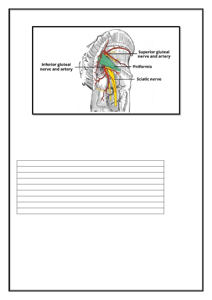
Piriformis
The piriformis muscle is a key landmark in the gluteal region. It is the most
superior of the deep muscles.
Attachments:
Originates from the anterior surface of the sacrum. It then
travels inferolaterally, through the greater sciatic foramen, to insert into the
greater trochanter of the femur.
Actions
: Lateral rotation and abduction.
Innervation
: Nerve to piriformis.
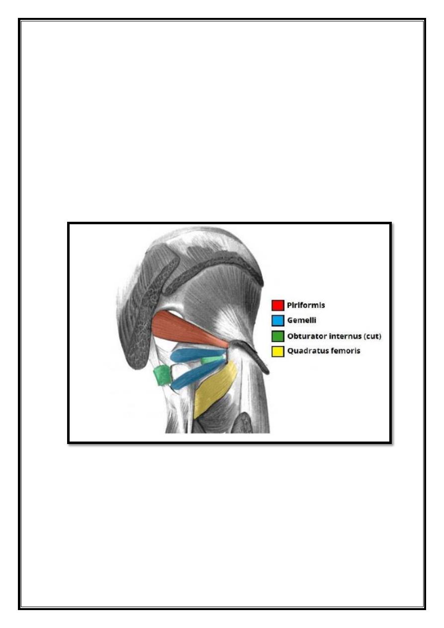
Obturator Internus
Fig 12 – The deep muscles of the gluteal region.
The obturator internus forms the lateral walls of the pelvic cavity. In some texts,
the obturator internus and the gemelli muscles are considered as one muscle – the
triceps coxae.
Attachments
: Originates from the pubis and ischium at the obturator foramen.
It travels through the lesser sciatic foramen, and attaches to the greater trochanter
of the femur.

8
Actions
: Lateral rotation and abduction.
Innervation
: Nerve to obturator internus.
The Gemelli – Superior and Inferior
The gemelli are two narrow and triangular muscles. They are separated by the
obturator internus tendon.
Attachments
: The superior gemellus muscle originates from the ischial spine,
the inferior from the ischial tuberosity. They both attach to the greater trochanter
of the femur.
Actions
: Lateral rotation and abduction.
Innervation
: The superior gemellus muscle is innervated by the nerve to
obturator internus, the inferior gemellus is innervated by the nerve to quadratus
femoris.
Quadratus Femoris
The quadratus femoris is a flat, square-shaped muscle. It is the most inferior of the
deep gluteal muscles, located below the gemelli and obturator internus.
Attachments
: It originates from the lateral side of the ischial tuberosity, and
attaches to the quadrate tuberosity on the intertrochanteric crest.
Actions
: Lateral rotation.
Innervation
: Nerve to quadratus femoris.
9
Fig 13 – The piriformis as an anatomical landmark in the gluteal
region.
Thigh
Contents
1. Iliopsoas
2. Quadriceps Femoris
o 2.1 Vastus Lateralis
o 2.2 Vastus Intermedius
o 2.3 Vastus Medialis
o 2.4 Rectus Femoris
3. Sartorius
4. Pectineus
The musculature of the thigh can be split into three sections; anterior, medial and
posterior. Each compartment has a distinct innervation and function. The muscles
in the anterior compartment of the thigh are innervated by the femoral nerve (L2-
L4), and as a general rule, act to extend the leg at the knee joint. There are three
major muscles in the anterior thigh – the pectineus, sartorius and quadriceps
femoris. In addition to these, the end of the iliopsoas muscle passes into the
anterior compartment.
10
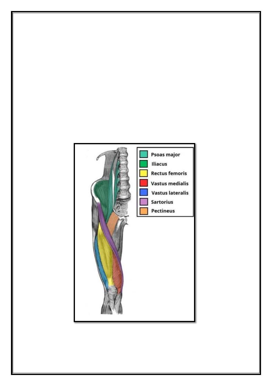
Iliopsoas
The iliopsoas is actually two muscles, the psoas major and the iliacus. They
originate in different areas, but come together to form a tendon, hence why they
are commonly referred to as one muscle.
Unlike many of the anterior thigh muscles, the iliopsoas does not extend the leg at
the knee joint.
Attachments
: The psoas major originates from the lumbar vertebrae, and the
iliacus originates from the iliac fossa of the pelvis. They insert together onto the
lesser trochanter of the femur.
Actions
: Flexes the thigh at the hip joint.
Innervation
: The psoas major is innervated by anterior rami of L1-3, while the
iliacus is innervated by the femoral nerve.
Fig 14 – The muscles of the anterior thigh.

11
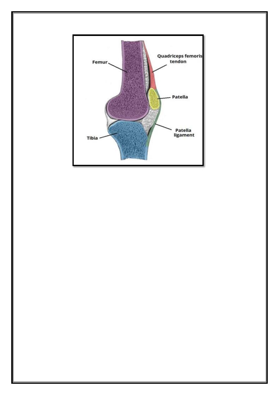
Quadriceps Femoris
The quadriceps femoris consists of four individual muscles; three vastus muscles
and the rectus femoris. They form the main bulk of the thigh, and collectively are
one of the most powerful muscles in the body. The muscles that form the
quadriceps femoris unite proximal to the knee and attach to the patella via the
quadriceps tendon. In turn, the patella is attached to the tibia by the patella
ligament. The quadriceps femoris is the main extensor of the knee.
Vastus Lateralis
Proximal attachment:
Originates from the greater trochanter and the lateral lip
of linea aspera.
Actions:
Extends the knee joint and stabilizes the patella.
Innervation:
Femoral nerve.
Vastus Intermedius
Proximal attachment:
Anterior and lateral surfaces of the femoral shaft.
Actions:
Extends the knee joint and stabilizes the patella.
Innervation:
Femoral nerve.
Vastus Medialis
Proximal attachment
: The intertrochanteric line and medial lip of the linea
aspera.
Actions
: Extends the knee joint and stabilizes the patella, particularly due to its
horizontal fibers at the distal end.
Innervation
: Femoral nerve.
Rectus Femoris
Attachments
: Originates from the ilium, just superior to the acetabulum. It
runs straight down the leg (the Latin for straight is rectus), and attaches to the
patella by the quadriceps femoris tendon.
Actions
: The only muscle of the quadriceps to cross both the hip and knee
joints. It flexes the thigh at the hip joint, and extends at the knee joint.
Innervation
: Femoral nerve
12
Fig 15 – The femur, tibia and patella of the knee joint.
Sartorius
The sartorius is the longest muscle in the body. It is long and thin, running across
the thigh in a inferomedial direction. The sartorius is positioned more superficially
than the other muscles in the leg.
Attachments
: Originates from the anterior superior iliac spine, and attaches to
the superior, medial surface of the tibia.
Actions
: At the hip joint, it is a flexor, abductor and lateral rotator. At the knee
joint, it is also a flexor.
Innervation
: Femoral nerve.

13
Fig 16 – Cross section of the distal thigh. The iliopsoas and pectineus
muscles originate and attach in the proximal thigh, and hence are not
included in this diagram.
Pectineus
The pectineus muscle is a flat muscle that forms the base of the femoral triangle.
It has a dual innervation, and thus can be considered a transitional muscle
between the anterior thigh and medial thigh compartments.
Attachments
: It originates from the pectineal line on the anterior surface of the
pelvis, and attaches to the pectineal line on the posterior side of the femur, just
inferior to the lesser trochanter.
Actions
: Adduction and flexion at the hip joint.
Innervation
: Femoral nerve. May also receive a branch from the obturator
nerve.
14
Medial compartment of the thigh
Contents
1. Muscles of the Medial Thigh
o 1.1 Adductor Magnus
o 1.2 Adductor Longus
o 1.3 Adductor Brevis
o 1.4 Obturator Externus
o 1.5 Gracilis
The muscles in the medial compartment of the thigh are collectively known as the
hip adductors. There are five muscles in this group; gracilis, obturator externus,
adductor brevis, adductor longus and adductor magnus. All the medial thigh
muscles are innervated by the obturator nerve, which arises from the lumbar
plexus. Arterial supply is via the obturator artery.
Muscles of the Medial Thigh
Adductor Magnus
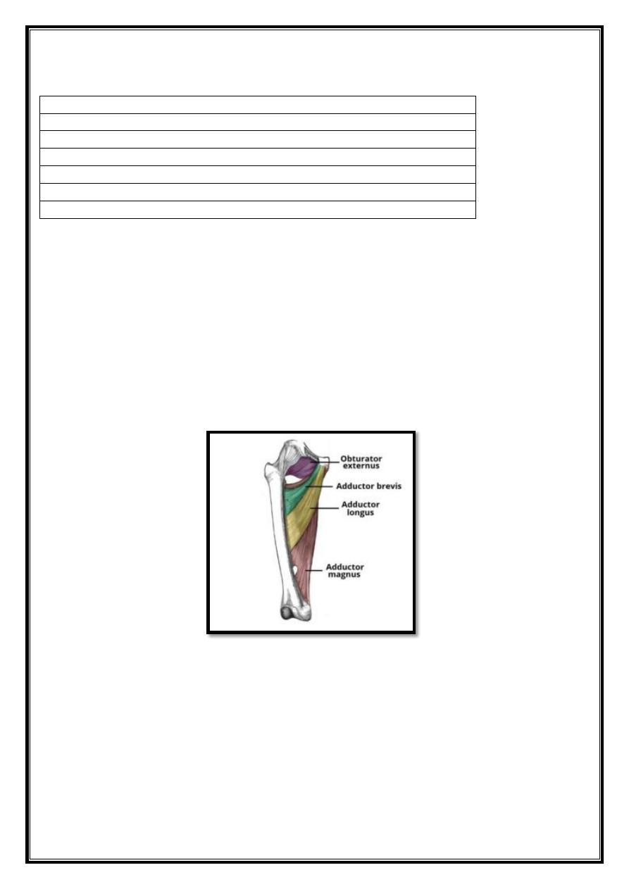
Fig 17 – Muscles of the medial thigh. The overlying muscles in the
anterior compartment have been removed.
15
The adductor magnus is the largest muscle in the medial compartment. It lies
posteriorly to the other muscles. Functionally, the muscle can be divided into two
parts; the adductor part,and the hamstring part.
Attachments
- Adductor part – Originates from the inferior rami of the pubis and the rami of
ischium, attaching to the linea aspera of the femur.
- Hamstring part – Originates from the ischial tuberosity and attaches to the
adductor tubercle and medial supracondylar line of the femur.
Actions
: They both adduct the thigh. The adductor component also flexes the
thigh, with the hamstring portion extending the thigh.
Innervation
: Adductor part is innervated by the obturator nerve (L2-L4), the
hamstring part is innervated by the tbial component of the sciatc nerve (L4-S3).
Adductor Longus
The adductor longus is a large, flat muscle. It partially covers the adductor brevis
and magnus.
The muscle forms the medial border of the femoral triangle.
Attachments
: Originates from the pubis, and expands into a fan shape, attaching
broadly to the linea aspera of the femur
Actions
: Adduction of the thigh.
Innervation:
Obturator nerve (L2-L4)
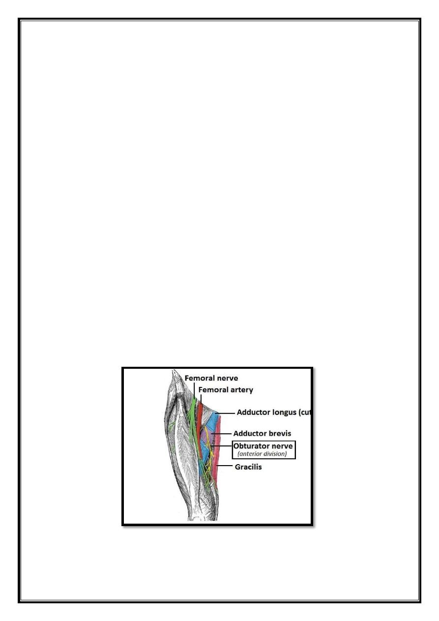
Adductor Brevis
Fig 18– View of the medial thigh, with the course of the obturator
nerve highlighted

16
The adductor brevis is a short muscle, lying underneath the adductor longus. It lies
in between the anterior and posterior divisions of the obturator nerve. Therefore, it
can be used as an anatomical landmark to identify the aforementioned branches.
Attachments
: Originates from the body of pubis and inferior pubic rami. It
attaches to the linea aspera on the posterior surface of the femur, proximal to the
adductor longus.
Actions
: Adduction of the thigh.
Innervation
: Obturator nerve (L2-L4).
Obturator Externus
This is one of the smaller muscles of the medial thigh, and it is located most
superiorly.
Attachments
: It originates from the membrane of the obturator foramen, and
adjacent bone. It passes under the neck of femur, attaching to the posterior aspect
of the greater trochanter.
Actions
: Adduction and lateral rotation of the thigh.
Innervation
: Obturator nerve (L2- L4).
Gracilis
The gracilis is the most superficial and medial of the muscles in this
compartment. It crosses at both the hip and knee joints. It is sometimes
transplanted into the hand or forearm to replace a damaged muscle.
Attachments
: It originates from the inferior rami of the pubis, and the body of
the pubis.Descending almost vertically down the leg, it attaches to the medial
surface of the tibia,between the tendons of the sartorius (anteriorly) and the
semitendinosus (posteriorly).
Actions
: Adduction of the thigh at the hip, and flexion of the leg at the knee.
Innervation
: Obturator nerve (L2-L4).
17
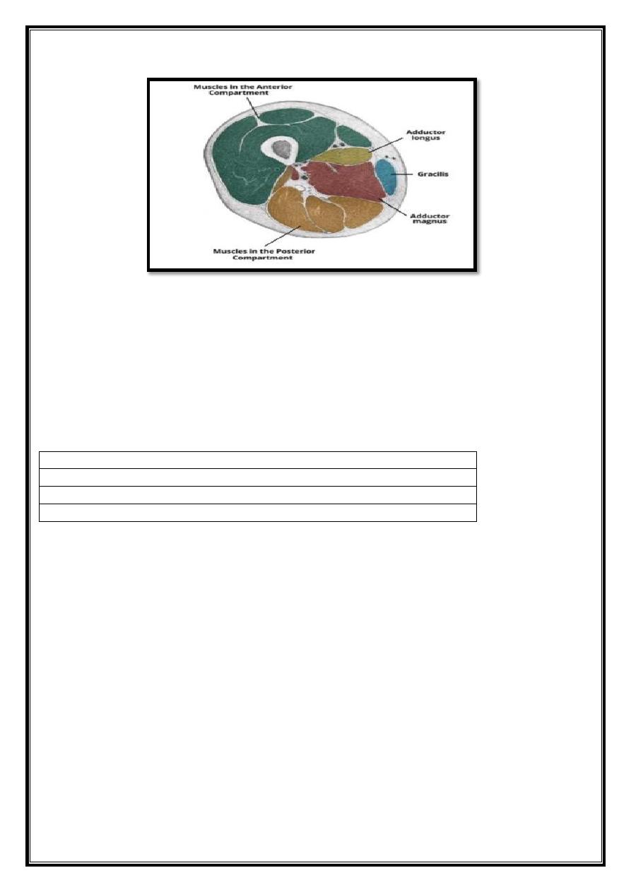
Fig 19 – Cross section of the inferior thigh, showing some of the
medial thigh muscles. The adductor brevis and obturator externus
attach superiorly in the thigh, and so are not visible in this cross-
section.
Posterior compartment of the thigh
Contents
1. Muscles in the Posterior Compartment
o 1.1 Biceps Femoris
o 1.2 Semitendinosus
o 1.3 Semimembranosus
The muscles in the posterior compartment of the thigh are collectively known as
the hamstrings. They consist of the biceps femoris, semitendinosus and
semimembranosus,
which form prominent tendons medially and laterally at the back of the knee. As
group, these muscles act to extend at the hip, and flex at the knee. They are
innervated by the sciatic nerve (L4-S3).
Muscles in the Posterior Compartment
The muscles located within the posterior compartment of the thigh are the biceps
femoris, semitendinosus and semimembranosus.
Note: The hamstring portion of the adductor magnus has a similar action to these
muscles, but is located in the medial thigh.
18
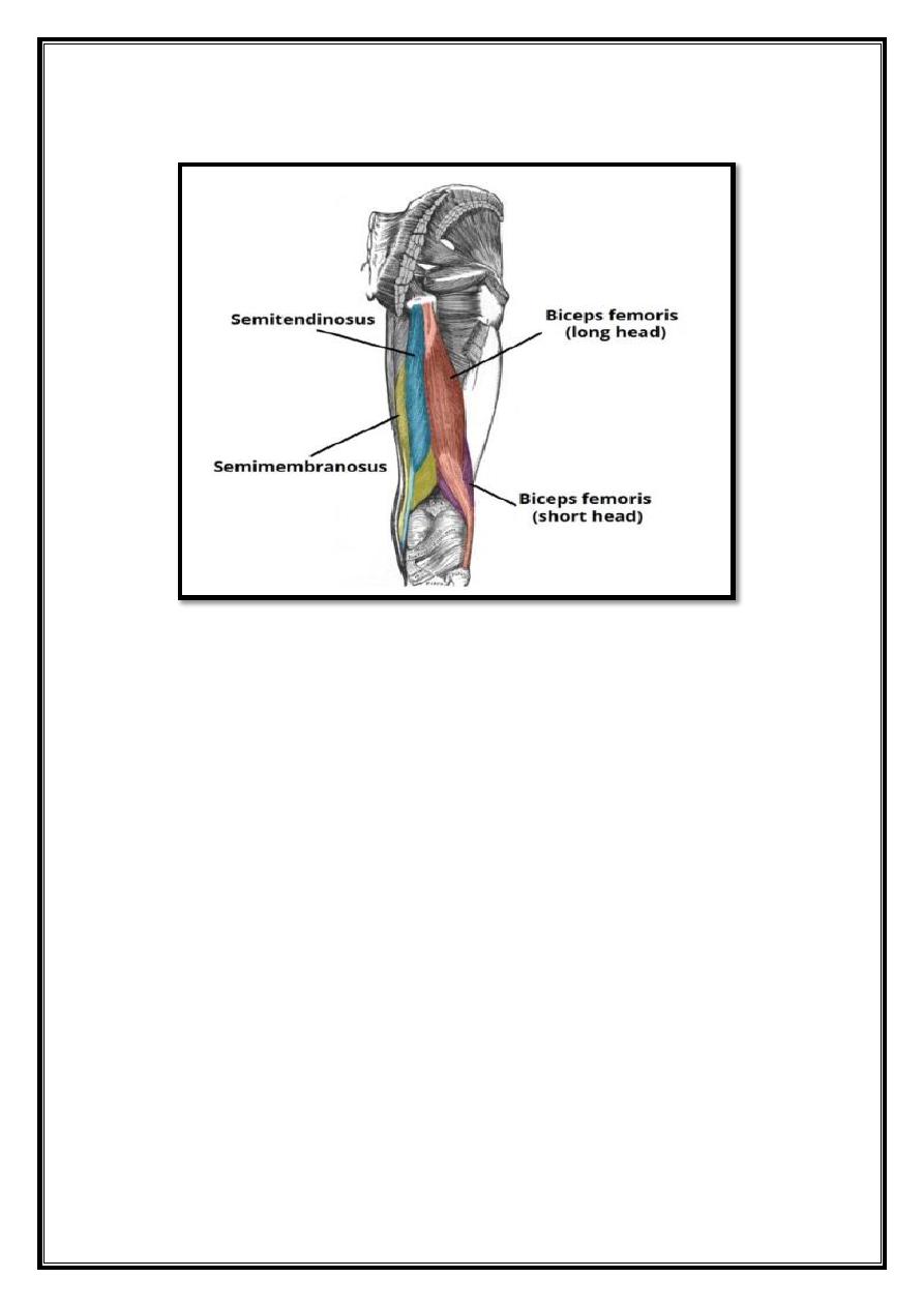
Biceps Femoris
Fig 20 – The muscles of the posterior thigh (right).
Like the biceps brachii in the arm, the biceps femoris muscle has two heads – a
long head and a short head. It is the most lateral of the muscles in the posterior
thigh – the common tendon of the two heads can be felt laterally at the posterior
knee.
Attachments
: The long head originates from the ischial tuberosity of the pelvis.
The short head originates from the linea aspera on posterior surface of the femur.
Together, the heads form a tendon, which inserts into the head of the fibula.
Actions
: Main action is flexion at the knee. It also extends the thigh at the hip,
and laterally rotates at the hip and knee.
Innervation
: Long head innervated by the tibial part of the sciatic nerve, whereas
the short head is innervated by the common fibular part of the sciatic nerve.
19
Semitendinosus
The semitendinosus is a largely tendinous muscle. It lies medially to the biceps
femoris, and covers the majority of the semimembranosus.
Attachments
: It originates from the ischial tuberosity of the pelvis, and attaches
to the medial surface of the tibia.
Actions
: Flexion of the leg at the knee joint. Extension of thigh at the hip.
Medially rotates the thigh at the hip joint and the leg at the knee joint.
Innervation
: Tibial part of the sciatic nerve.
Semimembranosus
The semimembranosus muscle is flattened and broad. It is located underneath the
semitendinosus.
Attachments
: It originates from the ischial tuberosity, but does so more
superiorly than the semitendinosus and biceps femoris. It attaches to the medial
tibial condyle.
Actions
: Flexion of the leg at the knee joint. Extension of thigh at the hip.
Medially rotates the thigh at the hip joint and the leg at the knee joint.
Innervation
: Tibial part of the sciatic nerve.
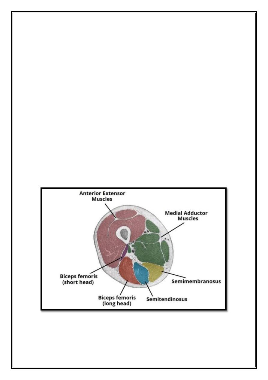
Fig 21 – Cross section of the left thigh, showing the position of the
major muscle groups.