


1
The cornea
Gross Anatomy:
The cornea is the anterior continuation of the sclera. It is a transparent
structure, its function is to focus the light rays on the retina, it represents the most
important refractive power with (43) diopter.
Applied anatomy:
The cornea consists of the five following layers: (from anterior to posterior)
1- The epithelium: is stratified (multiple cell layers), squamous and non-
keratinized when if damaged, it regenerates without any scarring.
2- Bowman's layer:
Is the Acellular superficial layer of the stroma, which scars when damaged?
3- The stroma: makes up 90% of the corneal thickness. It is composed of
regularly oriented layers of collagen fibrils whose spacing is maintained by
glycosaminoglycan ground substance.
4- Descemet's membrane: it is composed of a fine latticework of collagen
fibrils. It is the basement membrane of the endothelial cells.
5- The endothelium:
It consists of a single layer of hexagonal cells. It plays a vital role in maintaining
corneal transparency & deturgescence , as water composes 70% of cornea that
makes it relatively dehydrated as in other body tissue the water composes 98%
of them, so active pump mechanism that removes the fluid that leaks to stroma.
With age, the number of endothelial cells decreases and they are non-
regenerative cells, therefore the neighboring cells enlarge to fill the space.
The cornea is transparent for the following reasons:
1- The epithelium is not keratinized.
2- The stroma is regularly oriented.
3- The endothelium has active pump, it pushes the fluid into aqueous and it acts
as barrier to prevent entrance of aqueous inside the cornea.
4- The corneal nerves are unmyelinated.
5- It contains NO blood vessels.
6- It contains NO pigments (as melanin).
Signs of corneal diseases:

2
1- Epithelial signs:
a- Punctate epithelial erosions (PEE):
Are tiny, slightly depressed, epithelial defects (micro-ulcers seen by slit-
lamp), which stain with fluorescein.
e.g.(causes): vernal keratoconjunctivitis, poorly fitting contact lens, dry eyes,
decreases corneal sensation (as in trigeminal nerve palsy or after herpes
simplex viral keratitis), exposure to ultraviolet (during welding, *do not treat
pain by xylocaine as it is toxic to the eye), corneal exposure, toxicity from drops
(as those of aminoglycosides).
b- Punctate epithelial keratitis (PEK):
Is the hallmark of viral infections, it is characterized by granular, opalescent,
swollen epithelial cells .
c- Epithelial oedema:
It is a sign of endothelial decompensation or severe and acute or sudden
elevation of intraocular pressure (as that occurring in glaucoma as IOP is raised
leading to oedema that affects the vision ).
d- Filaments:
Small, comma-shaped mucus strands lined with epithelium (one end attached
to the epithelial cornea and the other is free).
Causes:
Keratoconjunctivitis sicca (dry eye), recurrent erosion syndrome, eye
patching, corneal exposure, diminished corneal sensation and herpes zoster
ophtrhalmicus.
e- Pannus:
Is an inflammatory or degenerative sub-epithelial in growth of fibrovascular
tissue from limbus.
2- Stromal signs:
a- Stromal infiltration:
Focal areas of active stromal inflammation composed of accumulations
leucocytes and cellular debris. These focal areas are granular, gray-white
opacities within the stroma.
Causes:
i- Non-infectious (Antigen sensitivity): e.g. contact lens wear and marginal
keratitis.
ii- Infectious keratitis: e.g. bacteria, viruses, fungi and protozoa.
b- Stromal oedema:
Causing disturbance of regularly arranged collagen fibers (or fibrils) that
affects eye vision by disturbance of corneal transparency.
Causes:

3
Keratoconus (bulging of the cornea making the cornea have conical shape,
causing stretching Descemet’s membrane leading to rupture it and influx of the
aqueous inside the stroma) and surgical damage to the corneal endothelium.
c- Vascularization:
Causes: Wide variety of corneal disorders, e.g. microbial keratitis, chemical
burns, trauma, TB, syphilis and autoimmune keratoconjunctivitis (cicatricial
pemphigoid and stevens-johnson syndrome).
3- Descemet's membrane signs:
a- Breaks:
Causes: Corneal enlargement, birth trauma, keratoconus and glaucoma (in
children). It leads to influx of aqueous causing stromal edema.
b- Folds (Striate keratopathy):
Causes: Surgical trauma, ocular hypotony, stromal inflammation and edema.
* Normal IOP is 10-21mmHg, if it is less than 6 then it is hypotony, while if it is
more than 21 it is ocular hypertension.
Microbial keratitis
1- Bacterial Keratitis:
Predisposing factors:
- Bacteria capable of penetrating intact epithelium include Neisseria gonorrhea
and H. influenza.
- Other bacteria are capable of producing keratitis only after compromisation
of epithelial integrity with the following factors:
a- Contact lens wear: is the most common predisposing factor in patients
with previously normal eye therefore meticulous lens hygiene therefore vital.
b- Pre-existing corneal disease: such as trauma (usually after surgery of
cataract), exposure keratopathy and diminished corneal sensation.
c- Other factors: chronic blepharoconjunctivitis, chronic dacryocystitis, dry
eyes, topical steroid therapy and hypavitaminosis A.
Clinical Features:
Symptoms:
Foreign body sensation, photophobia, blurring of vision, pain, eyelid oedema
and discharge.
Signs:
- Conjunctival and circumcorneal injection (almost always with severe anterior
uveitis ).
- Epithelial defects associated with an infiltrate around the margin and base.
- Enlargement of the infiltrate associated with stromal oedema.
- Secondary sterile anterior uveitis with hypopyon.

4
- Progressive ulceration may lead to corneal perforation and bacterial
endophthalmitis (involvement of all intraocular tissues).
Differential diagnosis of bacterial keratitis:
a- Fungal keratitis.
b- Acanthamoeba keratitis.
c- Stromal necrotic herpes simplex keratitis..
d- Sterile inflammatory corneal infiltrates associated with contact lens wear.
Treatment:
a- Topical antibiotics:
- Initial instillation of fortified antibiotic is at hourly intervals.
- If response is favorable, frequency is decreased to 2-hourly during waking
hours.
- Then fortified drops can be replaced by weaker commercial preparations,
which are then tapered and eventually discontinued.
b- Oral ciprofloxacin (750mg twice daily):
- Copious secreted in the tears.
- Lipid soluble and has excellent intraocular penetration.
c- Atropine:
- To prevent the formation of posterior synechiae (adhesions between papillary
margin and lens).
- Reduce pain from ciliary spasm and uveitis.
d- Steroid therapy:
It is controversial, the potential benefits of topical steroids in reducing stromal
necrosis and scaring should be weighed against decreased fibroblast activity
and increased risk of perforation.
We can use it only when cultures become sterile and there is clear evidence of
improvement (7-10 days after initial treatment).
2- Fungal keratitis:
Rare infection but have devastating effects, the most common pathogens are:
- Filamentous fungi (Aspergillus and Fusarium species): Infection occurs after
trauma by wood usually.
- Candida albicans: Usually infects immuno-compromised patients.
Clinical features:
Symptoms:
- Gradual onset of foreign body sensation.
- Photophobia.
- Blurred vision (due to opacification of cornea, whether due to epithelial or
stromal oedema) and discharge (mucopurulent).
* Progression is much slower and less painful than in bacterial keratitis.
Signs:

5
- A grayish, stromal infiltration with indistinct margin.
- Surrounding, satellite, feathery, finger-like lesions (extensions).
- Hypopyon (pus in the anterior chamber).
* There is always some sort of iritis associating keratitis.
Treatment:
a- Topical treatment:
Filamentous: Natamycin 5%, and may add Amphotericin 0.15%.
Candida: Imidazole 1% or Flucytosine 1%.
b- Systemic antimycotics: E.g. Ketoconazole (tablets) or Itraconazole in
severe keratitis or endophthalmitis.
c- Therapeutic penetrating keratopathy: In unresponsive cases (if there is
resistant infection).
4- Viral keratitis:
a- Herpes simplex keratitis:
Basic concepts:
- HSV is a DNA virus, which infects only human.
- Infection with HSV is common; up to 90% of the population is seropositive
for HSV-1 antibodies although most infections are sub-clinical.
- HSV-1 predominantly causes infection above the waist (face, lips and eyes).
- HSV-2 typically causes venereally acquired infection below the waist
(genital herpes).
- Rarely HSV-2 may be transmitted to the eye through infected genital
secretions, either venereal or at birth.
i- Primary infection:
Usually in early childhood through droplet (the most common route) or direct
inoculation. It may be sub-clinical or may cause mild fever, malaise and URT
infection. In immuno-compromised subject, the infection may become
generalized and life threatening.
ii- Recurrent disease:
- Following primary infection, the virus travels up to the ganglion (trigeminal
"Gasserian" for HSV-1 and spinal for HSV-2), where it lies in a latent state.
- This latent state may subsequently reverse and the virus reactivates
replicates & travels down to its target tissue causing recurrent disease (genital
herpes, herpes labials & herpes keratitis).
Primary ocular infection:
- Typically occurs in children between ages of 6 months- 5 years, and may be
associated with generalized symptoms.
Signs:
}
for 6 weeks
6
- Skin vesicles typically involve the lids and periorbital area.
- Acute, unilateral, follicular conjunctivitis associated with lymphadenopathy.
- Keratitis is uncommon.
Treatment:
- Aciclovir (Zovirax
®
) eye ointment five times a day for three weeks to prevent
keratitis.
Recurrent ocular disease (Epithelial keratitis):
* the virus invades the epithelium or the stroma, and we will
deal with the epithelium only as it is the commonest one)
Presentation:
- Occurs at any age.
- Mild discomfort.
- Watering eye.
- Blurring of vision.
Signs: (in chronological)
- punctuate epithelial keratitis-PEK- (coarse punctuate or stellate pattern).
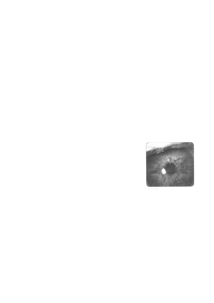
- Central desquamation results in a linear-branching
(dendritic) ulcer.
- Decreased corneal sensation (as it involves the
nerves).
- Anterior stromal infiltration under the ulcer.
- Progressive centrifugal (from the center outwards)
enlargement may result in a large epithelial defect
with a geographical or amoeboid configuration,
especially in the context of injudicious topical steroid
therapy.
- Following healing, there are persistent linear-branching shapes, which
represent waves of healing epithelial cells.
Differential diagnosis of dendritic ulceration:
(pseudo-dendritic
ulceration)
a- Herpes Zoster keratitis.
b- Healing cornel abrasion.
c- Soft contact lens wear.
d- Acanthamoeba keratitis.
e- Toxic keratopathies (kertitis medicamentosa).
Treatment of Herpes simplex epithelial keratitis:

7
a- Topical: without treatment, 50% resolves spontaneously, with treatment,
the cure rate is 95%.
Aciclovir 3% ointment, five times daily for 2 weeks.
or Ganciclovir 0.15% (Virgan
®
) gel: it is a new preparation which is used
five times daily and is as effective as Aciclovir.
b- Debridement:
Used in dendritic but not geographic ulcers in patients who are: non-
compliant, allergic to drugs, when antiviral agents are not available, resistant
cases. Drugs are ideally used after debridement. Cure rate is above 50% and
below 95%.