
1
Bones of lower limb
1
st
stage
Dr.Kalid Ali Zayer
Femur
The femur is the only bone in the thigh and the longest bone in the body.
It acts as the site of origin and attachment of many muscles and ligaments, and
can be divided into three areas; proximal, shaft and distal.
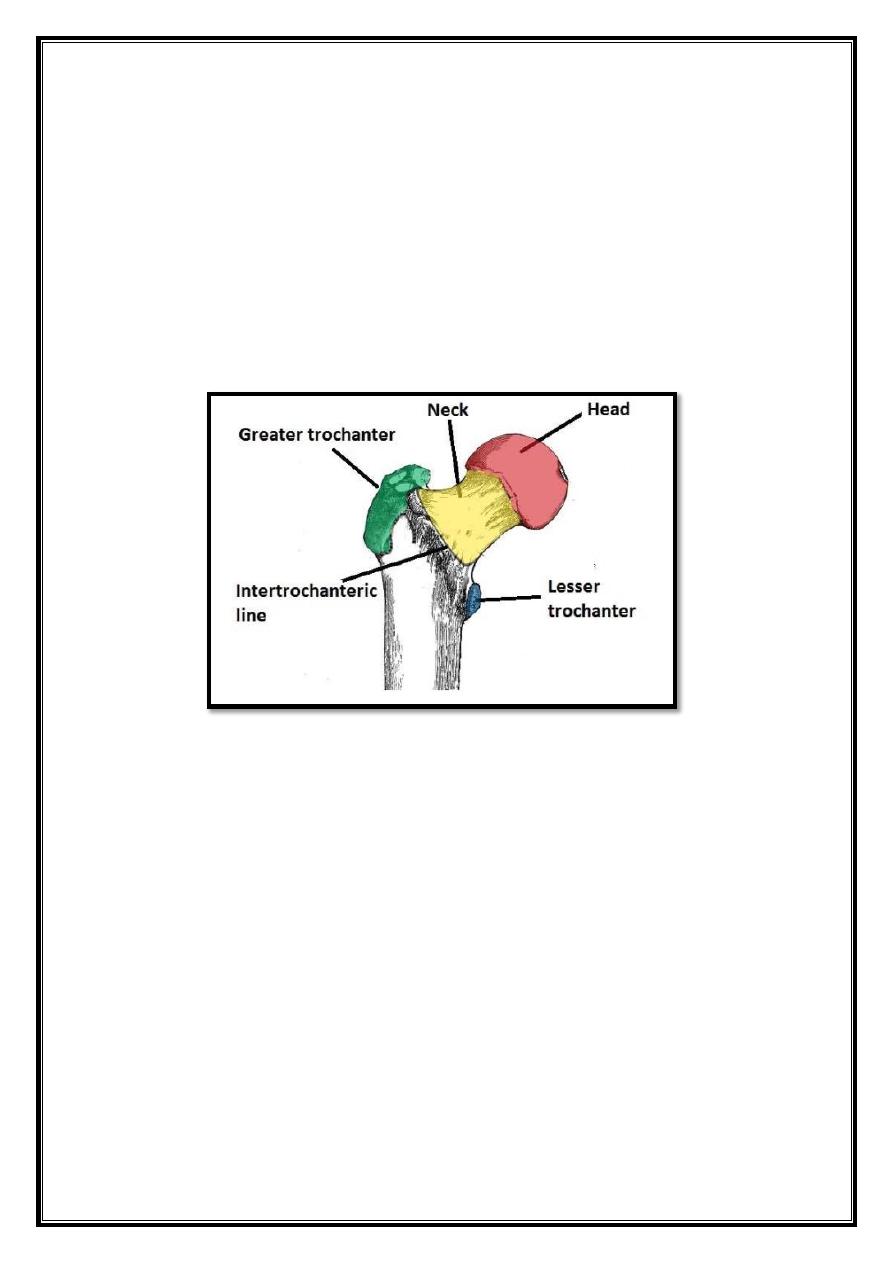
Proximal
The proximal area of the femur forms the hip joint with the acetabulum of the
pelvis. It consists of a head and neck, and two bony processes – the greater and
lesser trochanters.
There are also two bony ridges connecting the two trochanters; the
intertrochanteric line anteriorly and the trochanteric crest posteriorly.
Head
– articulates with the acetabulum of the pelvis to form the hip joint.
It has a smooth surface, covered with articular cartilage (except for a small
depression – the fovea – where ligamentum teres attaches).
Neck
– connects the head of the femur with the shaft. It is cylindrical,
projecting in a superior and medial direction. It is set at an angle of 135 degrees to
the shaf. This angle of projection allows for an increased range of movement at the
hip joint.
Greater trochanter
– the most lateral palpable projection of bone that
originates from the anterior aspect, just lateral to the neck.
-
It is the site of attachment for many of the muscles in the gluteal region,
such as gluteus medius, gluteus minimus and piriformis. The vastus lateralis
originates from this site.
Lesser trochanter
– smaller than the greater trochanter. It projects from
the posteromedial side of the femur, just inferior to the neck-shaft junction.
-
It is the site of attachment for iliopsoas (which can avulse the lesser
trochanter).
Intertrochanteric line
– a ridge of bone that runs in an inferomedial
direction on the anterior surface of the femur, connecting the two trochanters
together. After it passes the lesser trochanter on the posterior surface, it is known
as the pectineal line.
Contents
1. Proximal
2. The Shaft
3. Distal
2
- It is the site of attachment for the iliofemoral ligament (strong ligament
of the hip joint).
-The hip joint capsule also attaches anteriorly at this point.
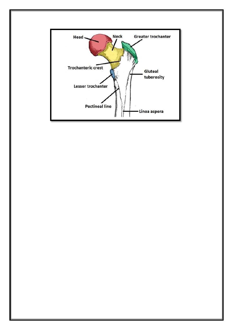
Intertrochanteric crest
– similar to the intertrochanteric line, this
is a ridge of bone that connects the two trochanters together. It is located on the
posterior surface of the femur.
There is a rounded tubercle on its superior half, this is called the quadrate
tubercle, which is where quadratus femoris attaches.
Fig 1 – The anterior surface of the proximal right femur.
3
Fig 2 – The posterior surface of the right femur.
The Shaft
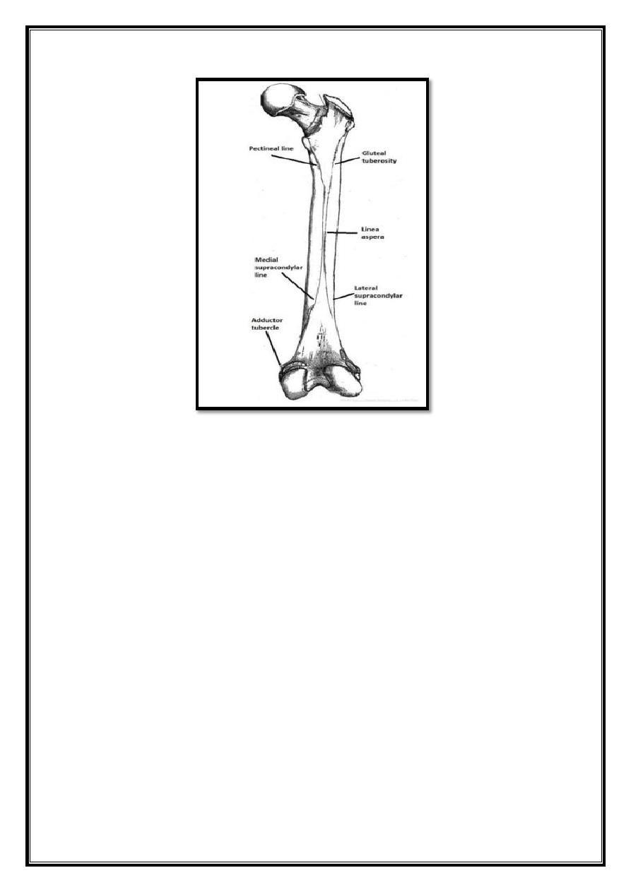
The shaft of the femur descends in a slight medial direction. This brings the knees
closer to the body’s center of gravity, increasing stability. A cross section of the
shaft in the middle is circular but flattened posteriorly at the proximal and distal
aspects.
On the posterior surface of the femoral shaft, there are roughened ridges of bone,
these are called the linea aspera (Latin for rough line). This splits inferiorly to
form the medial and lateral supracondylar lines. The flat popliteal surface lies
between them.
Proximally, the medial border of the linea aspera becomes the pectineal line. The
lateral border becomes the gluteal tuberosity, where the gluteus maximus
attaches.
Distally, the linea aspera widens and forms the floor of the popliteal fossa, the
medial and lateral borders form the medial and lateral supracondylar lines. The
medial supracondylar line stops at the adductor tubercle, where the adductor
magnus attaches.
4
Fig 3 – Posterior surface of the right femoral shaft.
Distal
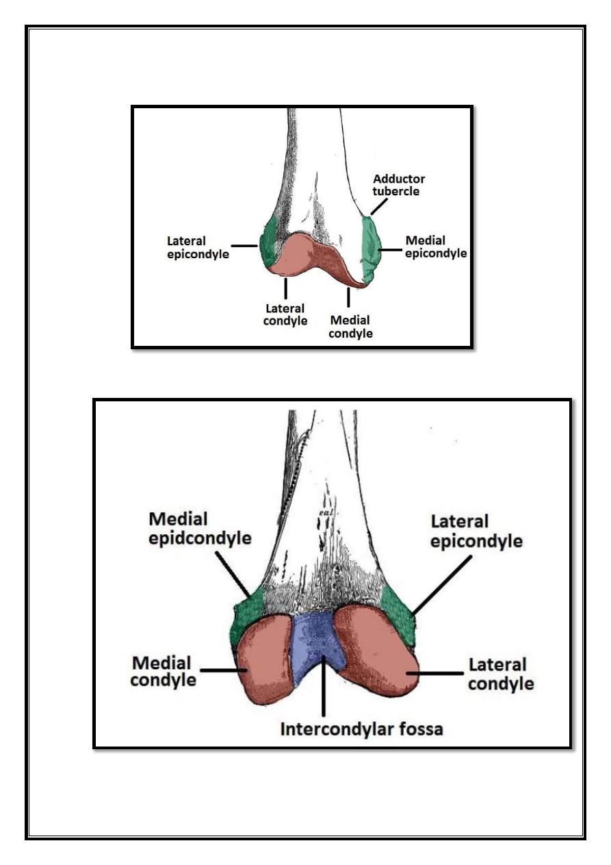
The distal end is characterized by the presence of the medial and lateral condyles,
which articulate with the tibia and patella to form the knee joint.
Medial and lateral condyles
– rounded areas at the end of the femur. The
posterior and inferior surfaces articulate with the tibia and menisci of the knee,
while the anterior surface articulates with the patella. The more prominent lateral
condyle helps prevent the natural lateral movement of the patella; a flatter
condyle is more likely to result in dislocation.
Medial and lateral epicondyles
– bony elevations on the non-articular areas of
thecondyles.
The
medial
epicondyle
is
the
larger
of
the
two.
o The medial and lateral collateral ligaments of the knee originate from their
respective epicondyles.
Intercondylar fossa
– a depression found on the posterior surface of the femur,
it lies in between the two condyles. It contains two facets for attachment of
internal knee ligaments; the anterior cruciate ligament (ACL) attaches to the
medial aspect of the lateral condyle and the posterior cruciate ligament (PCL) to
the lateral aspect of the medial condyle.
5
Fig 4 – Anterior surface of the distal right femur.
Fig 5 – Posterior surface of the distal right femur.
6
Patella
Contents
1. Bony Landmarks
2. Functions
The patella (knee-cap) is located at the front of the knee joint, within the
patellofemoral groove of the femur. Its superior aspect is attached to the
quadriceps tendon, and inferior aspect to the patellar ligament.
It is classified as a sesamoid type bone due to its position within the quadriceps
tendon, and is the largest sesamoid bone in the body.
Bony Landmarks
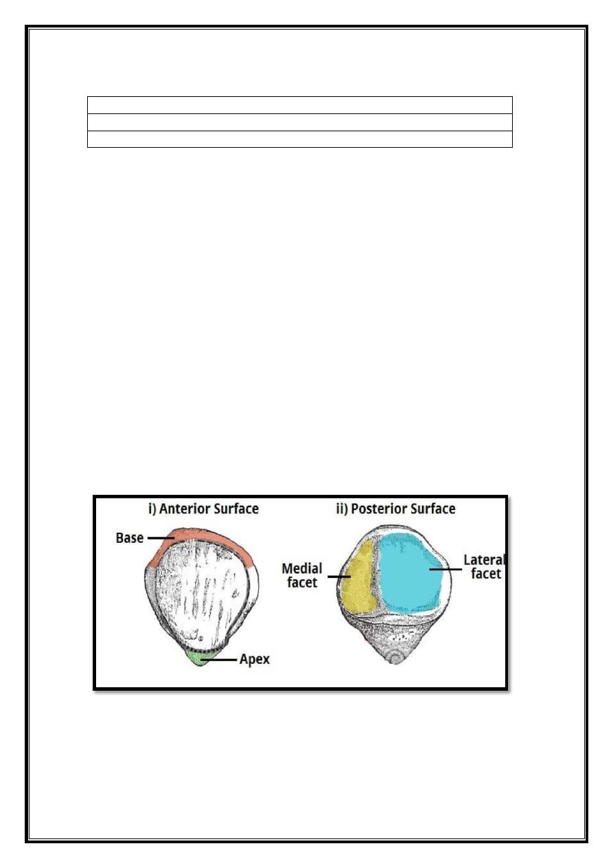
The patella has a triangular shape, with anterior and posterior surfaces. The apex
of the patella is situated inferiorly, and is connected to the tibial tuberosity by the
patella ligament.
The base forms the superior aspect of the bone, and provides the attachment area
for
the quadriceps tendon.
The posterior surface of the patella articulates with the femur, and is marked by
two facets:
Medial facet
– articulates with the medial condyle of the femur.
Lateral facet
– articulates with the lateral condyle of the femur.
Fig 6 – Anterior and posterior surfaces of the patella.

7
Functions
The patella has two main functions:
1.
Leg extension
– Enhances the leverage that the quadriceps tendon
can exert on the femur, increasing the efficiency of the muscle.
2.
Protection
– Protects the anterior aspect of the knee joint from
physical trauma.