Rheumatology L-3

It ‘s a chronic systemic autoimmune inflammatory disease affecting all joints covered by synovium leading to destructive polyarthritis
causes severe joint destruction
is a systemic disease with systemic damageleads to disability
Is associated with significant costs
Is an immune mediated disease driven by inflammatory cytokines

It can be disabling and painful condition , that can lead to substantial loss of function and mobility if not adequately treated.
The process involves an inflammatory response of the capsule around the joints(synovium) secondary to swelling of synovial cells, excess synovial fluid, and the development of fibrous tissue(pannus) in the synovium.
The pathology of the disease process often lead to destruction of articular cartilage& ankylosis(fusion) of the joints .
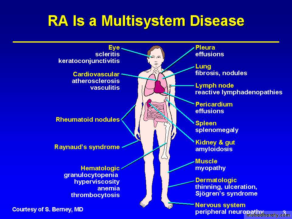
RA can also produce diffuse inflammation in the lungs , the membrane around the heart(pericardium),the membrane around the lungs(pleura),and the sclera, in addition to nodular lesions in the subcutaneous tissues.
Autoimmunity plays a role in the pathology of RA, and RA is a systemic autoimmune disease.
It is an autoimmune disease , higher in monozygotic twins &first degree relatives with HLA association , for example 50-75% of Caucasians have HLA-DR4, DR1 in Indians & DW15 in Japanese. DR4 is associated with more erosive disease.
Female gender & cigarette smoking are risk factors. There is an increased risk in breast feeding female.
Pathology
The inflammation and thickening of the synovial membranes cause irreversible damage to the joint capsule and articular cartilage as these structures replaced by scar like tissue called Pannus.
It is much more common than osteoarthritis which is associated with aging. It primarily affects young& middle age groups. Children are affected by a similar disorder called juvenile RA.
presentation
70% insidious onset (weeks to mnths)10% acute (fulminant onset)
20% sub acute onset
CLINICAL FEATURES& DIAGNOSIS
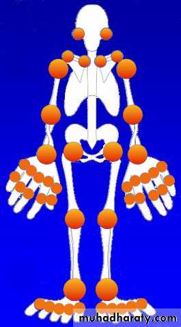
Tendon and bursa involvement are frequent and often clinically dominant in the early disease. RA can affect any joint, but has a predilection for metacarpophalangeal, proximal interphalangeal and metatarsophalangeal joints, as well as the wrists and knees.Articular and periarticular manifestations include joint swelling and tenderness to palpation ,with morning stiffness and sever motion impairment in the involved joints.
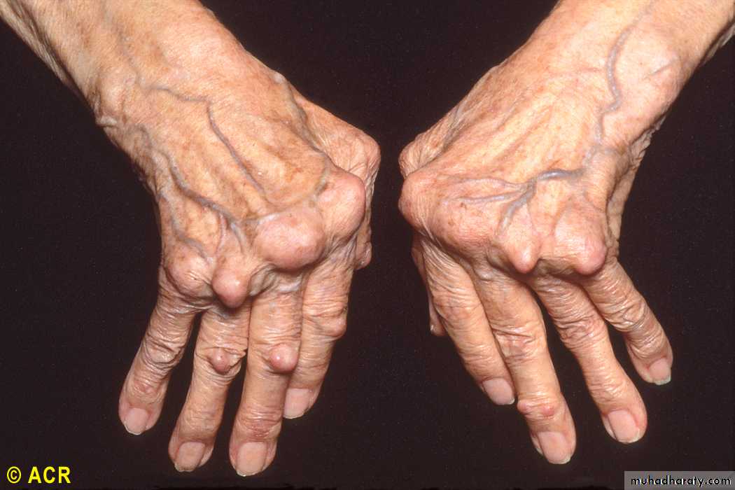
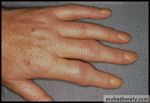
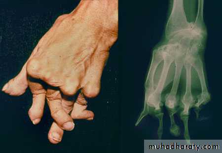
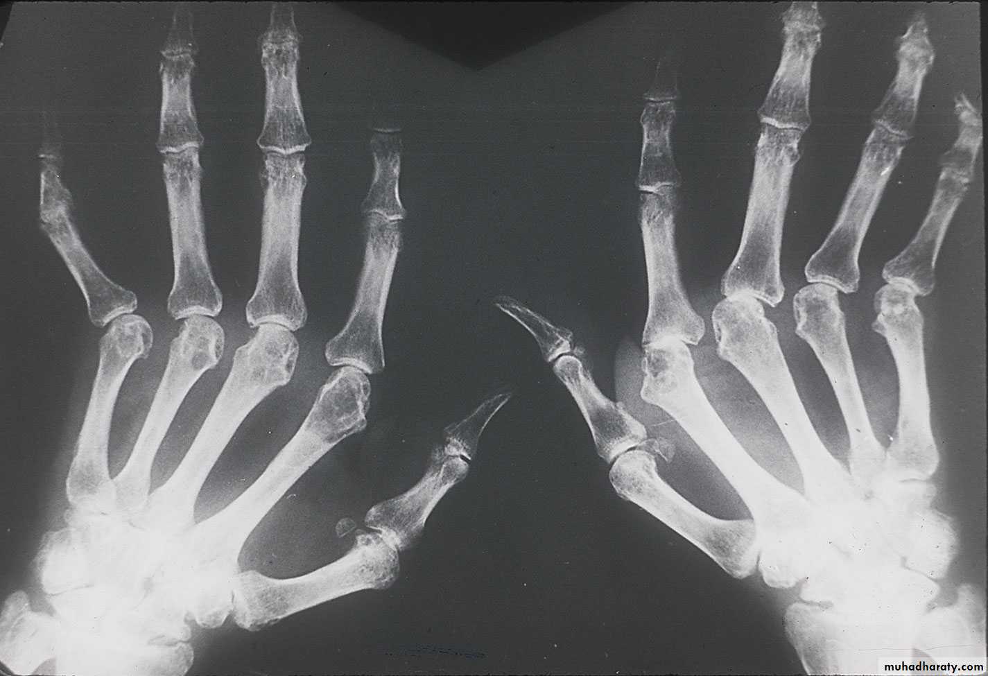
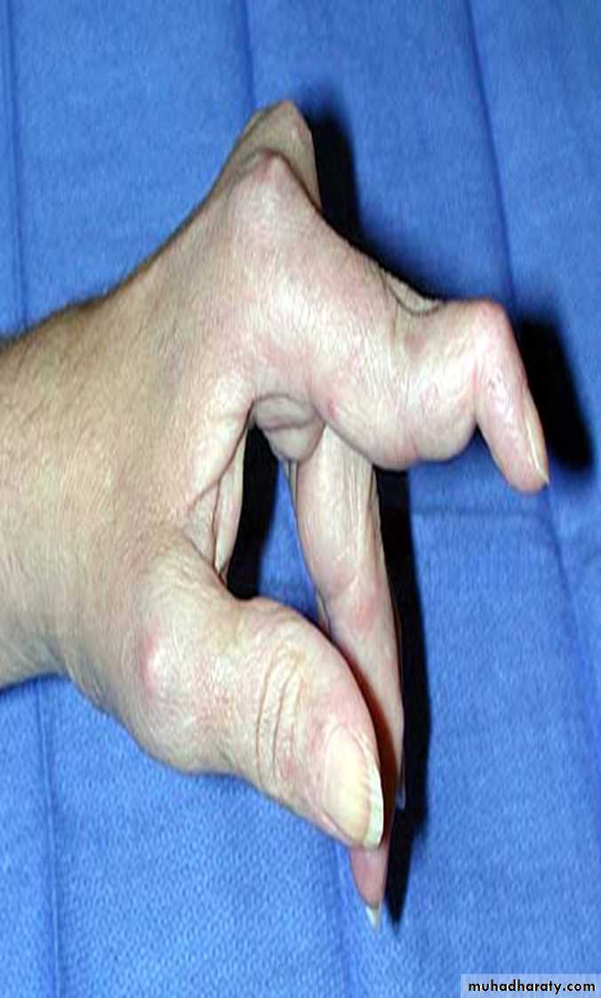
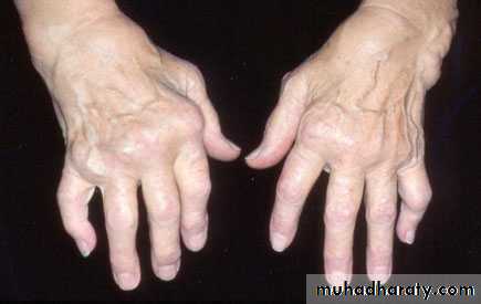
HAND JOINTS
Provide a good reflection of disease activity. The typical features are symmetrical swelling of MCP& PIP joints. They are hot, tender & have stress pain on passive movement.Specific hand abnormalities include swan neck deformity, button hole (boutonnière) deformity& Z- deformity of thumb. Other abnormalities include :
dorsal subluxation of ulnar styloid of the wrist , trigger finger, cock up deformity of the toe, flatfoot, Baker's(popliteal) cyst which can be mistaken for DVT, but past history of joint disease & Doppler U/S can establish the diagnosis of DVT.
Are more common in longstanding RA, but may occur at presentation specially in men.
Most features are due to serositis, granuloma, nodule formation or vasculitis:
1-systemic = fever, weight loss, fatigue, susceptibility to infection
2-MSK =muscle wasting, tenosynovitis, bursitis, osteoporosis3- hematological= anemia, thrombocytosis, eosinophilia.
Causes of anemia in RA:*anemia of chronic disease.
*megaloblastic anemia due to folate deficiency or associated pernicious anemia.
*Felty's syndrome.
*drugs: NSAIDs causing iron deficiency anemia; bone marrow suppression caused by gold& cytotoxic drugs.4-lymphatic= splenomegaly, Felty's syndrome
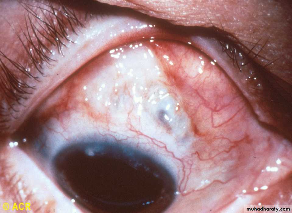
5-nodules= sinuses, fistula6-ocular= episcleritis, scleritis, scleromalacia, keratoconjunctivitis sicca
7-vasculitis= digital arteritis, ulcers, pyoderma gangerenosum, mononeuritis multiplex, visceral arteritis
8-cardiac= pericarditis, myocarditis, endocarditis, conduction defect, coronary vasculitis, granulomatous arteritis
9-pulmonary= nodules, pleural effusion, fibrosing alveolitis, bronchiolitis, Caplan's syndrome(RA with multiple pulmonary nodules & pneumoconiosis)
10-neurological= cervical cord compression, compression neuropathies, peripheral neuropathy, mononeuritis multiplex
• 11-amyloidosis= nephrotic syndrome
Felty's syndrome
The association of splenomegaly, leucopenia, neutropenia(hypersplenism)& leg ulcers with RA. Splenectomy ameliorates hypersplenism.It occurs in 1% of RA patients over the age of 50 , more common in female than male with long standing RA & in deforming but inactive disease..
Such patients have normochromic normocytic anemia, thrombocytopenia& abnormal liver function tests. It is very common in African Americans& such patients are susceptible to infections because of neutropenia
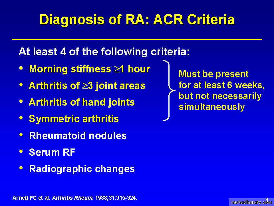
Diagnosis of RA: 2010 ACR Criteria
Diagnostic Tools in Rheumatoid Arthritis
Rheumatoid factorAnti-CCP antibodies
Plain X-rayMRI
Ultrasound
.INVESTIGATIONS
1- clinical criteria.2- acute phase response high ESR& CRP
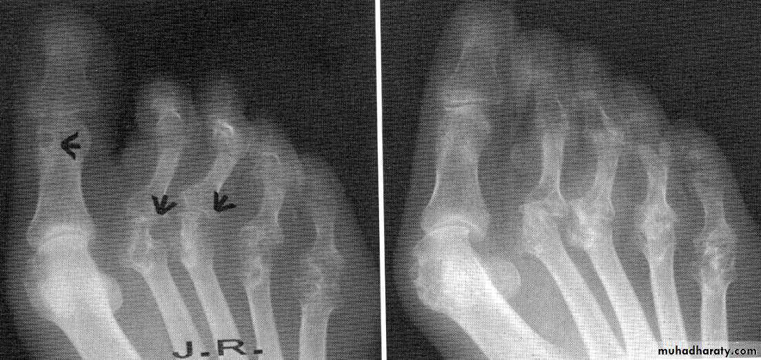
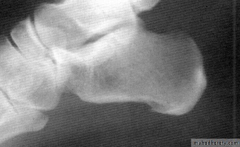
3- X-ray: the typical radiographic changes of RA are periarticular osteopenia& marginal non proliferative erosions.
4-serological tests : rheumatoid factor(RF) is usually positive in patients with seropositive RA. RF has low specificity & sensitivity. It is an IgM antibody directed against Fc fragment of human IgG. It occurs in a wide variety of diseases & also in some normal adults.
It has prognostic marker, a high titer at presentation is associated with poorer prognosis. Therefore, it has no rule in diagnosis, since it may appear years after presentation.
10-20% of patients with RA are seronegative
5- What Is Anti-CCP?
Anti-CCP, which stands for anti-cyclic citrullinated peptide antibody, is a blood test which helps in a diagnosis of rheumatoid arthritis.When Is It Appropriate to Have the Anti-CCP Test?
Anti-CCP is a very useful test to order during the diagnostic evaluation of a person who may have rheumatoid arthritis. If present in such a patient at a moderate to high level, it not only confirms the diagnosis but also may indicate that the patient is at increased risk for joints damage. Low levels of this antibody are less significant. In the past, doctors relied on another antibody, the rheumatoid factor (RF) to help confirm a diagnosis.
6-- examination of synovial fluid: is the most helpful laboratory procedure; the fluid is inflammatory with >10.000 WBC (PMN cells).
*Antinuclear Abs are common but not diagnostic.
Differential diagnosis
1-SLE
2- Lyme disease-
3-Viral arthritis
4-polymyalgia rheumatica
5- hypothyroidism
6-paraneoplastic syndromes
Copyright © 1972-2004 American College of Rheumatology Slide Collection. All rights reserved.
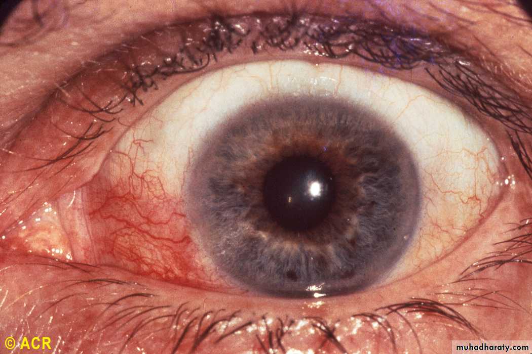
Rheumatoid arthritis: episcleritis
Copyright © 1972-2004 American College of Rheumatology Slide Collection. All rights reserved.
Rheumatoid arthritis: scleromalacia perforans
Together we can work to prevent this: