Brain imaging
INTERNAL MEDICINE DEPARTMENTSUPERVISOR: DR.ABBAS FADHEL
DONE BY :ZAHRAA ABBAS ALI
Computed tomography: CT
▪ A collimated X-ray beam moves synchronously across a slice of brain between 2 mm and 13 mm thick.▪ attenuation (density) between bone, brain and CSF enable recognition of normal and infarcted tissue, tumour, extravasated blood, and oedema .
Enhancement with i.v. contrast media delineates areas of increased blood supply. CT imaging of the spinal cord (myelography) and cerebral ventricles (ventriculography) is occasionally performed
▪ CT is safe apart from occasional systemic reactions to contrast; the irradiation involved is small.
CT scanning demonstrates :
▪ cerebral tumours▪ intracerebral haemorrhage and infarction
▪ subdural and extradural haematoma
▪ free blood in the subarachnoid space (subarachnoid haemorrhage)
▪ lateral shift of midline structures and displacement/enlargement of the ventricular system
▪ cerebral atrophy
▪ CT imaging, within its limitations, shows that the brain is anatomically normal, and gives reassurance .
Limitations of CT
▪ Lesions under 1 cm diameter may be missed .
▪ Lesions with attenuation close to that of bone may be missed if near the skull .
▪ Lesions with attenuation similar to that of brain are poorly imaged (e.g. MS plaques, isodense subdural haematoma(
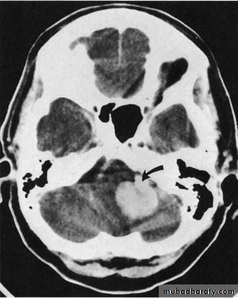
▪ CT sometimes misses lesions within the posterior fossa .
▪ Cooperation is required - a general anaesthetic is occasionally needed .
The cardinal signs of an abnormality on a CT scan are:
• Abnormal tissue density• Mass effect
• Enlargement of the ventricles.
Abnormal tissue density
• Abnormal tissue may be of higher or lower density than the normal surrounding brain.• High density is seen with recent haemorrhage, calcified lesions, and areas of contrast enhancement
• Low density is usually due to neoplasms or infarcts, or to oedema, which commonly surrounds neoplasms, infarcts, haemorrhages and areas of inflammation.
Abnormal tissue density
• Oedema characteristically shows finger-like projections and does not enhance with intravenous contrast medium.
• As a rule, it is not possible to diagnose the nature of a mass based on attenuation values alone; an exception is lipoma which, because it contains fat, has a value of approximately (- 100 Hounsfield units).
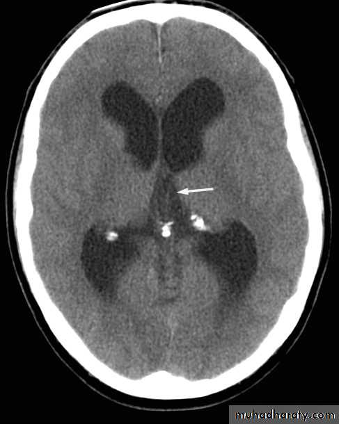
Mass effect
• The lateral ventricles should be examined to see if they are displaced or compressed.• Shift of midline structures, such as the septum pellucidum , the third ventricle, or the pineal, is a common finding with intracranial masses.
Enlargement of ventricles
There are two basic mechanisms which cause the cerebral ventricles to enlarge:• Obstruction to the CSF pathway, either within the ventricular system (non-communicating hydrocephalus) or over the surface of the brain (communicating hydrocephalus)
• Secondary to atrophy of brain tissue
Magnetic resonance imaging: MRI
▪ The hydrogen nucleus is a proton whose electrical charge creates a local electrical field. These protons are aligned by sudden strong magnetic impulses. Protons are then imaged with radiofrequency waves at right angles to their alignment. The protons resonate and spin, then revert to their normal alignment. As they do so, images are made at different phases of relaxation, known as T1, T2, T2 'STIR', diffusion-weighted imaging (DWI) and other sequences. Gadolinium is used as an intravenous contrast medium .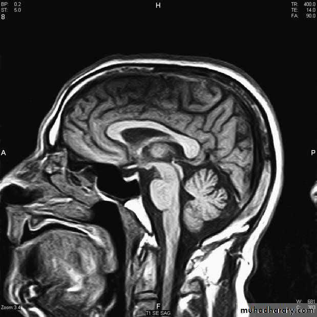
Advantages of MRI
▪ High quality soft tissue images .▪ MR distinguishes between brain white and grey matter .
▪ Pituitary imaging .
▪ MRI has greater resolution than CT (around 0.5 cm(
▪ No radiation is involved .
▪ Magnetic resonance angiography (MRA) images blood vessels without contrast .
▪ Useful for posterior fossa and temporal lobe .
Limitations of MRI are principally
▪ time and cost. Imaging one region takes about 20 minutes.▪ Patients do need to cooperate.
▪ Claustrophobia is an issue but 'open' machines are available.
▪ Patients with pacemakers or with metallic bodies in the brain cannot be imaged.
MRA images blood flow, not the vessel anatomy.
▪ MR imaging for some days after lumbar puncture frequently shows diffuse meningeal enhancement with gadolinium .
Doppler studies B-mode and colour ultrasound are valuable in the detection of stenosis of the carotid arteries
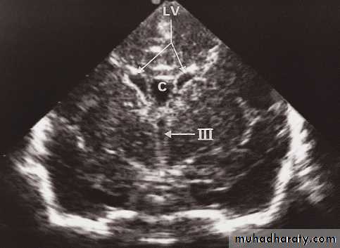
Neurosonography
• Simple to scan the heads of neonates and young babies to obtain images of the ventricular system and the adjacent brain.• Scanning is best done through an open fontanelle where there is no bone to impede the transmission of ultrasound.
• Little discomfort is caused to the baby and the procedure is readily carried out even on ill babies in intensive care units.
• Neurosonography has proved particularly useful in detecting intracerebral haemorrhage and the ventricular dilatation that may follow. It has also been used to demonstrate the presence and cause of other forms of hydrocephalus and congenital abnormalities of the brain.
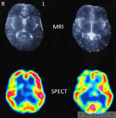
Radio-isotope (isotope brain scan ,SPECT,PET)
These techniques image function more than anatomy by tracking the uptake and metabolism of radioisotopes.Disadvatages :
Poor spatial resolution .
Ionising radiation.
Expensive.
Not widely available.