
1
Nerves of the upper limb
1
st
stage
Dr.Kalid Ali Zayer
Brachial plexus
Contents
1. Roots
2. Trunks
3. Divisions
4. Cords
5. Major Branches
o
5.1 Musculocutaneous Nerve
o
5.2 Axillary Nerve
o
5.3 Median Nerve
o
5.4 Radial Nerve
o
5.5 Ulnar Nerve
6. Minor Branches
The brachial plexus is a network of nerve fibers that supplies the skin and musculature
of the upper limb. It begins in the root of the neck, passes through the axilla, and runs
through the entire upper extremity. The plexus formed by the anterior rami (divisions)
of cervical spinal nerves C5, C6, C7 and C8, and the frst thoracic spinal nerve, T1. The
brachial plexus divided into five parts; roots, trunks, divisions, cords and branches .
There are no functional differences between these divisions; they simply used
to aid explanation of the brachial plexus.
Roots
At each vertebral level, paired spinal nerves arise. They leave the spinal cord via the
intervertebral foramina of the vertebral column. Each spinal nerve then divides into an
anterior and a posterior ramus.
The roots of the brachial plexus formed by the anterior rami of spinal nerves C5-T1 (the
posterior divisions innervate the skin and musculature of the intrinsic back muscles).
After their formation, these nerves pass between the anterior and medial scalene
muscles to enter the base of the neck.
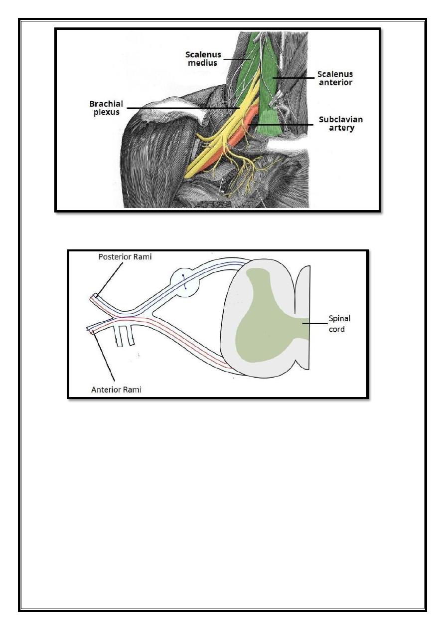
2
Fig 1 – Proximal portion of the brachial plexus, in the neck.
Fig 2 – The spinal cord outlow at each vertebral level. The anterior rami of vertebral
levels C5- C8 and T1 make up the roots of the brachial plexus.
Trunks
At the base of the neck, the roots of the brachial plexus converge to form three trunks.
These structures named by their relative anatomical location:
Superior trunk
– a combinaton of C5 and C6 roots.
Middle trunk
– contnuaton of C7.
Inferior trunk
– combinaton of C8 and T1 roots.
The trunks traverse laterally, crossing the posterior triangle of the neck.

3
Divisions
Each trunk divides into two branches within the posterior triangle of the neck. One
division moves anteriorly (toward the front of the body) and the other posteriorly
(towards the back of the body). Thus, they known as the anterior and posterior divisions.
We now have three anterior and three posterior nerve fibers. These divisions leave the
posterior triangle and pass into the axilla. They recombine into the cords of the brachial
plexus.
Cords
Once the anterior and posterior divisions have entered the axilla, they combine to form
three
cords, named by their position relative to the axillary artery.
The lateral cord formed by:
The anterior division of the superior trunk
The anterior division of the middle trunk
The posterior cord formed by:
The posterior division of the superior trunk
The posterior division of the middle trunk
The posterior division of the inferior trunk
The medial cord formed by:
The anterior division of the inferior trunk.
The cords give rise to the major branches of the brachial plexus.
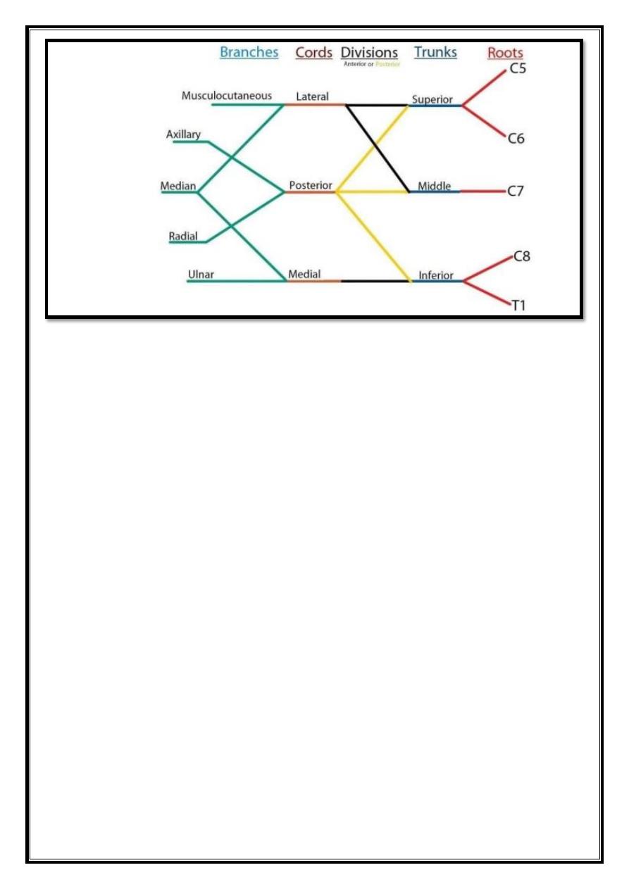
4
Fig 3 – Diagrammatic representation of the brachial plexus. For simplicity, the smaller
branches of the brachial plexus not shown. The posterior divisions shown in yellow
and anterior divisions in black.
Major Branches
In the axilla and the proximal aspect of the upper limb, the three cords give rise to five
major branches. These nerves continue into the upper limb to provide innervation to the
muscles and skin present.
Musculocutaneous Nerve
Roots: C5, C6, C7.
Motor Functions: Innervates the brachialis, biceps brachii and coracobrachialis
muscles.
Sensory Functions: Gives off the lateral cutaneous branch of the forearm, which
innervates the lateral half of the anterior forearm, and a small lateral portion of the
posterior forearm.
5
Fig 4 – The derivation of the musculocutaneous nerve from the brachial plexus
6
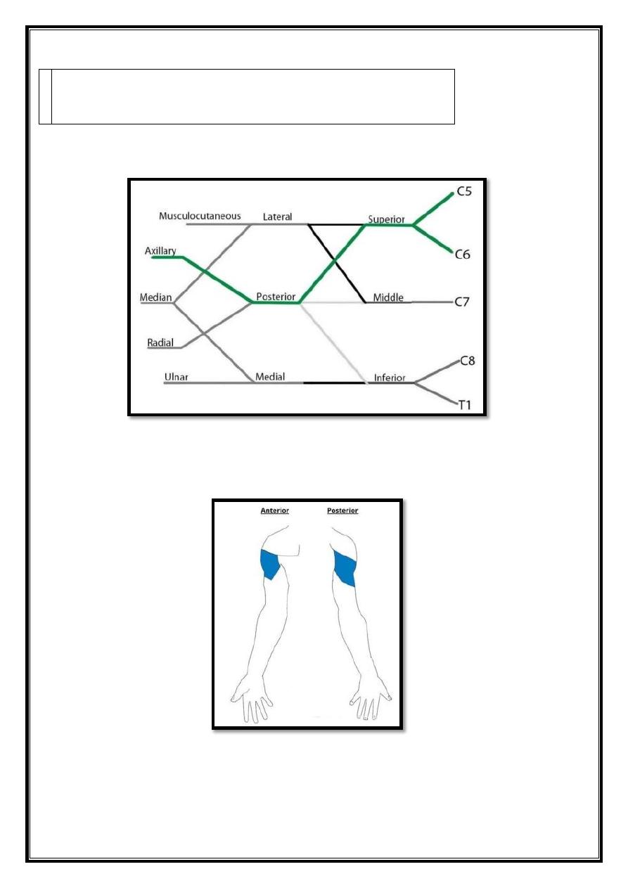
Axillary Nerve
Roots
: C5 and C6.
Motor Functions
: Innervates the teres minor and deltoid
muscles.
Sensory Functions
: Gives off the superior lateral cutaneous nerve of arm, which
innervates the inferior region of the deltoid (“regimental badge area”).
Fig 5 – The derivation of the axillary nerve from the brachial plexus.
7
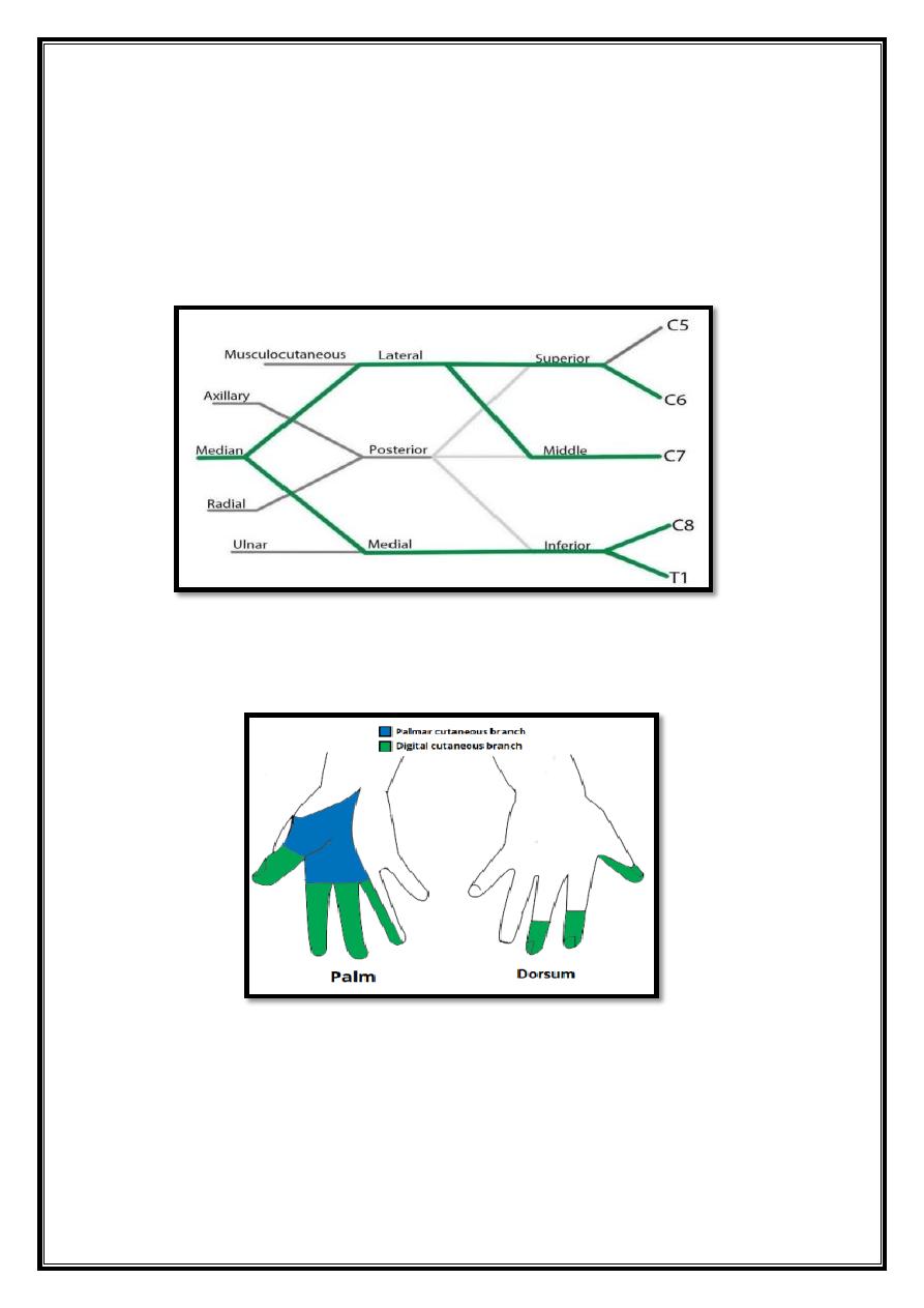
Median Nerve
Roots: C6 – T1. (Also contains fibers from C5 in some individuals).
Motor Functions: Innervates most of the flexor muscles in the
forearm, the thenar muscles, and the two lateral lumbricals associated
with the index and middle fingers.
Sensory Functions: Gives off the palmar cutaneous branch, which innervates the
lateral part of the palm, and the digital cutaneous branch, which innervates the lateral
three and a half fingers on the anterior (palmar) surface of the hand.
Fig 6 – The derivation of the median nerve from the brachial plexus.
8
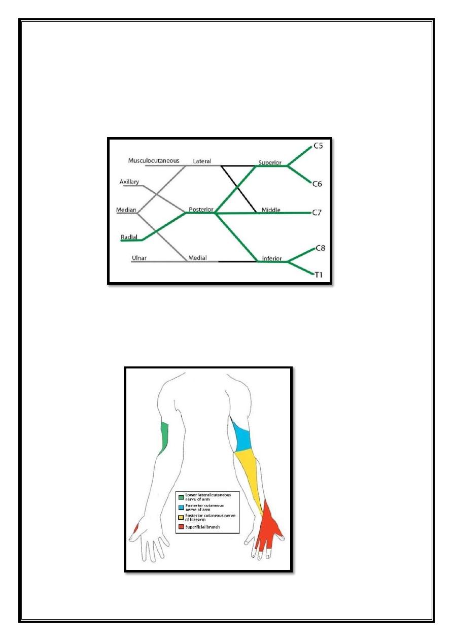
Radial Nerve
Roots: C5 – T1.
Motor Functions: Innervates the triceps brachii, and the muscles in the
posterior compartment of the forearm (which are primarily, but not
totally, extensors of the wrist and fingers).
Sensory Functions: Innervates the posterior aspect of the arm and forearm, and the
posterolateral aspect of the hand.
Fig 7 – The derivation of the radial nerve from the brachial plexus.
9
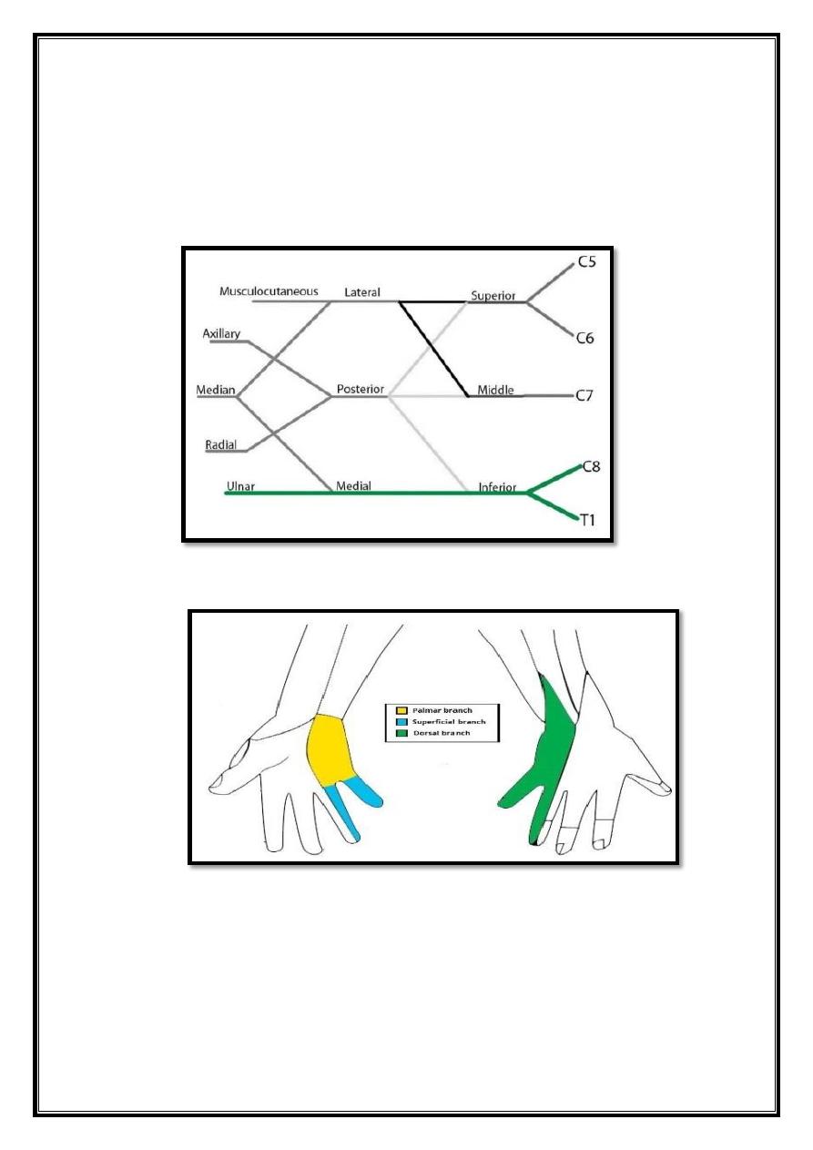
Ulnar Nerve
Roots: C8 and T1.
Motor Functions: Innervates the muscles of the hand (apart from the
thenar muscles and two lateral lumbricals), flexor carpi ulnaris and
medial half of flexor digitorum profundus.
Sensory Functions: Innervates the anterior and posterior surfaces of the medial one
and half fingers, and associated palm area.
Fig 8 – The derivation of the ulnar nerve from the brachial plexus.

10
Minor Branches
Number of smaller nerves arise in addition to the five major branches of the brachial
plexus.
They listed below:
Roots
Trunks
Lateral cord
Medial cord
Posterior cord
Dorsal
scapular
nerve
Long
thoracic
nerve
Suprascapular
nerve
Nerve to
subclavius
Lateral
pectoral
nerve
Medial pectoral
nerve
Medial cutaneous
nerve of arm
Medial cutaneous
nerve of forearm
Superior
subscapular nerve
Thoracodorsal
nerve
Inferior
subscapular nerve