1
Arterial supply to the upper limb
Contents
1. Subclavian Artery
2. Axilla: Axillary Artery
3. Upper Arm: Brachial Artery
4. Forearm: Radial and Ulnar Arteries
5. Hand: Superficial and Deep Palmar Arches
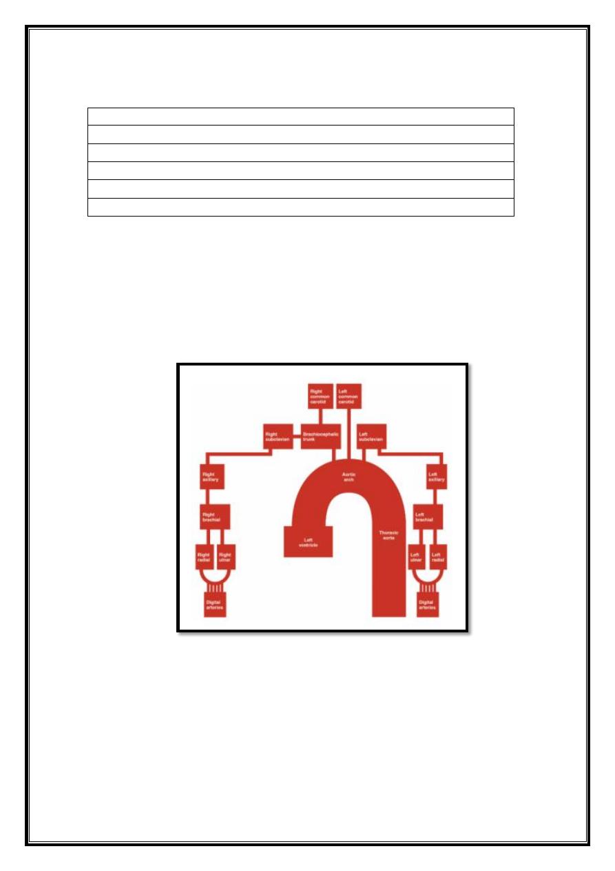
The arterial supply to the upper limb delivered via five main vessels
(proximal to distal):
1. Subclavian artery.
2. Axillary artery.
3. Brachial artery.
4. Radial artery.
5. Ulnar artery.
Fig 13 – Schematic demonstrating the arterial supply to the
upper limb.
2
Subclavian Artery
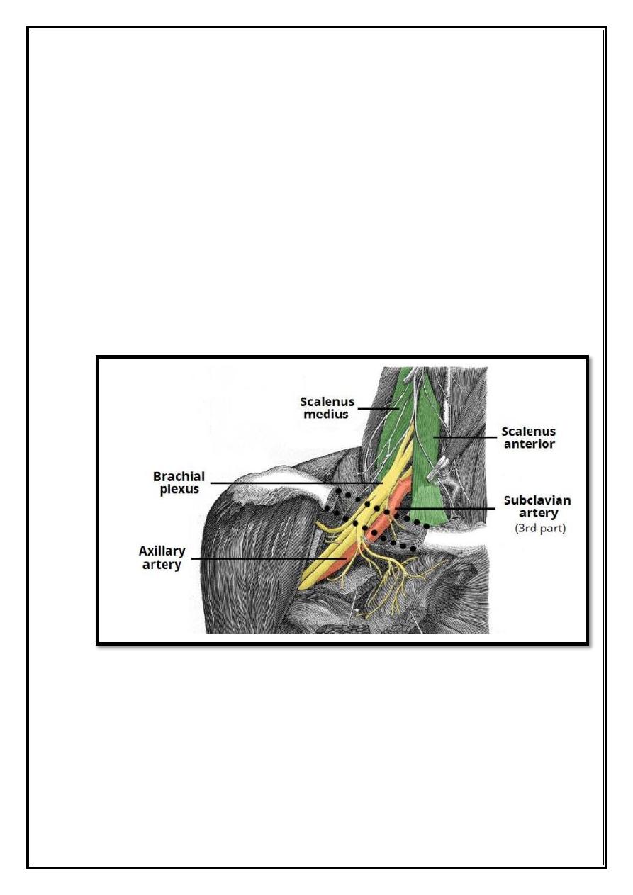
The arterial supply to the upper limb begins as the subclavian artery. On the
right, the subclavian artery arises from the brachiocephalic trunk. On the left, it
branches directly from the arch of aorta. The subclavian artery travels laterally
towards the axilla. It can divided into three parts based on its position relative to
the anterior scalene muscle:
First part
– origin of the subclavian artery to the medial border of the anterior
scalene.
Second part
– posterior to the anterior scalene.
Third part
– lateral border of anterior scalene to the lateral border of the first
rib.
At the lateral border of the first rib, the subclavian artery enters the axilla – and is
renamed the axillary artery.
Fig 14 – Anatomical course of the subclavian artery. When the vessel
crosses the first rib (not shown), it is renamed the axillary artery.
3
Axilla: Axillary Artery
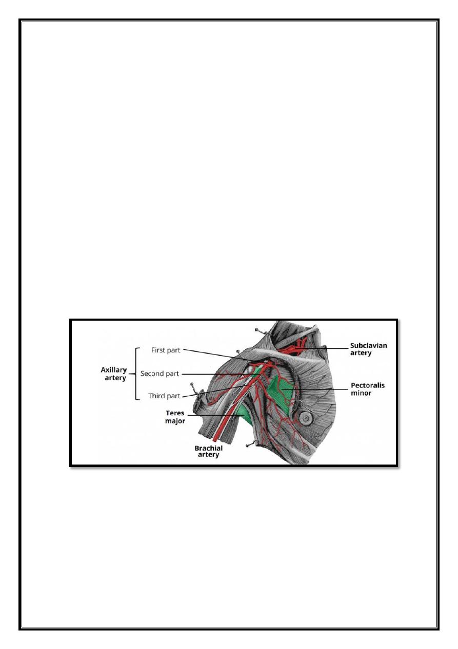
The axillary artery lies deep to the pectoralis minor and enclosed in the axillary
sheath (a fibrous layer that covers the artery and the three cords of the brachial
plexus).
Importantly, the artery can divided into three parts based on its position relative
to the pectoralis minor muscle:
First part – proximal to pectoralis minor.
Second part – posterior to pectoralis minor
Third part – distal to pectoralis minor
The main branches of the axillary artery include:
First Part:
Superior thoracic artery
Second Part:
Thoracoacromial artery & Lateral thoracic artery.
Third Part:
Subscapular artery, Anterior and posterior circumflex arteries
The anterior and posterior circumflex humeral arteries form an anastomotic
network around the surgical neck of the humerus and can damaged in cases of
fracture.
At the lower border of the teres major muscle, the axillary artery is renamed the
brachial artery.
Fig 15 – The axillary artery has three parts, named according to its
position relative to the
pectoralis minor muscle.
4
Upper Arm: Brachial Artery
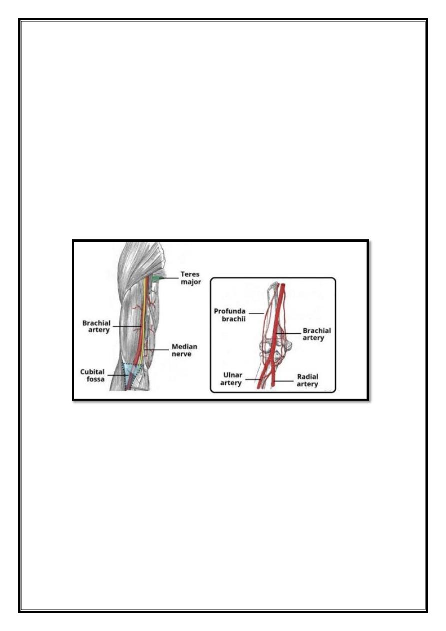
The brachial artery is a continuation of the axillary artery past the lower border
of the teres major. It is the main supply of blood for the arm.
Immediately distal to the teres major, the brachial artery gives rise to the
profunda brachii (deep artery), which travels with the radial nerve in the radial
groove of the humerus and supplies structures in the posterior aspect of the upper
arm (e.g. triceps brachii). The profunda brachii terminates by contributing to an
anastomotic network around the elbow joint.
The brachial artery proper descends the arm. As it moves through the cubital
fossa, underneath the brachialis muscle, the brachial artery terminates by
bifurcating into the radial and ulnar arteries.
Fig 16 – The anatomical course and major branches of the brachial
artery. Note its relation to the median nerve as it descends the
Forearm.

5
Venous system of the upper limb
Contents
1. Superficial Veins
1.1. Basilic Vein
1.2. Cephalic Vein
2. Deep Veins.
The venous system of the upper limb drains deoxygenated blood from the
arm, forearm and
hand. It can subdivided into the superficial system and the deep system.
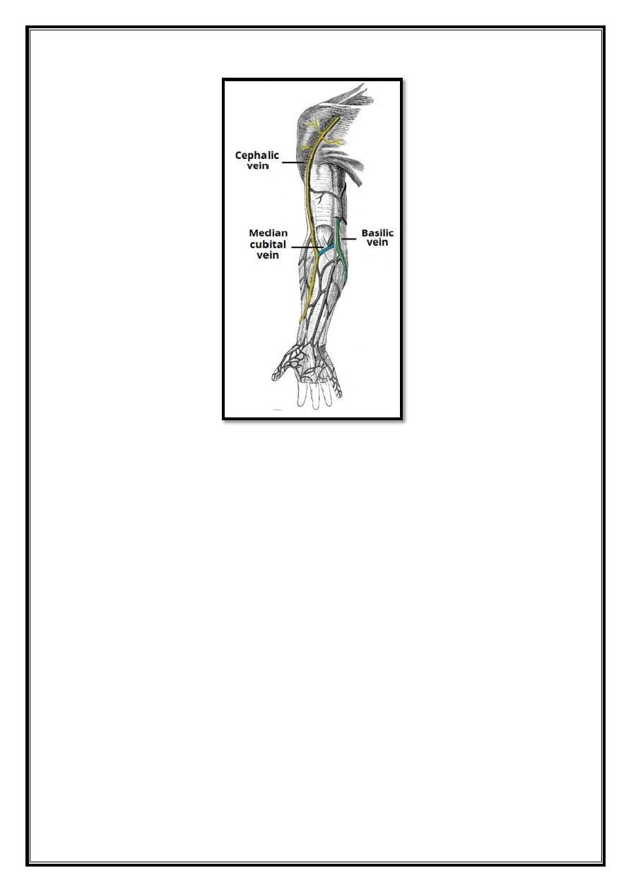
Superficial Veins
The major superficial veins of the upper limb are the cephalic and basilic veins.
They are located within the subcutaneous tissue of the upper limb.
Basilic Vein
The basilic vein originates from the dorsal venous network of the hand and
ascends the medial aspect of the upper limb. At the border of the teres major, the
vein moves deep into the arm.
Here, it combines with the brachial veins from the deep venous system to form the
axillary vein.
Cephalic Vein
The cephalic vein also arises from the dorsal venous network of the hand. It
ascends the antero-lateral aspect of the upper limb, passing anteriorly at the elbow.
At the shoulder, the cephalic vein travels between the deltoid and pectoralis major
muscles (known as the deltopectoral groove), and enters the axilla region via the
clavipectoral triangle. Within the axilla, the cephalic vein empties into axillary
vein.
The cephalic and basilic veins connected at the elbow by the median cubital vein.
6
Fig 19 – The superficial veins of the upper limb.
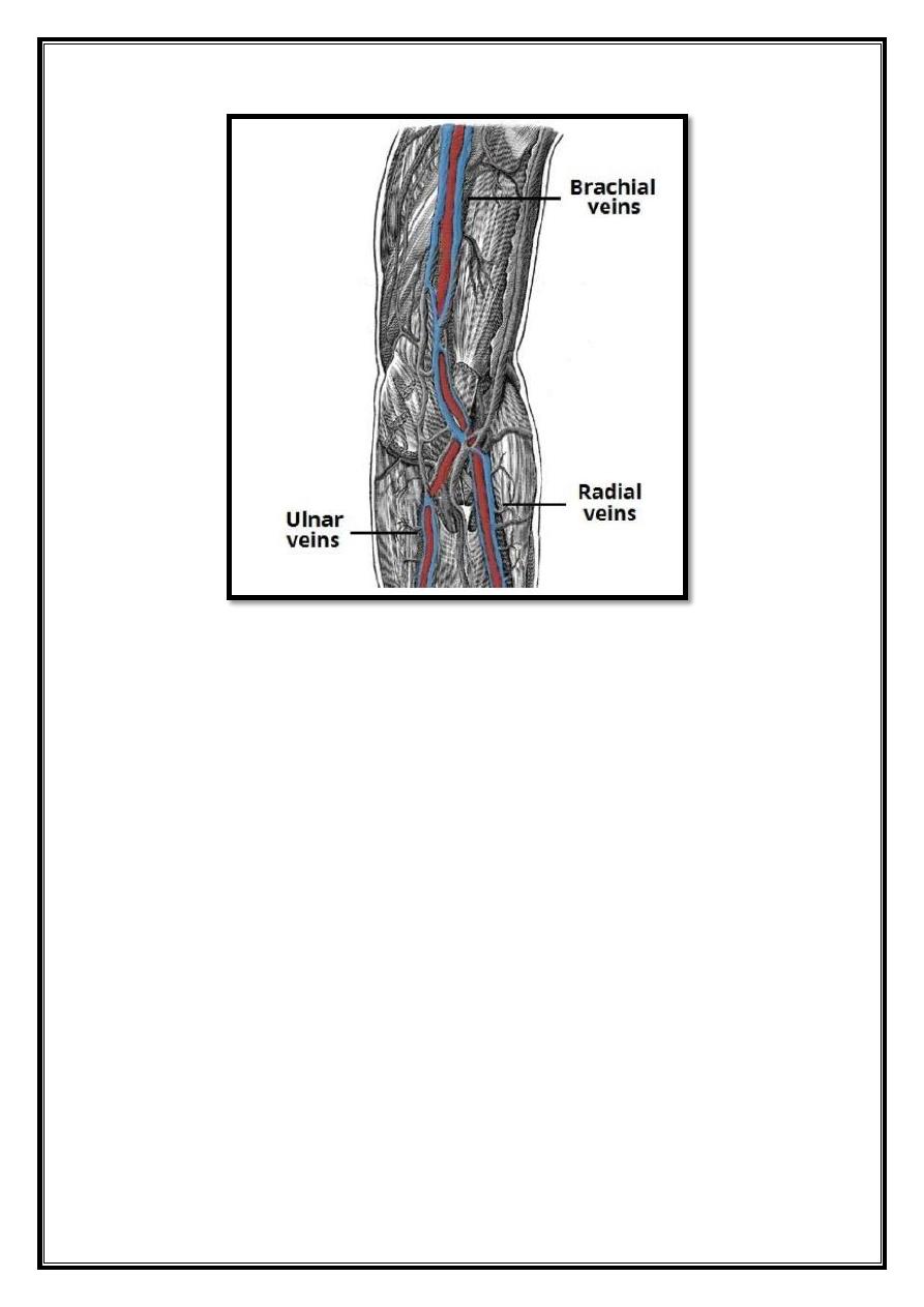
Deep Veins
The deep venous system of the upper limb situated underneath the deep fascia. It
formed by paired veins, which accompany and lie either side of an artery. In the
upper extremity, the deep veins share the name of the artery they accompany.
The brachial veins are the largest, and are situated either side of the brachial
artery.
Perforating veins run between the deep and superficial veins of the upper limb,
connecting the two systems.
7
Fig 20 – The major deep veins of the upper limb.