1
Area of upper limb
1
st
stage
Dr.Kalid Ali Zayer
Axilla
Contents
1. Borders
2. Contents
3. Passageways Exiting the Axilla
The axilla is the name given to an area that lies underneath the glenohumeral
joint, at the junction of the upper limb and the thorax. It is a passageway by
which neurovascular and muscular structures can enter and leave the upper limb.
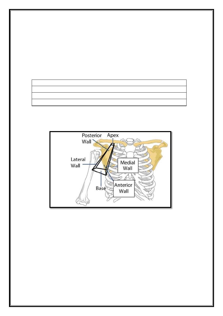
Fig 1 – Anterior view of the right axilla region. Note the pyramidal
shape, with six borders (or sides).
Borders
The overall 3D shape of the axilla looks slightly like a pyramid. It consists of four
sides, an open apex and base:
1. Apex
– Also known as, the axillary inlet, lateral border of the first rib, superior
border of scapula, and the posterior border of the clavicle form it.
2. Lateral wall
– formed by intertubercular groove of the humerus.
3. Medial wall
– consists of the serratus anterior and the thoracic wall (ribs and
intercostals muscles).
2
4. Anterior wall
– contains the pectoralis major, the underlying pectoralis minor,
and the subclavius muscles.
5. Posterior wall
– formed by the subscapularis, teres major and latissimus
dorsi.The size and shape of the axilla region varies with arm abduction. The apex
decreases in size
most markedly when the arm is fully abducted – leaving the contents of the axilla
at risk of
compression.
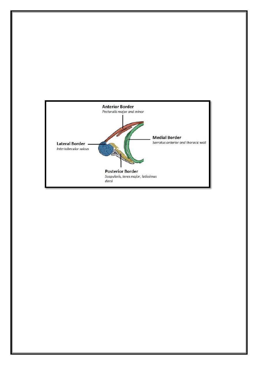
Fig 2 – Transverse section of the axilla region.
Contents
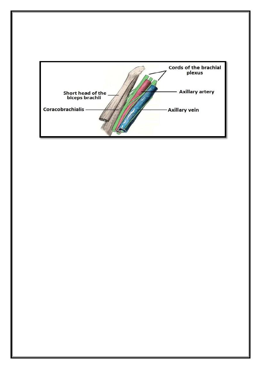
The contents of the axilla region include muscles, nerves, vasculature and
lymphatics:
1. Axillary artery (and branches)
– the main artery supplying the upper limb. It
commonly referred as having three parts; one medial to the pectoralis minor,
one posterior to pectoralis minor, and one lateral to pectoralis minor. The
medial and posterior parts travel in the axilla.
2. Axillary vein (and tributaries)
– the main vein draining the upper limb, its
two largest tributaries are the cephalic and basilic veins.
3. Brachial plexus (and branches)
– a collection of spinal nerves that form the
peripheral nerves of the upper limb.
4. Axillary lymph nodes
– they filter lymphatic fluid that has drained from the
upper limb and pectoral region. Axillary lymph node enlargement is a non-
specific indicator of breast cancer.
3
5. Biceps brachii (short head) and coracobrachialis
– these muscle tendons
move through the axilla, where they attach to the coracoid process of the
scapula.
Fig 3 – Contents of the axilla region.
Passageways Exiting the Axilla
Structures leave the axilla by three main routes.
1. The main route of exit is immediately inferiorly and laterally, into the upper
limb. The majority of contents of the axilla region leave by this method.
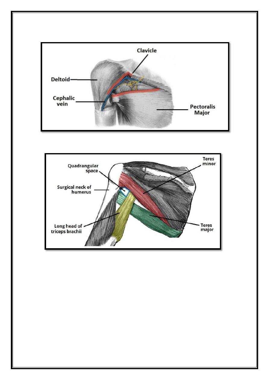
2. Another pathway is via the quadrangular space. This is a gap in the posterior
wall of the axilla, allowing access to the posterior arm and shoulder area.
Structures passing through include the axillary nerve and posterior circumflex
humeral artery (a branch of the axillary artery.
3. The last passageway is the clavipectoral triangle, which is an opening in the
anterior wall of the axilla. It is bounded by the pectoralis major, deltoid, and
clavicle. The cephalic vein enters the axilla via this triangle, while the medial and
lateral pectoral nerves leave.
4
Fig 4 – Boundaries and contents of the clavipectoral triangle
Fig 5 – Posterior view of the shoulder region, showing the
quadrangular space. The
subscapularis muscle lies anteriorly, and so cannot be seen.
5
Cubital fossa
Contents
1. Borders
2. Contents
The cubital fossa is an area of transition between the anatomical arm and the
forearm. It is located as a depression on the anterior surface of the elbow joint.
Borders
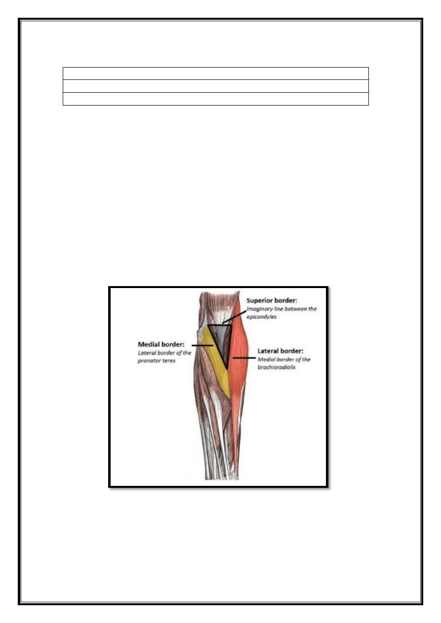
The cubital fossa is triangular, and thus has three borders:
1. Lateral border
– medial border of the brachioradialis muscle.
2. Medial border
– lateral border of the pronator teres muscle.
3. Superior border
– hypothetical line between the epicondyles of the humerus.
The floor of the cubital fossa formed proximally by the brachialis, and distally by
the supinator muscle. The roof consists of skin and fascia, and reinforced by the
bicipital aponeurosis. Within the roof runs the median cubital vein, which can
accessed for venipuncture.
6 – Anterior view of the superficial forearm, showing the borders
of the cubital fossa.
6
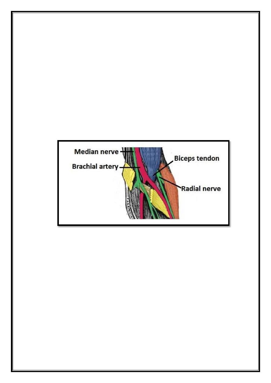
Contents
The contents of the cubital fossa include vessels, nerves and the biceps tendon
(lateral to medial):
Radial nerve
– this not always considered part of the cubital fossa, but is in the
vicinity, passing underneath the brachioradialis muscle. As it does so, the radial
nerve divides into its deep and superficial branches.
Biceps tendon
– runs through the cubital fossa, attaching to the radial tuberosity,
just distal to the neck of the radius.
Brachial artery
– supplies oxygenated blood to the forearm. It bifurcates into the
radial and ulnar arteries at the apex of the cubital fossa.
Median nerve
– leaves the cubital between the two heads of the pronator teres. It
supplies the majority of the flexor muscles in the forearm.
Fig 7 – Medial to lateral, the contents of the cubital fossa