1
Muscles of upper limb
1
st
stage
Dr.Kalid Ali Zayer
Muscles of Pectoral region
Contents
1. Pectoralis Major
2. Pectoralis Minor
3. Serratus Anterior
4. Subclavius
The pectoral region is located on the anterior chest wall. It contains four muscles that exert a
force on the upper limb; the pectoralis major, pectoralis minor, serratus anterior and
subclavius.
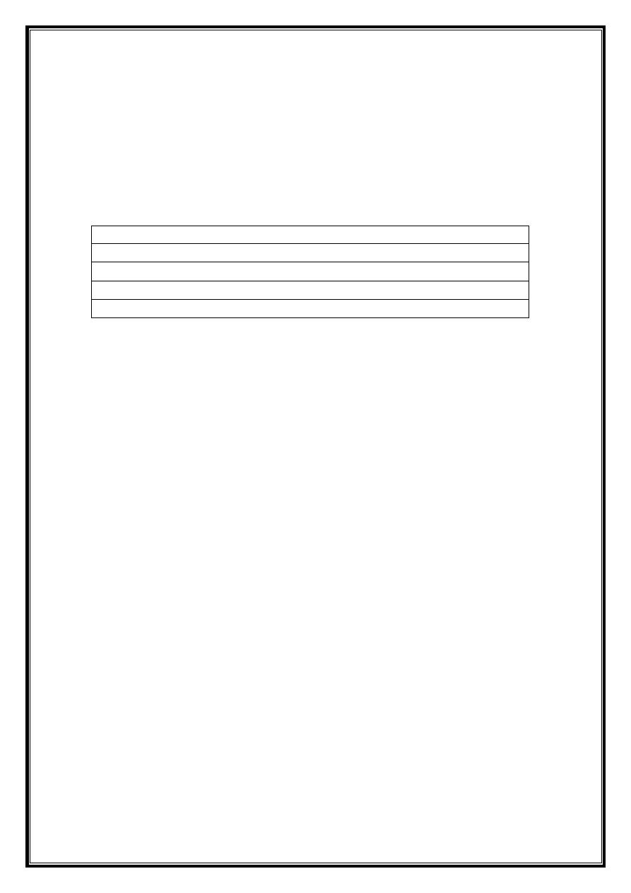
1. Pectoralis Major
The pectoralis major is the most superficial muscle in the pectoral region. It is large and fan
shaped, and is composed of a sternal head and a clavicular head:
Attachments
: The distal attachment of both heads is into the intertubercular sulcus of
the humerus.
o Clavicular head – originates from the anterior surface of the medial clavicle.
o Sternocostal head – originates from the anterior surface of the sternum, the superior six
costal cartilages and the aponeurosis of the external oblique muscle.
Function:
Adducts and medially rotates the upper limb, and draws the scapula
anteroinferiorly.The clavicular head also acts individually to flex the upper limb.
Innervation:
Lateral and medial pectoral nerves.
2
Fig 1 – The sternal and clavicular heads of the pectoralis major.
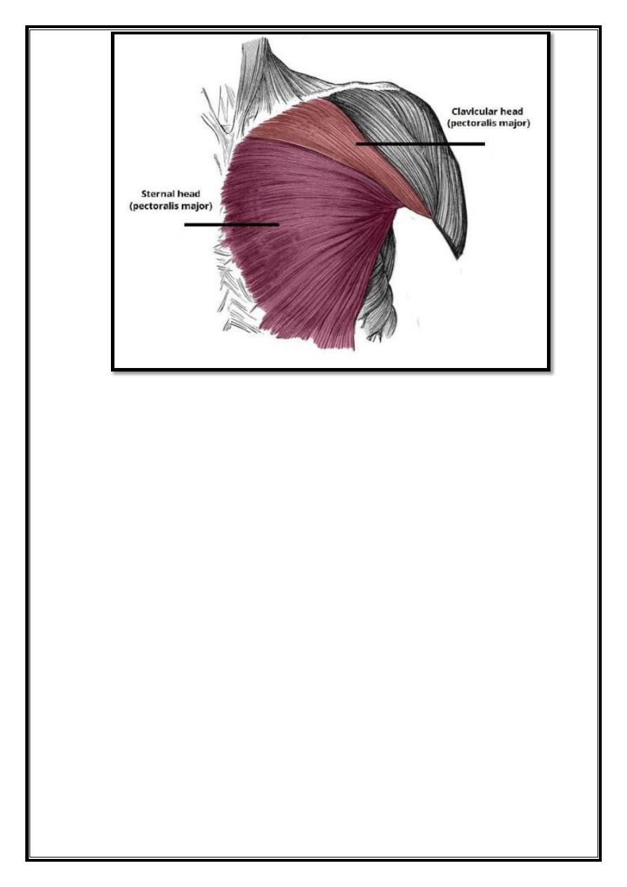
2. Pectoralis Minor
The pectoralis minor lies underneath its larger counterpart muscle, pectoralis major. Both of
these muscles form part of the anterior wall of the axilla region.
Attachments:
Originates from the 3rd-5th ribs, and inserts into the coracoid process of the
scapula.
Function:
Stabilizes the scapula by drawing it anteroinferiorly against the thoracic wall.
Innervation:
Medial pectoral nerve.
3. Serratus Anterior
The serratus anterior is located more laterally in the chest wall, and forms the medial border of
the axilla region.
Attachments:
The muscle consists of several strips, which originate from the lateral aspects
of ribs 1-8. They atach to the costal (rib facing) surface of the medial border of the scapula.
Function:
Rotates the scapula, allowing the arm to be raised over 90 degrees. It
also holds the scapula against the ribcage.
Innervation:
Long thoracic nerve.
3
Fig 2 – The serratus anterior and pectoralis minor muscles.
4. Subclavius
The subclavius is small muscle, which is located directly underneath the clavicle, running
horizontally.
It affords some minor protection to the underlying neurovascular structures (e.g in cases of
clavicular fracture or other trauma).
Attachments:
Originates from the junction of the first rib and its costal cartilage, inserting
into the inferior surface of the middle third of the clavicle.
Function:
Anchors and depresses the clavicle.
Innervation:
Nerve to subclavius.
Muscles of the shoulder region
The muscles of the shoulder are associated with movements of the upper limb. They produce
the characteristic shape of the shoulder, and can divided into two groups:
Extrinsic
– originate from the torso, and attach to the bones of the shoulder
(clavicle, scapula or humerus).
Intrinsic
– originate from the scapula and/or clavicle, and attach to the humerus.
Note: other muscles that act on the shoulder joint (the muscles of the pectoral region, and the
upper arm).

4
The extrinsic muscles
of the shoulder originate from the trunk, and attach to the bones of the
shoulder – the clavicle, scapula or humerus. They are located in the back, and also known as
the superficial back muscles.
The muscles organized into two layers – a superficial layer and a deep layer.
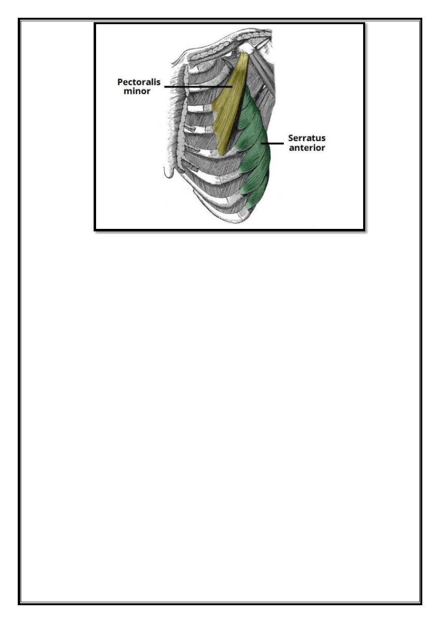
Superficial
There are two superficial extrinsic muscles – the trapezius and latissimus dorsi.
1. Trapezius
The trapezius is a broad, flat and triangular muscle. The muscles on each side form a trapezoid
shape. It is the most superficial of all the back muscles.
Attachments
: Originates from the skull, nuchal ligament and the spinous processes of C7-
T12. The fibers attach to the clavicle, acromion and the scapular spine.
Innervation:
Motor innervation is from the accessory nerve. It also receives proprioceptor
fibers from C3 and C4 spinal nerves.
Actions:
The upper fibers of the trapezius elevate the scapula and rotates it during abduction
of the arm.
The middle fibers retract the scapula and the lower fibers pull the scapula inferiorly.
2. Latissimus Dorsi
The latissimus dorsi originates from the lower part of the back, where it covers a wide area.
Attachments:
Has a broad origin – arising from the spinous processes of T6-T12, iliac crest,
thoracolumbar fascia and the inferior 3 ribs. The fibers converge into a tendon that attaches to
the intertubercular sulcus of the humerus.
Innervation:
Thoracodorsal nerve.
Actions:
Extends, adducts and medially rotates the upper limb.
Deep
There three muscles in this group – the levator scapulae and the two rhomboids. They
situated in the upper back, underneath the trapezius.
5
Fig 3 – The extrinsic muscles of the shoulder.
1. Levator Scapulae
The levator scapulae is a small strap-like muscle. It begins in the neck, and descends to
attach to the scapula.
Attachments:
Originates from the transverse processes of the C1-C4 vertebrae and
ataches to the medial border of the scapula.
Innervation:
Dorsal scapular nerve.
Actions:
Elevates the scapula.
2. Rhomboids
There are two rhomboid muscles – major and minor. The rhomboid minor situated
superiorly to the major.
6
a.
Rhomboid Major
Attachments:
Originates from the spinous processes of T2-T5 vertebrae. Ataches to
the medial border of the scapula, between the scapular spine and inferior angle.
Innervation:
Dorsal scapular nerve.
Actions:
Retracts and rotates the scapula.
b.
Rhomboid Minor
Attachments:
Originates from the spinous processes of C7-T1 vertebrae. Ataches to
the medial border of the scapula, at the level of the spine of scapula.
Innervation:
Dorsal scapular nerve.
Actions:
Retracts and rotates the scapula.
The intrinsic muscles
(also known as the scapulohumeral group) originate from the
scapula and/or clavicle, and attach to the humerus.
There are six muscles in this group – the deltoid, teres major, and the four rotator cuff
muscles (supraspinatus, infraspinatus, subscapularis and teres minor).
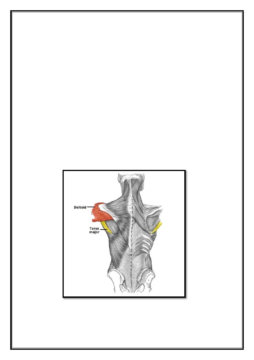
1. Deltoid
Fig 4 – The deltoid and teres major

7
The deltoid muscle is shape like the Greek letter delta – Δ. It can divided into an anterior,
middle and posterior part.
Attachments:
Originates from the scapula and clavicle, and attaches to the deltoid
tuberosity on the lateral surface of the humerus.
Innervation:
Axillary nerve.
Actions:
o Anterior fibers – flexion and medial rotation.
o Posterior fibers – extension and lateral rotation.
o Middle fibers – the major abductor of the arm (takes over from the supraspinatus, which
abducts the frst 15 degrees).
2. Teres Major
The teres major forms the inferior border of the quadrangular space – the ‘gap’ that the
axillary nerve and posterior circumflex humeral artery pass through to reach the
posterior scapula region.
Attachments:
Originates from the posterior surface of the inferior angle of the
scapula. It attaches to the medial lip of the intertubercular groove of the humerus.
Innervation:
Lower subscapular nerve.
Actions:
Adducts at the shoulder and medially rotates the arm.
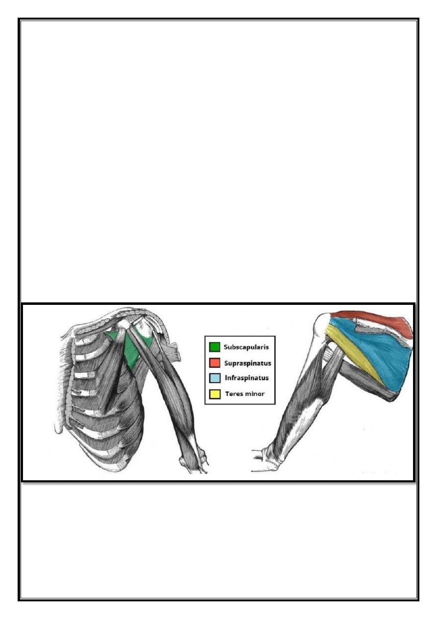
Rotator Cuff Muscles
The rotator cuff muscles are a group of four muscles that originate from the scapula and
attach to the humeral head. Collectively, the resting tone of these muscles acts to ‘pull’ the
humeral head into the glenoid fossa.
This gives the glenohumeral joint a lot of additional stability.
In addition to their collective function, the rotator cuff muscles also have their own individual
actions.
3. Supraspinatus
Attachments:
Originates from the supraspinous fossa of the scapula, attaches to the
greater tubercle of the humerus.
Innervation:
Suprascapular nerve.
Actions:
Abducts the arm 0-15o, and assists deltoid for 15-90o
8
4.
Infraspinatus
Attachments:
Originates from the infraspinous fossa of the scapula, attaches to the
greater tubercle of the humerus.
Innervation:
Suprascapular nerve.
Actions:
Laterally rotates the arm.
5.
Subscapularis
Attachments:
Originates from the subscapular fossa, on the costal surface of the
scapula. It attaches to the lesser tubercle of the humerus.
Innervation:
Upper and lower subscapular nerves.
Actions:
Medially rotates the arm.
6. Teres Minor
Attachments:
Originates from the posterior surface of the scapula, adjacent to its lateral
border. It attaches to the greater tubercle of the humerus.
Innervation:
Axillary nerve.
Actions:
Laterally rotates the arm.
Fig 5 – The rotator cuff muscles, which act to stabilize the shoulder joint.
9
Muscles of upper arm
Contents
1. Anterior Compartment
o
1.1 Biceps Brachii
o
1.2 Coracobrachialis
o
1.3 Brachialis
2. Posterior Compartment
o
2.1 Triceps Brachii
The upper arm is located between the shoulder joint and elbow joint. It contains four
muscles – three in the anterior compartment (biceps brachii, brachialis, coracobrachialis),
and one in the posterior compartment (triceps brachii).
Anterior Compartment
There are three muscles located in the anterior compartment of the upper arm – biceps brachii,
coracobrachialis and brachialis. They all innervated by the musculocutaneous nerve. A good
memory aid for this is BBC – biceps, brachialis, coracobrachialis.
Arterial supply to the anterior compartment of the upper arm is via muscular branches of the
brachial artery.
1. Biceps Brachii
The biceps brachii is a two-headed muscle. Although the majority of the muscle mass is
located anteriorly to the humerus, it has no attachment to the bone itself.
As the tendon of biceps brachii enters the forearm, a connective tissue sheet is given off
– the bicipital aponeurosis. This forms the roof of the cubital fossa and blends with the
deep fascia of the anterior forearm.
Attachments:
Longhead originates from the supraglenoid tubercle of the scapula, and
the short head originates from the coracoid process of the scapula. Both heads insert
distally into the radial
tuberosity and the fascia of the forearm via the bicipital aponeurosis.
Function:
Supination of the forearm. It also flexes the arm at the elbow and at the
shoulder.
Innervation:
Musculocutaneous nerve.
2. Coracobrachialis
The coracobrachialis muscle lies deep to the biceps brachii in the arm.
Attachments:
Originates from the coracoid process of the scapula. The muscle passes
through the axilla, and attaches the medial side of the humeral shaft, at the level of the
deltoid tubercle.
Function:
Flexion of the arm at the shoulder, and weak adduction.
Innervation:
Musculocutaneous nerve.
10
3. Brachialis
The brachialis muscle lies deep to the biceps brachii, and found more distally than the
other muscles of the arm. It forms the floor of the cubital fossa.
Attachments:
Originates from the medial and lateral surfaces of the humeral shaft
and inserts into the ulnar tuberosity, just distal to the elbow joint.
Function:
Flexion at the elbow.
Innervation:
Musculocutaneous nerve, with contributions from the
radial nerve.
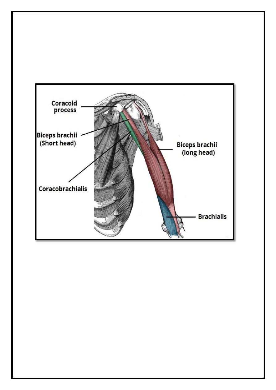
Fig 6 – The coracobrachialis, biceps brachii and brachialis muscles of the anterior upper
arm.
Posterior Compartment
The posterior compartment of the upper arm contains the triceps brachii muscle, which has
three heads. The medial head lies deeper than the other two, which cover it.
Arterial supply to the posterior compartment of the upper arm is via the profunda brachii
artery.
11
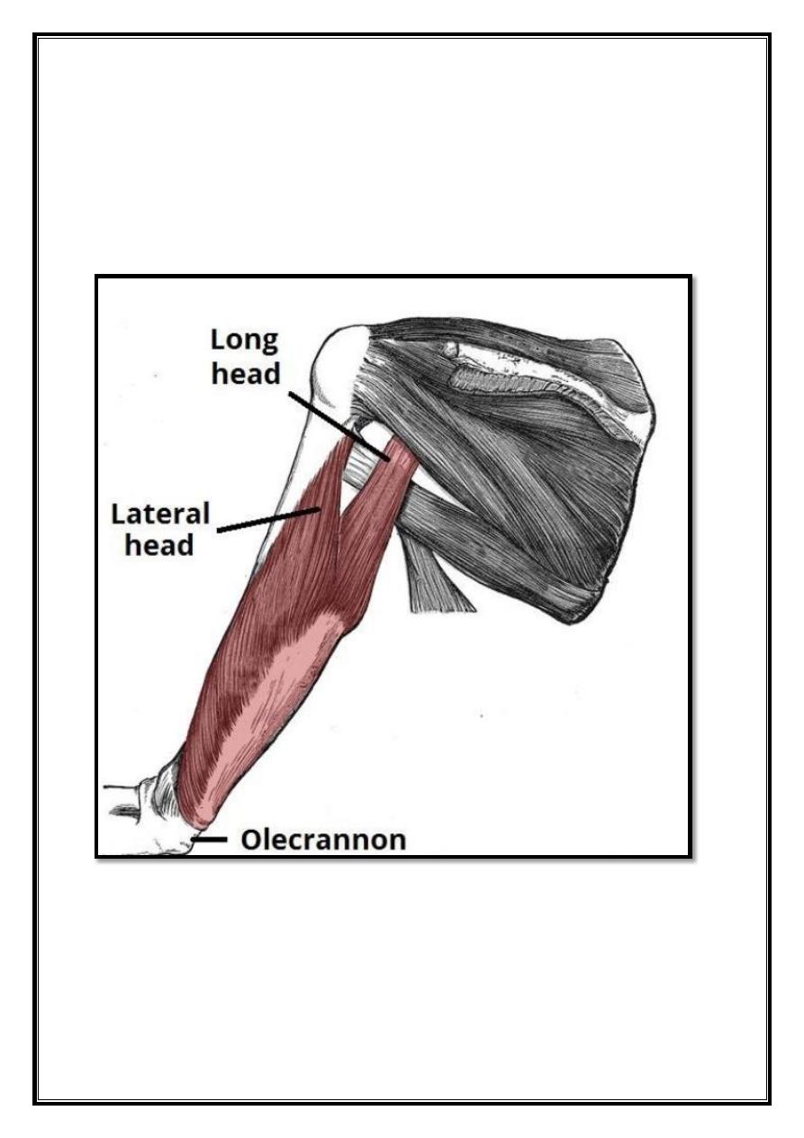
Triceps Brachii
Attachments:
Long head – originates from the infraglenoid tubercle. Lateral head –
originates from the humerus, superior to the radial groove. Medial head – originates from the
humerus, inferior to the radial groove. Distally, the heads converge onto one tendon and insert
into the olecranon of the ulna.
Function:
Extension of the arm at the elbow.
Innervation
: Radial nerve.
o Note: in some individuals, the axillary nerve innervates the long head
of the triceps brachii.
Fig 7 – The long and lateral heads of the triceps brachii.