Lec.5,6
Haemoflagellata
Trypanosomes :
Trypanosomes are hemoflagellate protozoa.
Two distinctly forms of genus
Trypansoma occur in humans. They cause African trypanosomiasis (or African
sleeping sickness) and American typanosomiasis respectively.
The complex Trypanosoma brucei have two subspecies that are morphologically
indistinguishable cause distinct disease patterns in humans:
T. b. gambiense causes West African sleeping sickness .
T. b. rhodesiense causes East African sleeping sickness.
The protozoan parasite, Trypanosoma cruzi, causes American typansosmiasis (or
Chagas' disease), that can be transmitted to humans by blood-sucking reduviid bugs.
Trypanosoma brucei
African trypanosomiasis – sleeping sickness
T. brucei gambiense (Gambian sleeping sickness) is seen in western and central
parts of equatorial Africa and T. brucei rhodesiense (Rhodesian sleeping sickness) in
east Africa . approximately 20.000 cases are reported each year. T. brucei gambiense
and T. brucei rhodesiense are similar in all aspect except their geographic distribution
and clinical manifestation . T. rhodesiense , which could infect man, in whom it
caused an acute disease; and T. gambiense, also infective to man but producing a
much more chronic disease. T. b. gambiense and T. b. rhodesiense parasites inhabit
the connective tissue. In man and other vertebrate hosts, these are found in the blood
stream, lymph nodes and cerebrospinal fluid.
Coll. Medicine \3rd stage
Medical Parasitology
Assist.prof.Dr. Amal kh.kh.

T. brucei gambiense T. brucei rhodesiense
Morphology :
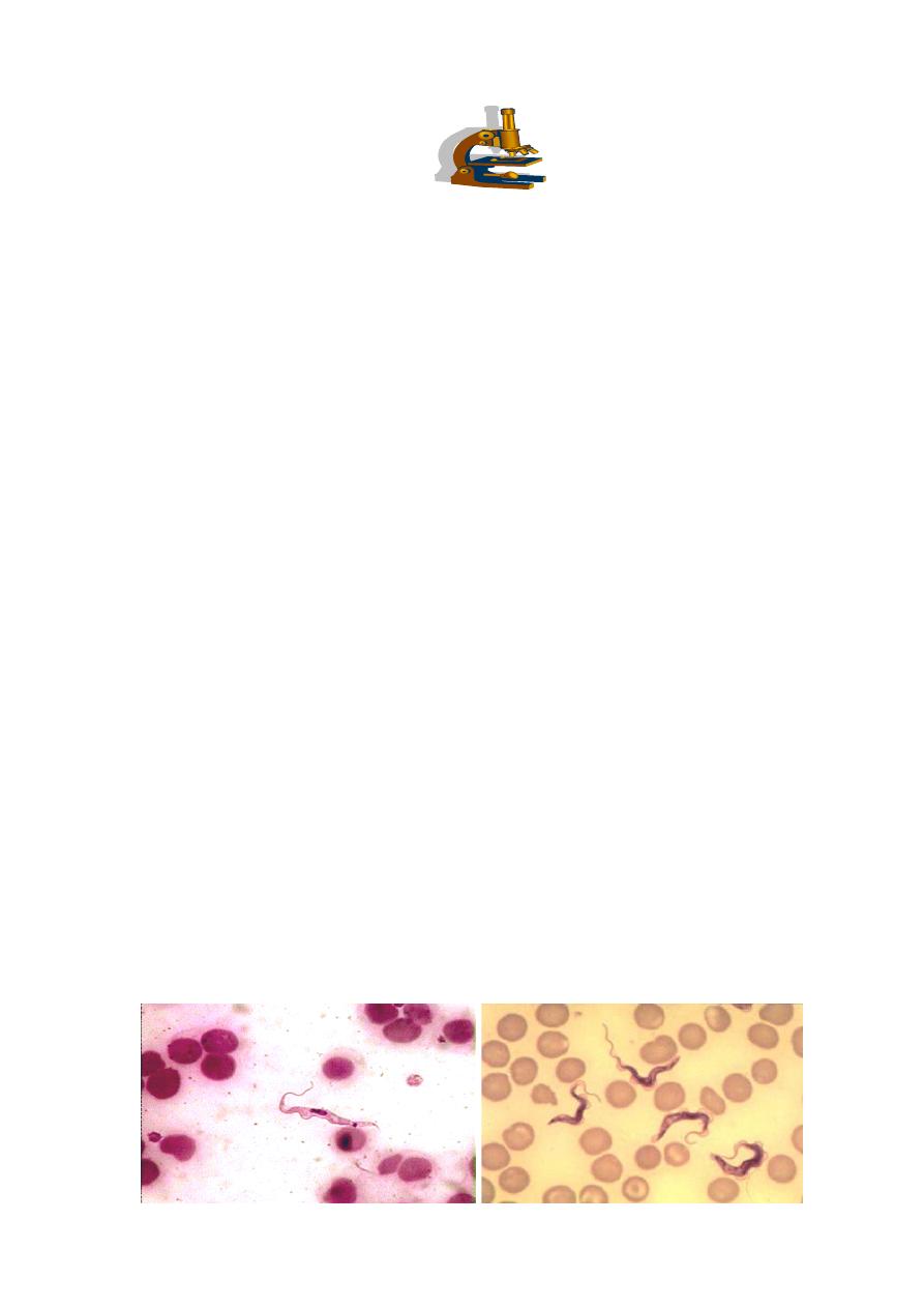
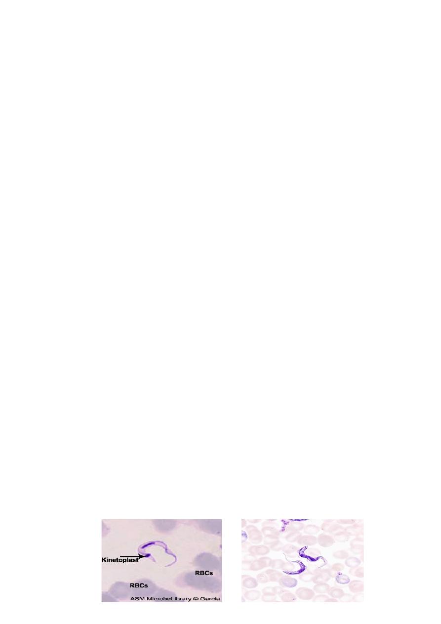
The parasite exists in two forms , trypomastigotes and epimastigotes .
trypomastigotes are seen in the vertebrates as well as in the insect host (see the next
diagram). trypomastigotes show two morphological variations . one of these is a
long , slender variant which is present extracellularly in blood , lymph and tissue
fluids . these variants multiply by longitudinal binary fission and survive in the tissue
of the vertebrate host due to their ability to change the outer variant surface
glycoprotein (VSG) coat . each VSG is immunogenic but antigenically different from
the preceding VSG and is changed every 8 to 10 days to evade the immune response .
in the insect vector , these long ,slender , trypomastigotes multiply in the mid – gut .
These long ,slender , trypomastigotes after some multiplication cycles convert into ,
short , stumpy , non dividing forms . in human and other vertebrates they are present
in blood, lymph and tissue fluid . these forms are infective to the insects host . the
short , stumpy , non dividing trypomastogotes are also seen in the salivary of the
insect hosts, where they called metacyclic trypomastigotes . these metacyclic
trypomastigotes are infective to human beings and other vertebrates .
The other forms of T. brucei gambiense and T. brucei rhodesiense are the
epimastigotes . epimastigotes are seen in the insect host . these forms multiply by
longitudinal fission in the salivary gland and ultimately produced metacyclic
trypomastigotes .
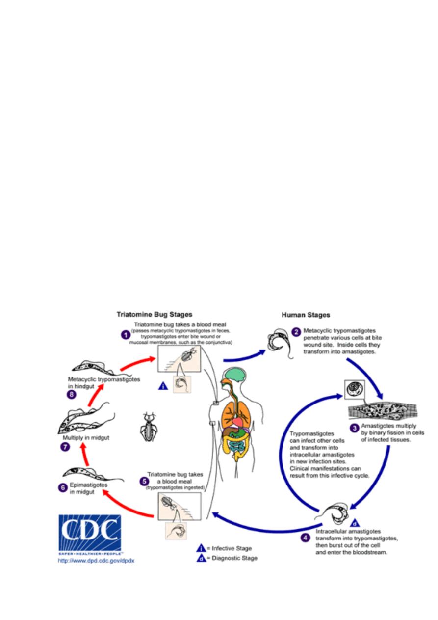
Life cycle :
T. brucei gambiense and T. brucei rhodesiense have a complex life cycle . they
required to two host to complete their life cycle . they do not have a mode of sexual
reproduction . human and other vertebrates are the vertebrate host and insects are the
invertebrate hosts . Human is only vertebrate host to T. brucei gambiense . human and
wild mammals are the vertebrate host of T. brucei rhodesiense. Tsetse flies (Glossina
sp) act as invertebrate hosts . Glossina palpalis for T. brucei gambiense , while
Glossina morsitans for T. brucei rhodesiense .
The infective form for human and other vertebrates are the metacyclic
trypomastigotes . they are introduced in the tissue of host by the bite of an infected
tsetse flies . metacyclic trypomastigotes convert into long, slender trypomastigotes
which is multiply locally as well as in blood . the short ,stumpy , non dividing
trypomastigotes formed after some multiplication cycles are sucked by the tsetse flies
during their blood meal . these forms reach the mid -gut and convert themselves into
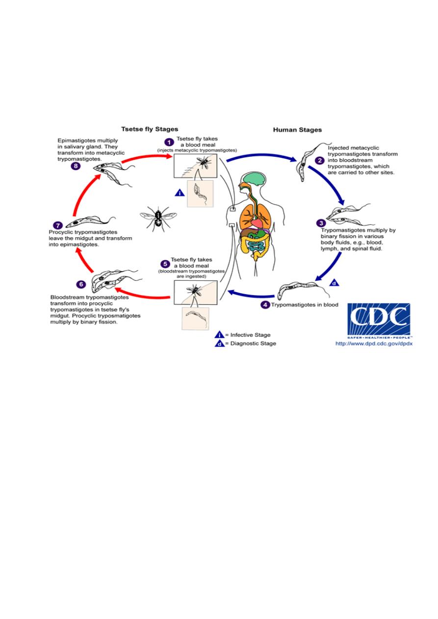
procyclic trypomastigotes . these procyclic trypomastigotes multiply in mid gut and
reach salivary gland where they change into epimastigotes. Epimastigotes, in turn,
multiply and produce metacyclic trypomastigotes . when these metacyclic
trypomastigotes reach a new vertebrate host , the life cycle of T. brucei gambiense and
T. brucei rhodesiense is completed (see the following diagram).
Pathogenesis and clinical manifestation :
Infection occurs in 3 stages: A Trypanosomal chancre can develop on the site of
inoculation. This is followed by a haematolymphatic stage with symptoms that
include fever, lymphadenopathy, and pruritus. In the meningoencephalitic stage ,
invasion of the central nervous system can cause headaches, somnolence, abnormal
behavior, and lead to loss of consciousness and coma. The course of infection is much
more acute with T. b. rhodesiense than T. b. gambiense .
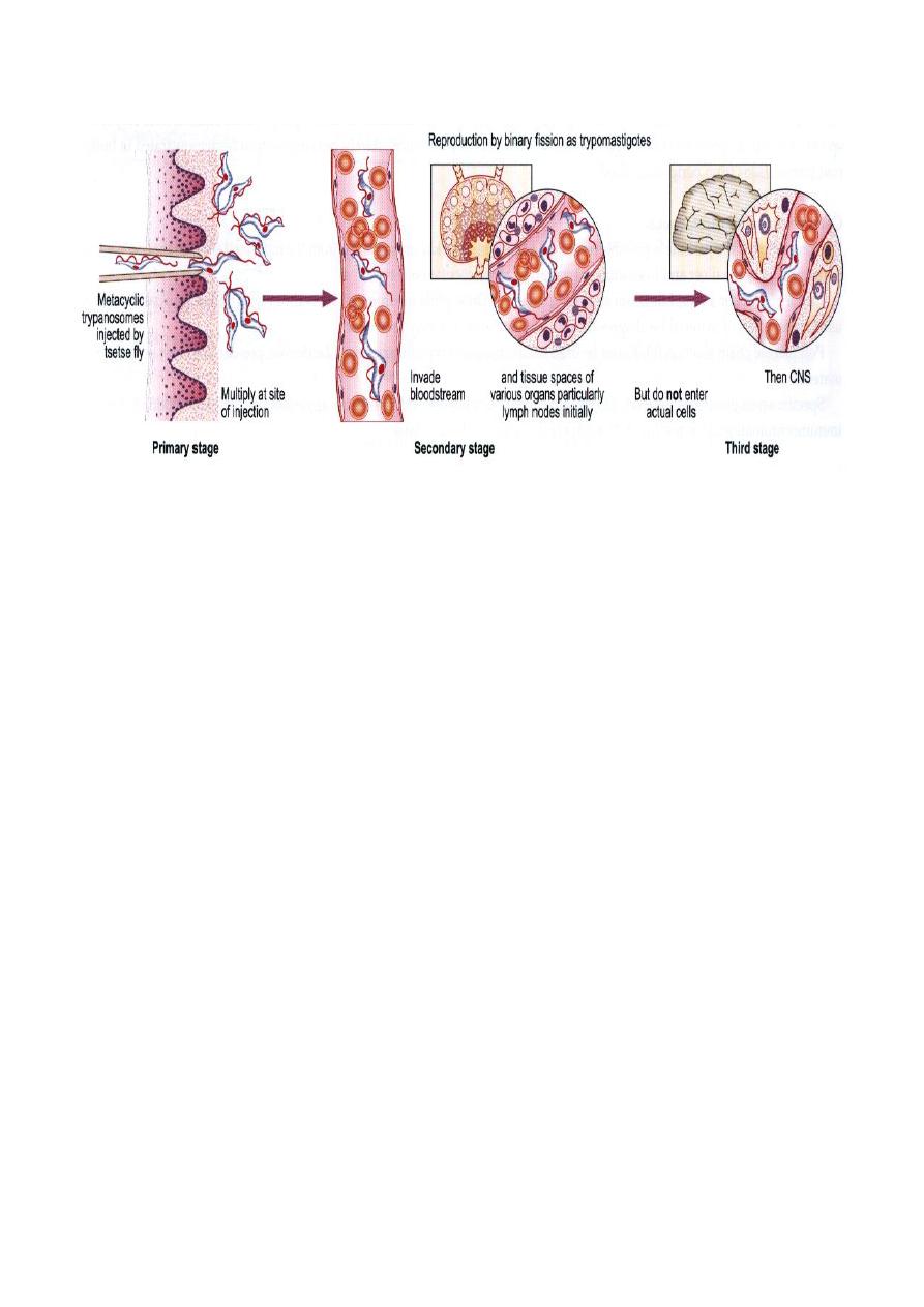
1. Chancre :
Trypansomal chancre is an acute inflammatory local response seen in a week or so
after the bite of infected tsetse fly. It is large, red and rubbery. It is more frequently
seen in Rhodesian trypanosomiasis. It shows an intense inflammatory infiltration,
vasodilatation and interstitial oedema. The chancre tissue is filled with parasites. A
painful trypansomal chancre appears within a few days at the site of bite and resolves
spontaneously within several weeks. It is characterized by erythema, swelling and
local tenderness.
2. Haematolymphatic stage :
In the early stage of the disease, after development of the chancre, infection of the
blood and lymph system results in a more or less acute febrile illness. Infected lymph
glands, especially those at back of the neck, may become very enlarged; the swollen
cervical glands constitute ”Winterbottom’s sign”, a classical diagnostic indication of
T. b. gambiense . Oedema, hepatosplenomegaly and tachycardia are other frequent
findings.
3. Meningoencephalitic stage:
More serious effects results from the penetration of the parasites into the CNS, which
may occur at any time from weeks (T. b. rhodesiense ) to years (T. b.gambiense ) after
initial infection. Here the parasites multiply in the blood vessels, tissue fluids and
cerebrospinal fluid (CSF). The outcome of the inflammatory process
(meningoencephalitis) is brain damage leading to somnolence , coma and, unless
treated, death in almost all cases.
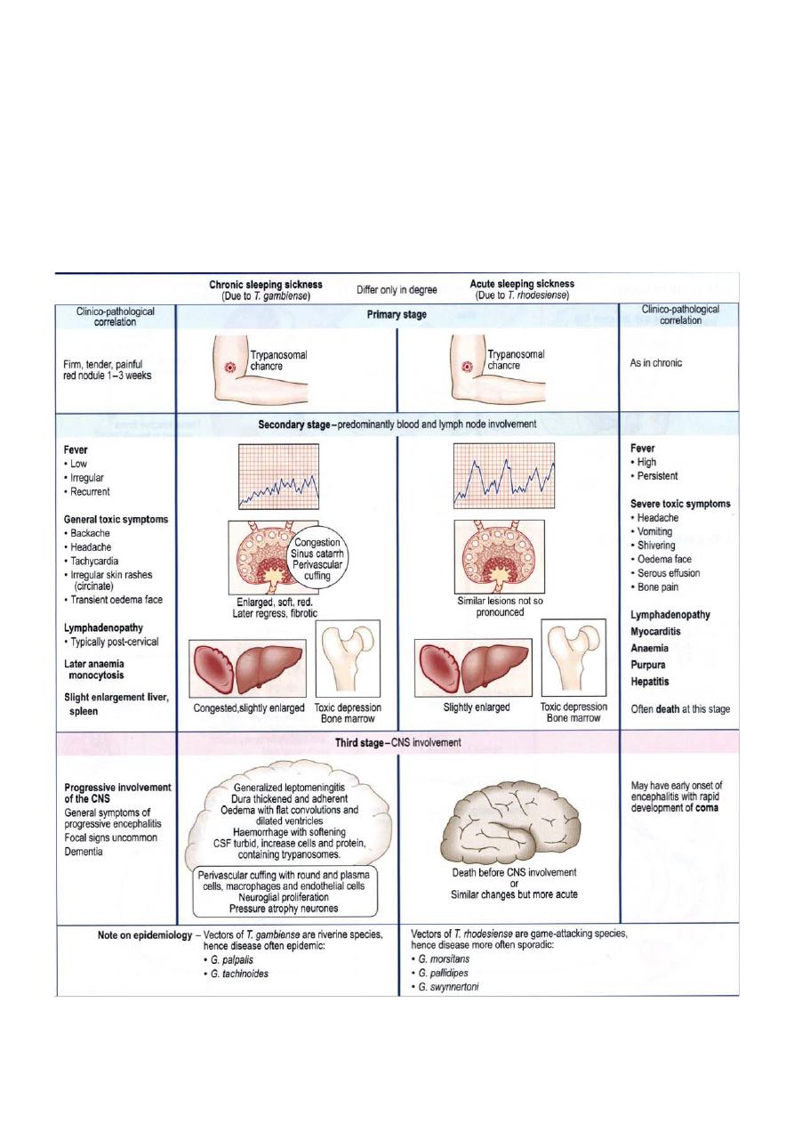
There are differences between the clinical manifestations of East African and West
African trypanosomiasis. In T. b. rhodesiense (East African trypanosomiasis), there is
usually little obvious glandular involvement and Winterbottom’s sign may not be
present; weight loss is rapid, and CNS is involved early. Untreated persons usually die
within 9 months to a year after onset of disease. The incubation period is commonly
short. In T. b. gambiense (West African trypanosomiasis) chronic CNS disease
developed.
Diagnosis:
The diagnosis rests upon demonstrating trypanosomes by microscopic examination of
chancre fluid, lymph node aspirates, blood, bone marrow, or, in the late stages of
infection, cerebrospinal fluid. A wet preparation should be examined for the motile
trypanosomes, and in addition a smear should be fixed, stained with Giemsa (or
Field), and examined. Concentration techniques can be used prior to microscopic
examination.
Treatment :
Treatment should be started as soon as possible and is based on the infected person’s
symptoms and laboratory results. The drug regimen depends on the infecting species
and the stage of infection. Pentamidine isethionate and suramin are the drugs of
choice to treat the hemolymphatic stage of West and East African Trypanosomiasis,
respectively. Melarsoprol is the drug of choice for late disease with central nervous
system involvement (infections by T.b. gambiense or T. b. rhodiense ).
Prevention :
1. avoiding areas harbouring tsetse flies .
2. using a protective clothing and insect repellants .
3. vaccines is not available.
Trypanosoma cruzi
Trypanosoma cruzi , causes Chagas Disease that can be transmitted to humans by
blood-sucking reduviid bugs . Chagas disease (South American trypansomiasis) is
commonly seen in the countries of South America , it is also occur in central part of
America.
Morphology :
T. cruzi exist in four forms amastigotes , promastigote , epimastigotes, and
trypomastigotes . Amastigote live in muscles of heart and skeletal system , nerve cell
and cell of reticulo- endothelial system. This form is multiplying for promastigote ,
epimastigotes, and trypomastigotes. Trypomastigotes appears in peripheral blood
from time to time and do not multiply in human. Trypanomastigotes are usually C-
shaped and slender .
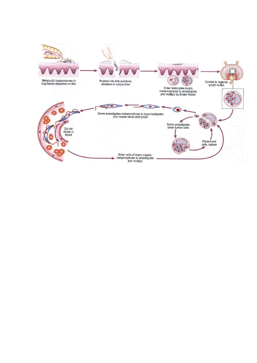
Life cycle :
T. cruzi has a complex life cycle . humans are the vertebrate hosts while reduviid
bugs (Triatoma infestans) are the invertebrate host. Infective stage of the parasite to
humans are the metacyclic trypomastigotes . metacyclic trypomastigotes are
deposited along with the feaces of the reduviid bugs near the bite wound . these
infective forms are then rubbed into the wound by the bitten person or transferred to
his conjunctiva through contaminated finger . these forms enter the cells of reticulo-
endothelial system and spread all over the body . in these cells metacyclic
trypomastigotes are converted to amastigote . Amastigotes multiply and developed
into trypomastigotes after passing through the successive stages of promastigotes and
epimastigotes . trypomastigotes are released in the blood .
When a reduviid bugs bites such a person , the trypomastigotes taken up along with
blood meal . trypomastigotes transformed into epimastigotes that multiply and
migrate to the hind gut . epimastigotes finally develop into the metacyclic
trypomastigotes . the life cycle is thus completed when a new vertebrate hosts are
infected with these metacyclic trypomastigotes . T. cruzi also acquired through
blood transfusion and placenta .
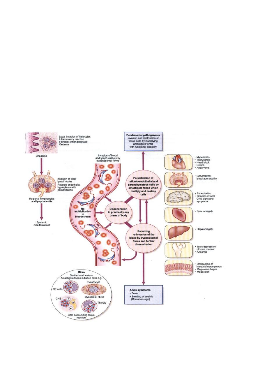
Pathogenesis and clinical features :
Trypomastigotes may induced a local inflammatory reaction and swelling at the
site of its entry . A Chagoma develops on skin while Rommana ُs sign develop as a
results of unilateral oedematous swelling of eyelids .
Intercellular multiplication of amastigotes damages the cells at various sites .
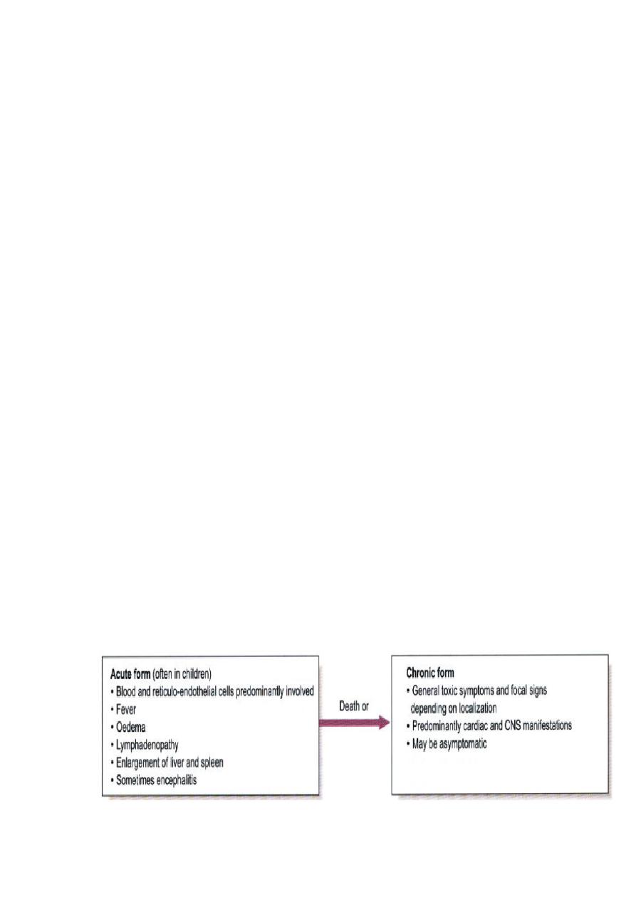
Chagas disease can be either acute or chronic . the incubation period is generally
from 1 to 2 weeks and the patient develops fever and generalized non pitting
oedema in the acute form. It last for 3 to 4 weeks and may end fatally with
myocardiatis or meningoencephalitis . the chronic form present with cardiological ,
neurological or visceral manifestation . complete heart block or brain damage may
cause sudden death.
Acute Chagas ’disease:
It occurs most commonly in infants and children. The first sign of illness occurs at
least 1 week after invasion by the parasites.
A local lesion (chagoma, palpebral edema) can appear at the site of inoculation.
Chagoma is localized swelling of the skin and contains intracellular amastigotes in
leucocytes and subcutaneous. When the parasite is inoculated in the conjunctiva, a
unilateral painless oedema of the palpebral and perioccular tissue develops in the eye.
It is called Romana’s sign and is the classical finding in the acute Chagas ’ disease.
The acute phase is usually asymptomatic, but can present with manifestations that
include fever, anorexia, lymphadenopathy, and mild hepatosplenomegaly; in severe
infection, myocarditis may developed. Most deaths in acute Chagas’ disease are due
to heart failure or meningoencephalitis. The acute stage lasts for 20-30 days.
Symptoms resolve in most of the patients who then enter into asymptomatic or
indeterminate stage of T. cruzi .
Chronic Chagas ’disease:
It is seen in older children and adults between 20-40 years of age. The symptomatic
chronic stage may not occur for years or even decades after initial infection; it may
also be seen in persons without any previous episode of acute disease. Its
manifestations include cardiomyopathy (the most serious manifestation); pathologies
of the digestive tract such as megaesophagus and megacolon; and weight loss.
Chronic Chagas’ disease and its complications can be fatal.
During the chronic phase, although signs may not be apparent, the
repeated cycle of
intracellular multiplication are continually destroying cells, not only those in which
the amastigotes multiply, but also neighbouring cells. An autoimmune mechanism is
probably involved. Neurons are particularly vulnerable to destruction. If the
intracellular groups of parasite are concentrated in parts of gastrointestinal tract,
especially in oesophagus or colon, peristalsis may be interfered with and the organ
may become hugely distended. This condition is indicated by the prefix mega; for
example megaoesophagus or megacolon
.
The unfortunate patient may be unable to
swallow and die of starvation. Megacolon may become so gross as to lead to rupture
of colon and death.
If the amastogote congregate in the heart muscle, and some strains are more prone to
do this than others, the ensuing neuronal and muscle destruction may gravely weaken
the heart wall, causing irreversible damage and leading to an early death from heart
attack.
Laboratory diagnosis :
1. Microscopic examination of stained blood film show the trypomastigotes .
2. Xenodiagnosis , non infected reduviid bug is allowed to feed on a suspected
patient then the faces of this bug are examined for trypomastigotes after 2
weeks.
Treatment and control :
Nifurtimox is the drug of choice , benznidazole is also useful. Prevention is
achieved by :
1. eradication of reduviid bug nets.
2. Construction of homes lacking cracks and crannies (the bug live there) .
Pathology of Trypanosoma cruzi

Lec.6
Class (3) :The Ciliated Protozoa
: The ciliate protozoa constitute a large number of species characterized by
1. having numerous short ectoplasmic threads or cilia, which are present in the
trophozoite.
2. Near the anterior end of the body there is a conical mouth, the cytostome, and
at the opposite end an anal opening, the cytopyge.
3. There are two types of nuclei, a larger, less dense macronucleus and nearby a
small, dense micronucleus ( or at times more than one ).
4. Multiplication is by transverse binary fission, with division of the cytoplasm
following that of the nuclei.
5. Many species also undergo conjugation, during which exchange of nuclei
occurs.
Balantidium coli:
Balantidium coli has a cosmopolitan distribution in hogs and is a common
parasites of man, it is found mostly in warm climates.
Morphology:
The organism has two stages, trophozoite and cyst.
-
Trophozoite : The trophozoite is the largest of the protozoa parasitizing man.
It is ovoidal, greenish-gray, covered with short cilia which are constantly in
motion during life, and has a vigorous forward movement as it plows through
even relatively thick liquid feces. The anterior end is somewhat conical and
the posterior end broadly rounded. To one side of the anterior tip there is a
funnel-shaped peristome, which leads into the cytostome , A minute
cytopyge is situated at the opposite end. One and at time two large pulsating
vacuoles are found within the cytoplasm. The body is covered with a
relatively tough pellicle. Somewhat posterior to the equator of the organism
there is an elongated kidney-shaped macronucleus and lying within the
concave side of the macronucleus a minute micronucleus.
-
Cyst : The cyst, spherical, is the resting and transfer stage. On encystation the
cilia are soon lost, though the marking on the cell surface remain.
Coll. Medicine \3rd stage
Medical Parasitology
Assist.prof.Dr. Amal kh.kh.
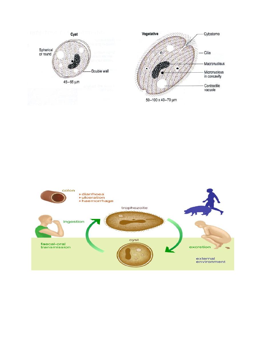
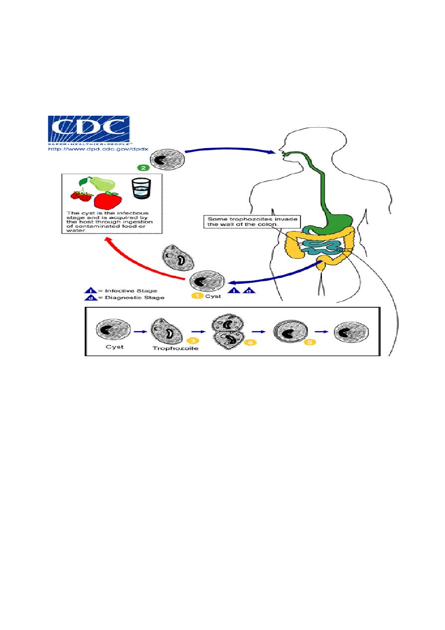
Route of transmission :
Transmission of parasite is by contaminated food and water by the cyst stage of
parasite through faecal – oral transmission . Trophozoite is not infective stage since it
is damage with gastric enzymes if it is enter through the oral route. Further
trophozoite encyst when release with stool and cannot persist for along time within
the external environment.
Life cycle :
Balantidium coli has simple life cycle . after ingestion of cyst stage by the suitable
host excystaion occur in small intestine to release the traophozoites . The natural
habitat of Balantidium coli trophozoite is the cecal level of the large intestine but the
parasite also occurs at all lower levels. It feeds on host cells, bacteria and other
nutritious substances in the tissues or lumen of the large bowel.
Asexual reproduction consist of transverse binary fission, in which the
micronucleus first divides, then the macronucleus, followed by the cytoplasm,
resulting in two daughter organisms. Although conjugation has been observed in
B.coli, this is not a common occurrence and apparently is not essential for its
propagation.
Pathology :
There is little if any evidence that Balantidium coli produces deep invasion of the
intestinal wall, although there may be superficial erosion of the mucosa. the mucosal
layer may be penetrated, with extensive submucosal destruction.
Since Balantidium coli is a much larger, sturdier organism than Entamoeba
histolytica, it produces a bigger opening in the intestinal mucosa as it enters the wall.
Moreover, its penetration seems to be accomplished more by boring action than by
lysis. Once established in the tissue it usually has no difficulty in penetration through
the muscularis mucosae into the submucosa, where it spreads out rapidly, causing
rapid destruction of the tissues; but unlike E. histolytica it rarely invades the muscular
coats and it has seldom been found in extra-intestinal tissues. While balantidial
lesions may develop at any level of the large intestine, they most commonly occur in
the cecal and sigmoid-rectal regions.

The symptoms in balantidiasis vary from fulminating, sometimes fatal dysentery
or profuse diarrhea to an essentially asymptomatic carrier state. In this respect the
parallel the broad spectrum of symptoms in amebic colitis.
Diagnosis and Treatment.:
Diagnosis is made on recovery of the characteristic trophozoites and cysts of
Balantidium coli in the stool. Care must be taken that the stool and the water or
physiologic saline solution employed in making fecal films or microscopic diagnosis
of this infection are not contaminated with free-living infusorians, otherwise these
ciliates may be mistaken for B. coli. Albendazole is the drug of choice.
Prevention :
1. Proper disposal of sewage .
2. Human stool should not be used as fertilizer.
3. Good personal hygiene .
4. Washing of raw vegetable and fruits before consumption and protection of food
from flies and cockroaches .
5. cyst is resistant to routine chlorination therefore drinking water should be
purified by either boiling , filtration or iodination.