Lec. : 4 Physiology
1
The Cardiovascular System
The Heart
The heart is a hollow, muscular organ about the size of a fist. It is responsible for
pumping blood through the blood vessels by repeated, rhythmic contractions. The
heart is composed of cardiac muscle, an involuntary muscle tissue that is found
only within this organ. The term "cardiac" (as in cardiology) means "related to the
heart” and comes from the Greek word kardia, for "heart." It has a four-chambered,
double pump and is located in the thoracic cavity between the lungs.
Myocardium
The myocardium is the muscular tissue of the heart. The myocardium is
composed of specialized cardiac muscle cells with an ability not possessed by
muscle tissue elsewhere in the body. Cardiac muscle, like other muscles, can

Lec. : 2 Physiology
contract, but it can also conduct electricity, like nerves. The blood to the
myocardium is supplied by the coronary arteries. If these arteries are occluded by
atherosclerosis and/or thrombosis, this can lead to angina pectoris or myocardial
infarction due to ischemia (lack of oxygen). Failure of the heart to contract
properly (for various reasons) is termed heart failure, generally leading to fluid
retention, edema, pulmonary edema, renal insufficiency, hepatomegaly, a
shortened life expectancy and decreased quality of life.
Pericardium
The pericardium is the thick, membranous sac that surrounds the heart. It
protects and lubricates the heart. There are two layers to the pericardium: the
fibrous pericardium and the serous pericardium. The serous pericardium is divided
into two layers; in between these two layers there is a space called the pericardial
cavity.
Epicardium
The layer next to the heart is the visceral layer, also known as the Epicardium.
This is the innermost layer and consists of connective tissue.
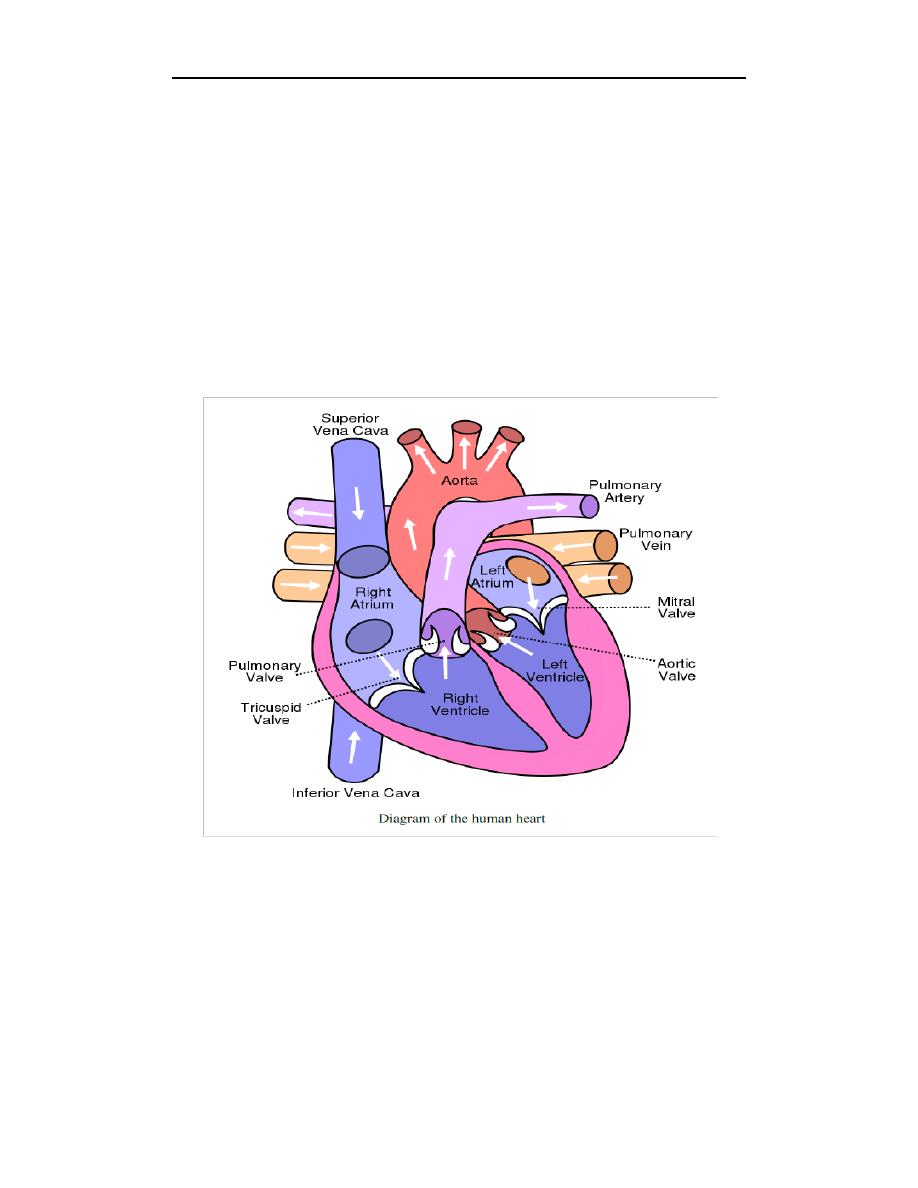
Heart Chambers
The heart has four chambers, two atria and two ventricles. The atria are smaller
with thin walls, while the ventricles are larger and much stronger.
Atrium
There are two atria on either side of the heart. On the right side is the atrium that
contains blood which is poor in oxygen. The left atrium contains blood which has
been oxygenated and is ready to be sent to the body. The right atrium receives de-
oxygenated blood from the superior vena cava and inferior vena cava. The left
atrium receives oxygenated blood from the left and right pulmonary veins.

Lec. : 4 Physiology
3
Ventricles
The ventricle is a heart chamber which collects blood from an atrium and pumps
it out of the heart. There are two ventricles: the right ventricle pumps blood into
the pulmonary circulation for the lungs, and the left ventricle pumps blood into the
systemic circulation for the rest of the body. Ventricles have thicker walls than the
atria, and thus can create the higher blood pressure. Comparing the left and right
ventricle, the left ventricle has thicker walls because it needs to pump blood to the
whole body. This leads to the common misconception that the heart lies on the left
side of the body.
Septum
The interventricular septum is the thick wall separating the lower chambers (the
ventricles) of the heart from one another.
Valves
The two atrioventricular (AV) valves are one-way valves that ensure that blood
flows from the atria to the ventricles, and not the other way. The two semilunar
(SL) valves are present in the arteries leaving the heart; they prevent blood from
flowing back into the ventricles. The sound heard in a heart beat is the heart valves
shutting. The right AV valve is also called the tricuspid valve because it has three
flaps. It is located between the right atrium and the right ventricle. The tricuspid
valve allows blood to flow from the right atrium into the right ventricle when the
heart is relaxed during diastole. When the heart begins to contract, the heart enters
a phase called systole, and the atrium pushes blood into the ventricle. Then, the
ventricle begins to contract and blood pressure inside the heart rises. When the
ventricular pressure exceeds the pressure in the atrium, the tricuspid valve snaps
shut. The left AV valve is also called the bicuspid valve because it has two flaps. It
is also known as the mitral valve due to the resemblance to a bishop's mitre
(liturgical headdress). This valve prevents blood in the left ventricle from flowing

Lec. : 2 Physiology
into the left atrium. As it is on the left side of the heart, it must withstand a great
deal of strain and pressure; this is why it is made of only two cusps, as a simpler
mechanism entails a reduced risk of malfunction. There are two remaining valves
called the Semilunar Valves. They have flaps that resemble half moons. The
pulmonary semilunar valve lies between the right ventricle and the pulmonary
trunk. The aortic semilunar valve is located between the left ventricle and the aorta.