Epithelium is a sheet of cells that covers the external surface of any solid
structure and the internal surface of any hollow tubular (e.g. lumen/cavities)
structure. Thus it serves as a barrier membrane separating the underlying
tissue from various external and internal environments.
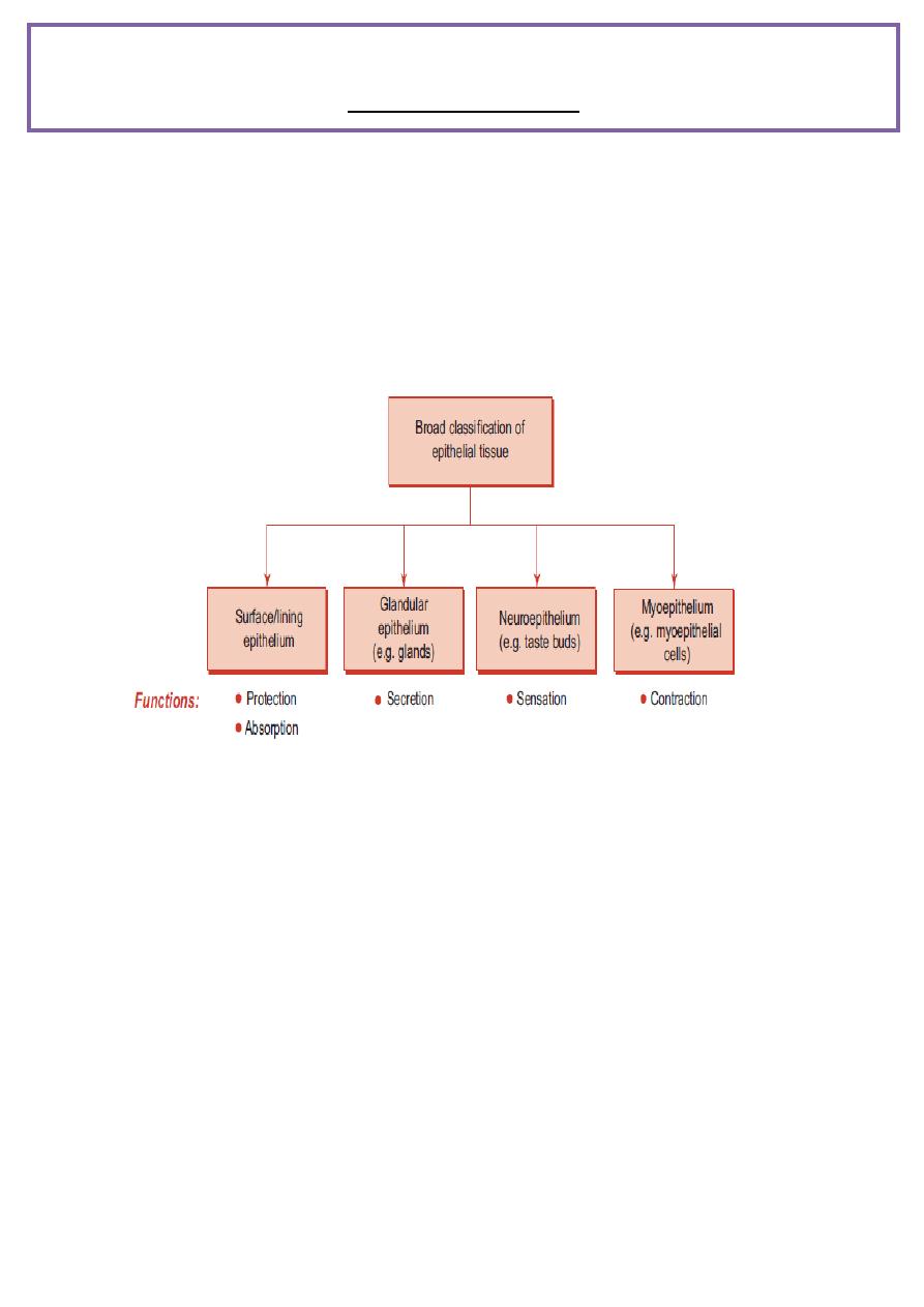
CLASSIFICATION OF EPITHELIAL TISSUE
On the basis of the function(s) performed, epithelial tissue can be broadly
classified into four types
SURFACE (OR) LINING EPITHELIUM
GENERAL FEATURES
Epithelium, the ‘cellular sheet’, is made of either single layer or many
layers of cells.
Epithelial cells are adherent to each other by means of junctional
complexes .
Very little intercellular material is found between the cells.
The deep surface (basal) of the epithelium rests on a basement
membrane, which separates it from the vascular connective tissue.
Basement membrane is made up of
(a) basal lamina (amorphous substance) – product of epithelium
(b) reticular lamina (reticular fibres) – product of connective tissue.
د
.
أديب عبد العالي االزبجي
Histology
Lecture :- 3
Epithelial Tissue

The superficial surface (apical) of the epithelium is free and exposed to
air or fluid and often shows modifications (i.e. presence of microvilli or
cilia) depending upon the function it is destined to perform.
No blood vessels nor lymphatics are found in the epithelium; nourishment
is provided by diffusion from the adjacent supporting tissues.
Epithelium has good regenerative capacity.
Its nuclear shape corresponds to cell shape (nuclei are oval in columnar
cells, round in cuboidal and polyhedral cells, and flat in squamous cells).
Epithelium invaginates/infolds and subsequently grows in the underlying
connective tissue, thus specialising as glands.
Epithelium may undergo morphological and functional changes from one
type to another type (metaplasia).
Functions: Protection, absorption, secretion, excretion, lubrication,
sensation and reproduction.
Epithelium is derived from all three germ layers (skin – ectoderm;
respiratory and digestive systems – endoderm; cardiovascular system –
mesoderm).
INTERCELLULAR JUNCTIONS (JUNCTIONAL COMPLEXES)
Epithelial cells are adherent to one another by the binding action of the
intercellular cell adhesion molecules (CAM) found
in the interval between the plasma membranes of adjacent cells.
The cell adhesion molecules are formed by glycoprotein and proteoglycan.
The quality of intercellular adhesion is increased in those epithelial cells
which are subjected to mechanical trauma (e.g. skin). Calcium ions are
important in maintaining this cellular cohesion.

1. Zonula occludens (tight junction)
This junction is located near the apical part of the cell, where the outer
surface of the plasma membrane of the cell fuses with that of the
neighbouring cell, obliterating the intercellular space completely.
It is in the form of a band or belt encircling the apical part of each cell.
It serves as a barrier device giving a sealing effect to the epithelium,
preventing passage of materials through the intercellular space from the
lumen of the viscus (e.g. intestine, urinary bladder).
2. Zonula adherens
This junction is present immediately below the zonula occludens and its
opposing plasma membranes are separated by a gap, 20 nm wide.
It also completely encircles the cell like zonula occludens.
It is characterized by the presence of dense plaque-like material on the
cytoplasmic surface of plasma membranes of the junction.
3. Macula adherens (desmosome) and hemidesmosome
Desmosomes are the third component of junctional complexes.
They are scattered over the lateral surfaces of epithelial cells in the
form of discs.
Hemidesmosomes are half desmosomes found on the basal surface of the
epithelial cell binding it to the subjacent basal lamina.
4. Gap junction (nexus)
Gap junction is seen on the lateral surface of the epithelial cells, where
adjacent plasma membranes are closely apposed.
Each junction contains numerous transmembrane protein channels
(connections) that permit the passage of inorganic ions and other small
molecules from the cytoplasm of one cell to another.

Surface modifications of epithelial cells
Functions
Surface
modifications
-Surface coat over the absorptive epithelium of small
intestine( rich in polysaccharides and also contains
proteins and hydrolytic enzymes) .
-Concentrates ions prior to absorption (intestine) .
-Acts as receptor sites for hormones and enzymes .
1. Glycocalyx
(cell coat/fuzzy
coat)
-finger-like projections of the plasma membrane
-Increase the surface area for absorption (intestine)
-Transport the absorbed material (by the microfilaments
in the central core)
-Participate in the digestion of carbohydrates
2.
Microvilli
(brush border/
striated border)
-Very long, thick microvilli, non-motile, may show branching
-Increase the surface area for absorption (epididymis)
-Help perception of stimuli (internal ear)
3. Stereocilia
-long hair-like projections of plasma membrane
-Beat towards one direction, thereby moving the
entangled particles from the surface(beat towards
pharynx in respiratory tract and towards uterus in uterine
tube)
4. Cilia
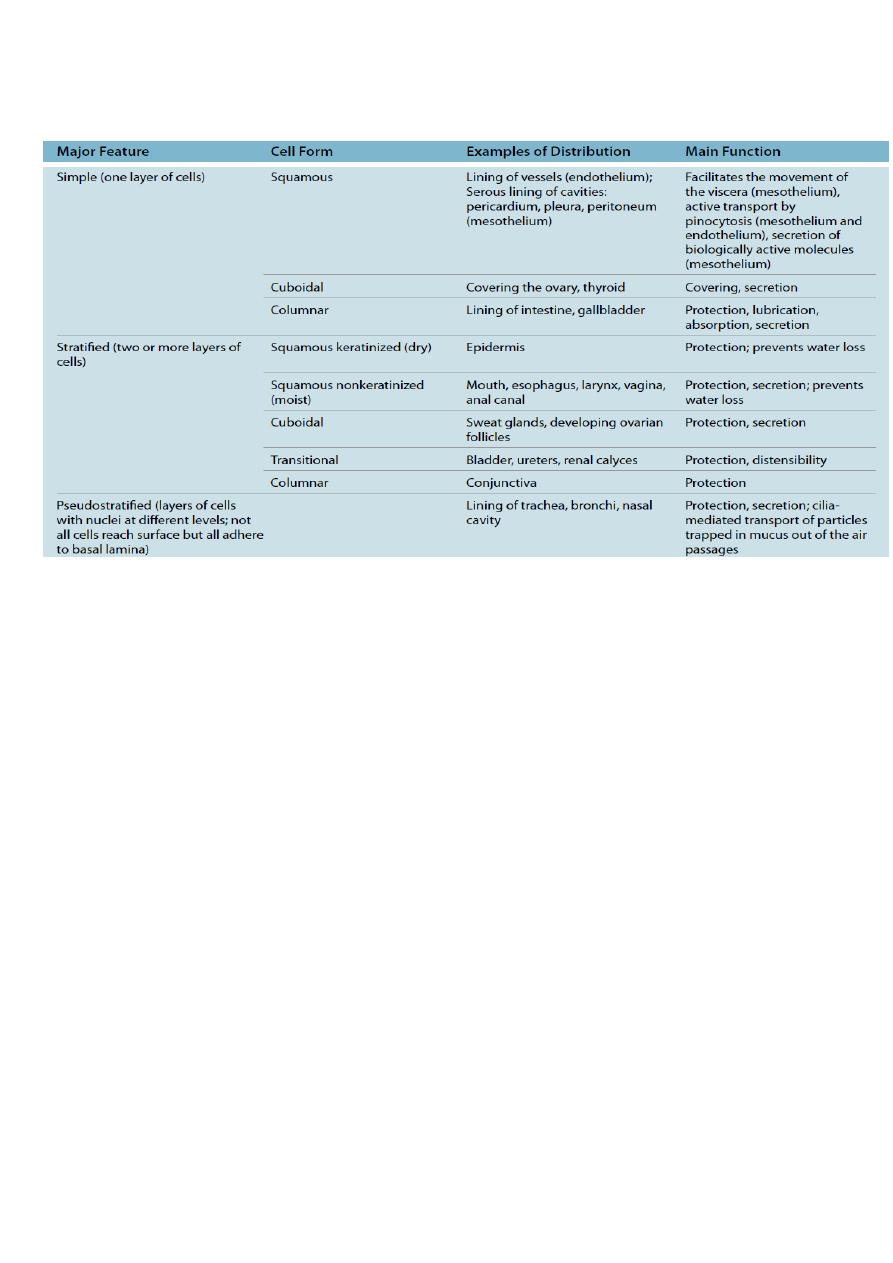
Classification of lining epithelium
Cell Death
Necrosis
Death of the cells due to tissue injury is called necrosis. Necrotic cells
swell and subsequently rupture resulting in formation of cell debris. This
induces an inflammatory response at the site of injury.
Apoptosis (Programmed Cell Death/Regulated Cell Suicide)
Cell division and differentiation are balanced by cell death during growth
and development of the organism.
Apoptosis is a central mechanism controlling multicellular development in
regulating the number of cells that mediate a particular activity (e.g.
separation of the developing digits during morphogenesis). It also ensures
that inappropriate or insufficient cells are eliminated.
The morphological changes exhibited by apoptotic cells are very different
from those seen in necrotic cells :
Apoptotic cells shrink.
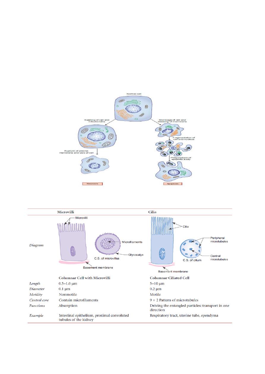
Their plasma membranes undergo blebbing without any loss (i.e. they are
intact).
Their nuclei and chromosomes fragment, forming apoptotic bodies.
Since the plasma membrane is intact, their intracellular contents are not
released into the extracellular environment, so
the inflammatory reactions are avoided.
Differences between microvilli and cilia