Lecture one Anatomy of neck
SkinThe lines of the skin run almost horizontally around the neck. Therefor the incision along a this lines will heal as a narrow scar
Superficial Fascia
The superficial fascia of the neck forms a thin layer that encloses the platysma muscle. Also embedded in it are the cutaneous nerves the superficial veins, and the superficial lymph nodes.
1-Platysma muscle
Its important muscular sheet embedded in the superficial fasciaOrigin : fascia covering pectorals major & deltoid
Insertion: lower border of mandible &skin of face
Nerve supply :-cervical br. of facial N
Action:-wrinkle of skin of neck
-depress of mandible
2-Superficial Veins
--External Jugular Vein
begins just behind the angle of the mandible by the union of the posterior auricular vein with the posterior division of the retromandibular vein. It descends obliquely across the sternocleidomastoid muscle and,drains into the subclavian vein
Branches
-Posterior auricular vein
-Posterior division of the retromandibular vein
-Transverse cervical vein
-Suprascapular vein
-Anterior jugular vein
--Anterior Jugular Vein
--Formed just below the chin, by the union of several small veins .It runs down the neck close to the midline. Just above the suprasternal notch, the veins of the two sides are united by a transverse trunk called the jugular arch.
--drain into the external jugular vein
3-Cutaneous nerves of neck
Arise from cervical plexusLesser occipital n from ant. Rami of C2 supply upper part of skin of inner surface of auricle & adjescent part of skin of scalp
Greet auricular n from ant. Rami of C2,C3 supply lower part of skin of inner surface of auricle , skin over parotid gl & skin over angle of mandible
Supraclavicular n from C3, C4 supply skin of post. Triangle , skin of tip of shoulder& skin of chest down to second rib
Transverse nerve of neck from C2,C3 supply of skin of ant. triangle
4-Superficial Lymph Nodes
Its lie along the external jugular vein superficial to the sternocleidomastoid muscle
- receive lymph vessels from the occipital and mastoid lymph nodes
- drain into the deep cervical lymph nodes
Deep Cervical Fascia
Its supports the muscles, the vessels, and the viscera of the neck .
In certain areas, it is condensed to form well-defined, fibrous sheets called the investing layer,
the pretracheal layer, the prevertebral layer. , carotid sheath
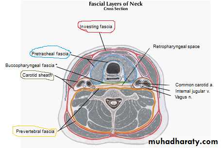
1-Investing Layer
Its is a thick layer that encircles the neck.(like collar ) It splits to enclose the trapezius and the sternocleidomastoid muscles.its form base of ant. & post. TrianglesAttachment –postlegemantum nuchae
--ant. hyoid bone , thyroid cartlage ,
--inf. maniberium , clavicle , acromian processes & spin of scapula
-- sup mandible , then splite to involve parotid gl. to attached to ext. audetory meatus
2-Pretracheal Layer
It is a thin layer that is attached above to the laryngeal cartilages It surrounds the thyroid and the parathyroid glands, forming a sheath for them, and encloses the infrahyoid muscles.
3-Prevertebral Layer
The prevertebral layer is a thick layer that passes like a septum across the neck
-- behind the pharynx and the esophagus
-- in front of the prevertebral muscles and the vertebral column ,extend from base of skull to third Thoracic vertebrate.
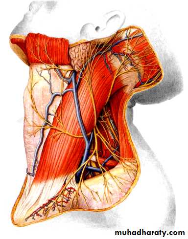
Axillary Sheath
The subclavian artery and the brachial plexus and the anterior rami of the cervical nerves emerge in the interval between the scalenus anterior and the scalenus medius muscles, they carry with them a sheath of the fascia, which extends into the axilla and is called the axillary sheath
Parotid f :- Its derived from investing f , have 2 layers superficial layer attach to zygomatic arch . Deep layer attach to petrous ,temporal, styloid and mastoid
Pharyngo –basilar f Tough f connect pharynx to base of skull
Bucco-parhngial f Cover muscles of pharynx
Supra plural membrane Cover apex of upper pole of lungs
Ligamentum Nuchae
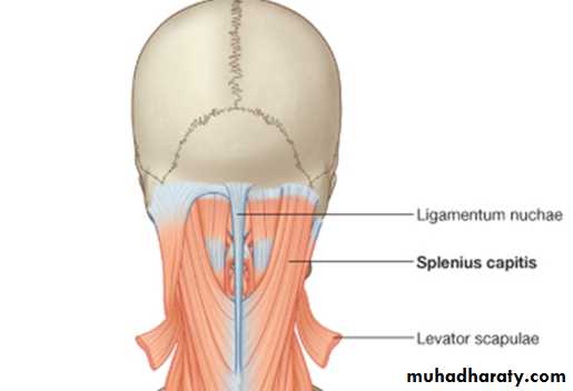
Its strong triangalar leg.
Its upper continuation of supraspinus & infraspinus legAttachement
Above (base)occipital protrberance and crest
below (apex)spine of C7
Ant. all cervical spines
Post. free
Clinical Significance of the Deep Fascia of the Neck
Acute InfectionsDental infections
lower molar teeth. The infection spreads medially from the mandible into the submandibular spaces and pushes the tongue forward and upward. Further spread downward may lead to edema of the vocal cords and airway obstruction.
( Ludwig's angina is an acute infection of the submandibular fascial space and is commonly secondary to dental infection)
Chronic Infection
Tuberculous TB of the deep cervical lymph nodes can result in liquefaction and destruction of one or more of the nodes. collar-stud abscess. The clinician is aware of the superficial abscess but must not forget the existence of the deeply placed abscess
Lecture tow Muscular Triangles of the Neck
The muscles of neck divided neck to several trianglesGenerally the neck divided into 2 large triangles ant. & post. Triangles by sternomastoid muscleand trapezius muscle forming the post. border of post triangles
Sternocleidomastoid Muscle
The origin have 2 heads Manubrium sterni & medial third of clavicle,
insertion Mastoid process of temporal bone and occipital bone ,
nerve supply Spinal part of accessory nerve and C2 and 3,
Action Two muscles acting together extend head and flex neck; one muscle rotates head to opposite side
Trapezius Muscle
Flat diamond-shaped muscle at back of neck and chest , formed from 3 parts upper , middle & lower fibers
Origin: Superior nuchal line, ext. occipital protuberance, lig. nuchae, spines of C7 – T12
Insertion: Lateral 1/3 of clavicle, acromion, spine of scapula
Functions:
have two effects: movement of the scapulae when the spinal origins are stable, and movement of the spine when the scapulae are stable. Its main function is to stabilize and move the scapula.
--(sup. fibers)elevation of scapula
-- ( --inf. fibers)depression of scapula
--(middle fibers)retraction of scapula --,
--(sup+ inf fibers).
superior rotation of glenoid fossa of scapula
Nerve supply the accessory nerve Cr 11
Posterior Triangle
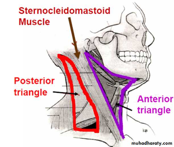
bounded posteriorly by the trapezius muscle, anteriorly by the sternocleidomastoid muscle, inferiorly by the clavicle .It is further subdivided by the inferior belly of the omohyoid muscle into :-
---a large occipital triangle above --- a small supraclavicular triangle below
Contains of post triangle:Nerves :- accessory nerve, cutaneous branches of cervical plexus, roots and trunks of brachial plexus. nerve to subclavius , dorsal scapular, suprascapular, long thoracic nerves
Vessles :- external jugular vein, transverse cervical , suprascapular vessels,
subclavian vein & artery,
Muscles Inferior belly of omohyoid
the floor of Post. Triangle formed by :-splenius capitis , levator scapulae & scalene muscles
Scalenus Muscles
Its 4 small muscles at root of neck , forming part of floor of post. triangle of neck
1-Scalenus Anterior Muscle
The scalenus anterior muscle is a key muscle in understanding the root of the neck .It is deeply placed and it descends almost vertically from the vertebral column to the first rib.
Origin Transverse processes of third, fourth, fifth, and sixth cervical vertebrae
Insertion First rib
Nerve supply C4, 5, and 6
Action Elevates first rib; laterally flexes and rotates cervical part of vertebral column
2-Scalenous medius
Largest of all scalenous mOrigin :post tubercle of all C. vertebrae
Insertion : 1st rib behind subclavian groove
N. supply:all cervical nn
Action flexion of neck &fixation of1st rib
3-Scalenous post.
Smallest of all scalenous
4-Scalenous minimous
v. Small m arise from transverse process of C7 inserted at plural memberan
Anterior Triangle
Anterior Trianglebounded
above by the body of the mandible
posteriorly by the sternocleidomastoid
anteriorly by the midline .
roof :……formed by:
platysma
investing layer of deep cervical fascia
The upper part of ant. Triangle (suprahyoid )subdivided by digastric m into diagastric & submental triangles
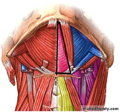
Digastric (submandibular) triangle
boundariesAbove ….lower border of mandible
Below & infront .anterior belly of Digastric muscle
Below & behind posterior belly of Digastric & stylohyoid m
Floor :
Anteriorly : ……..mylohyoid muscle
Posteriorly ………part of hyoglossus muscle
the Submental triangle
boundaries
Posterior (to the back) by the anterior belly of the digastricus,
Anterior (to the front) by the midline of the neck between the mandible and the hyoid bone;
Inferior (below) by the body of the hyoid bone
floor is formed by the mylohyoid.
Contents of the Submental triangle
1- submental lymph nodes2- beginning of the anterior jugular vein
Contents of Digastric triangle
submandibular salivary gland
submandibular LN lie on the surface of the submandibular salivary gland
facial artery deep to posterior end of submandibular salivary gland
facial vein lies superficial to submandibular salivary gland
hypoglossal nerve & nerve to mylohyoid muscle
The lower part of ant. triangle (infrahyoid ) also subdivided by omohyoid superior belly into the muscular triangle &the carotid triangle
-- Carotid triangle
boundary :Behind : …………..sternomastoid muscle
Infront & above : posterior belly of digastric m
Infront&below : superior belly of omohyoid m
Floor :
infont : hyoglossus muscle ( above ) and the thyrohyoid muscle (below)
Behind: the middle constrictor muscle of the pharynx (above ) and the inferior constrictor muscle of the pharynx (below )
-- Muscular triangle
bounderies
in front, by the median line of the neck from the hyoid bone to the sternum;
behind, by the anterior margin of the sternocleidomastoid;
above, by the superior belly of the omohyoid
Contents of Muscular triangle
A-infrahyoid muscles ( strap muscles)
They consist of 4 muscles arranged minto 2 layers :
1- superficial layer :-- sternohyoid &-- omohyoid
2- deep layer: -- sternothyroid & -- thyrohyoid
B- Lobe of thyroid gl . lying deep to sternohyoid &sternothyroid

Upper part of ant. Triangle ( suprahyoid )
digastric muscleconsists of two muscular bellies united by an intermediate rounded tendon.
Posterior belly longer than the anterior belly
arises from the mastoid notch
Insertion at Intermediate tendon is held to hyoid by fascial sling
Nerve supply Facial nerve
Anterior belly
Arise from Body of mandible have same insertion at Intermediate tendon is held to hyoid by fascial sling s held in connection with the side of the body and the greater cornu of the hyoid
N supply Nerve to mylohyoid
Action of both belly Depresses mandible or elevates hyoid bone
Mylohyoid muscle
is a paired muscle , flat and triangular, and is situated immediately superior to the anterior belly of the digastric muscle running from the mandible to the hyoid bone, forming the floor of the oral cavity of the mouth
arise from the mandible at the mylohyoid line, (which extends from the mandibular symphysis in front to the last molar tooth behind)
Insertion
--The posterior fibers pass inferomedially and insert at anterior surface of the hyoid bone.
-- The medial fibres of the two mylohyoid muscles unite in a midline raphe (where the two muscles intermesh)
N supply branch of the mandibular nerve through inf . Alvolaer N
Function its elevates the hyoid and the tongue. & support of floor of mouth This is particularly important during swallowing and speaking
The hyoglossus
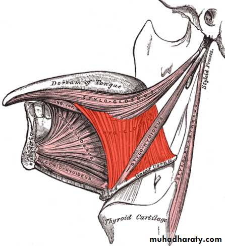
Its thin and quadrilateral muscle
, arises from the side of the body of hyoid bone and from the whole length of the greater cornu,and passes almost vertically upward to enter the side of the tongue, between the styloglossus and the inferior longitudinal muscle of the tongue.
It forms a part of the floor of submandibular triangle
The Styloglossusthe shortest and smallest of the three styloid muscles,
arises from the anterior and lateral surfaces of the styloid process near its apex, and from the stylomandibular ligament.
Insertion Passing downward and forward between the internal and external carotid arteries, it divides upon the side of the tongue near its dorsal surface, blending with the fibers of the Longitudinalis inferior in front of the Hyoglossus; the other, oblique, overlaps the Hyoglossus and decussates with its fibers.
N supply the Hypoglossal nerve (CN XII) like all muscles of the tongue except palatoglossus which is innervated by the Pharyngeal plexus of vagus nerve
Action draws up the sides of the tongue to create a trough for swallowing. As a pair they also aid in retracting the tongue
The geniohyoid muscle
is a narrow muscle situated superior to the medial border of the mylohyoid muscleIt arises from the inferior mental spine, on the back of the mandibular symphysis,
insertion its runs backward and slightly downward, to be inserted into the anterior surface of the body of the hyoid bone
N supply The geniohyoid muscle is innervated by fibres from the first cervical nerve travelling alongside the hypoglossal nerve. These fibers are called ansa cervicalis.
Action its brings the hyoid bone forward and upwards. This dilates the upper airway, assisting respiration.
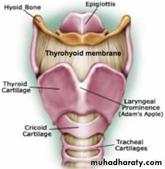
Hyoid bone
The hyoid bone (lingual bone)
The hyoid bone is U shaped and consists of a body and two greater and two lesser cornua , unlike other bones, not articulate to other bones by muscles or ligaments. The hyoid is anchored by muscles from all directions,
situated in the anterior midline of the neck between the chin and the thyroid cartilage . At rest, it lies at the level of the base of the mandible in the front and the (C3) behind.

Attachment
-to the skull by the stylohyoid ligament- to the thyroid cartilage by the thyrohyoid membrane.
-to the base of tongue and is suspended in position by muscles t
-to the mandible,
-to the styloid process of the temporal bone,
- to the sternum, and to the scapula
function is aids in tongue movement and swallowing.
The hyoid bone provides attachment to the muscles of the floor of the mouth and the tongue above, the larynx below, and the epiglutis and pharynx behind.
Styloid process
Is a slender projection of variable length
extends downward & forward from temporal bone.
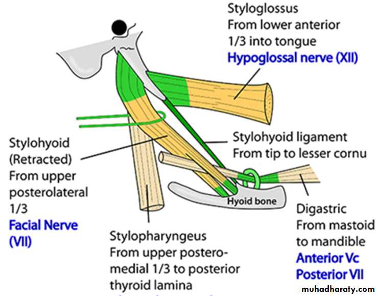
Gives origin to :
A- three muscles:stylohyoid,
styloglossus,
Stylopharyngeus
B-two ligaments :
stylohyoid
stylomandibular.
The mastoid
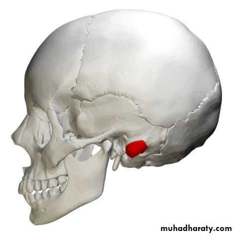
part of the temporal bone
*Its rough surface give attachment occipitalis , posterior auricular m&its tip give attachment of sterncledomastoid.
* it has openings for the transmission of blood vessels.( mastoid foramen);
* From its borders the mastoid part articulates with two other bones
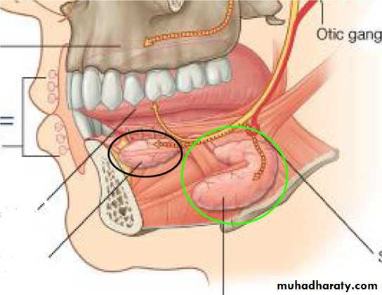
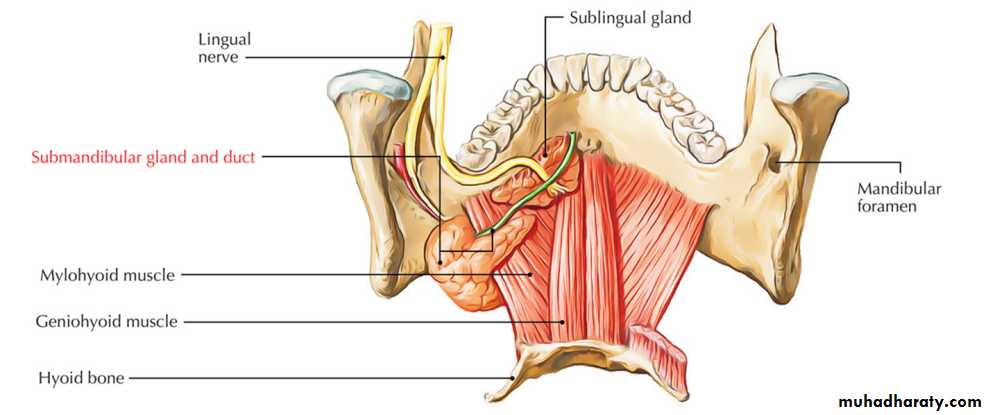
submandibular glands
The paired submandibular glands are major salivary glands located beneath the floor of the mouth. At diagastric triangle--wedge shape about 15 gr and produce 60–67% of saliva secretion;
Its divided into superficial (large) and deep lobes ( small), which are separated by the mylohyoid muscle ,together continue at post . free border of mylohyoid
submandibular duct arise from medial surface of superfecial lobe about 5 cm length, The excretory ducts are then crossed by the lingual nerve, and ultimately drain into the sublingual caruncles on either side of the lingual frenulum a
Blood supply. is supplied by sublingual and submental arteries branches from the facial and lingual arteries
drained by common facial and lingual veins
The lymphatics drain into submandibular LN and subsequently into jugulo - digastric lymph nodes
Nerve supply
Sensery fibers from lingual N through submandibular ganglion without relay while
Secretmotor fibers --parasympathetic and sympathetic
Parasympathetic via the chorda tympani, a branch of the facial nerve, that becomes part of the trigeminal nerve then synapsing on the submandibular ganglion.
-Increased parasympathetic activity promotes the secretion of saliva.
The sympathetic
--through the arteries that supply it.
Increased sympathetic activity decreasing the volume of secretions, and increase enzymatic secretions.
Note:- The submandibular gland accounts for 80% of all salivary duct stones because viscous nature of the saliva that it produces and the tortuous travel of the submandibular duct
Sublingual gland
They are the smallest, most diffuse, and the only unencapsulated major salivary glands. They provide only 3-5% of the total salivary volume
They lie anterior to the submandibular gland inferior to the tongue, as well as beneath the mucous membrane of the floor of the mouth
drained by 8-20 excretory ducts called the ducts of Rivinus
N supply :- branches from lingual N containing sensory fibers , sympathetic ¶sympathetic f
blood supply sublingual and submental arteries.
Lymph drainage the sublingual salivary gland drains into the submandibular LN
Lecture three Lower part of ant. Triangle (infrahyoid):-
Carotid Sheath
The carotid sheath is a local condensation of the prevertebral, the pretracheal, and the investing layers of the deep fascia
Attached –
-- above At base of skull surround caroted &jagular foramens-below blend with adventecia
of arch of aorta
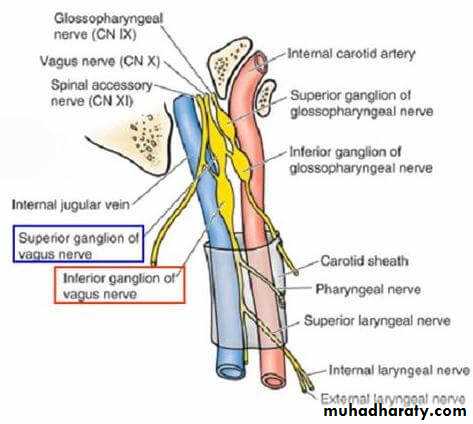
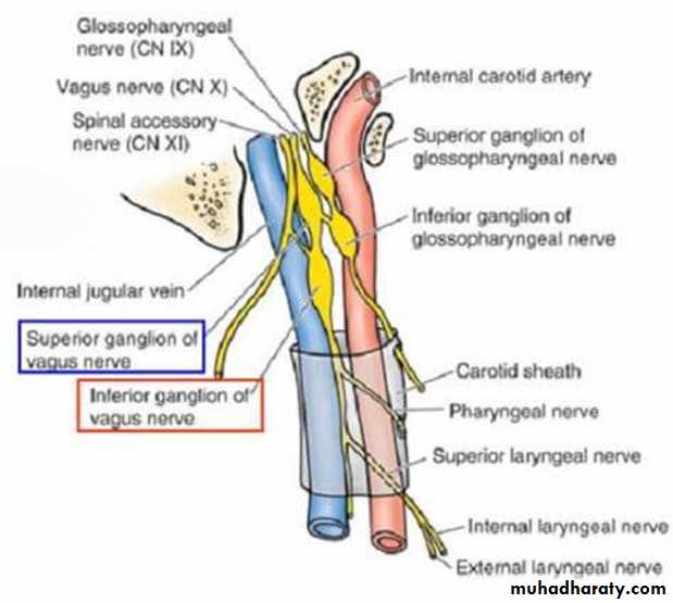
Contents of carotid sheath
a- in the upper part(above post belly of digastric m)
internal carotid artery
the internal jugular vein,
4 cranial NN (9,10,11,12 )
deep cervical lymph nodes .
b-in the lower part (below post belly of digastric m)
.common internal carotid arteries
the internal jugular vein,
Vegus between CCA&IJV)
Note:-
-- sympathetic chain embeded in post layer of sheath
-- 2 limbs of ansa cervicalis at ant. Wall
-- sheath thick over art. While thin over vein
Contents of the Carotid triangle
1- The carotid sheath and its contents :
2- the external carotid artery :gives most of its branches in the carotid triangle ( superior thyroid artery ,lingual artery ,facial artery , ascending pharyngeal artery and occipital artery )
3- hypoglossal nerve
4- Descendes cervicalis (C2,3) anterior to carotid sheath.
5- sympathetic trunk adherent to the posterior wall of carotid sheath
The Muscular triangle
1- superficial layer :sternohyoidOrigin : back of maniberium & back of clavicle
Insertion :- lower border of hyoid bone along midline
N supply :-ansa cervicalis
Action :-
1-depresion of hyoid bone after deglutision
2-fixation of hyoid bone during movement of tongue
omohyoid
Origin :- it has 2 bellis
1-sup. Belly —lower border of hyoid body lateral to sternohyoid
1-inf. Belly –upper border of scapula &suprascapular leg
Insertion :-intermediate tendon lies on IJV &held in position by facial sling connect it to clavicle
N supply ansa cervicalis Action like sternohyoid & pull deep facia around Ext. J
2- deep layer: sternothyroid
Origin back of meniberium sterni & costal cartlage
Insertion Oblique line on lamina of thyroid cartilage
N supply Ansa cervicalis; C1, 2, and 3
action Depresses larynx in second phase of diglution
thyrohyoid
Origin :- Oblique line on lamina of thyroid cartilage
Insertion :-Lower border of body of hyoid bone
N supply 1st cervical n C1(hypoglusal n)
action Depresses hyoid bone or elevates larynx
Ansa cervicalis
Its nerve loop formed of 2 descending nn & supply 3 infrahyoid mm
Site :- lies infront of carotid sheath
Formation : its formed by union of 2 descending nn
1-descending hypoglussi( sup . Limbe of ansa )
Its fibers aris from C1 join hypoglussal n then leave it its turn forwareds across carotid sheath
2- descending cervicalis ( inf. Limb of ansa):
Its fiber arise from C2,3 its began behind IJV then descend along its ant. Surface
to join descending hypoglussi infront caroted sheath( thus the ansa cervicalis
arise from C1,2 ,3)
Branches of ansa cervicalis
Aris from convixity of loop and supply 3 of infrahyoid mm (the two belly of omohyoid ,sterno hyoid &sternothyroid)
except thyrohyoid (from C1 through hypoglossal n)
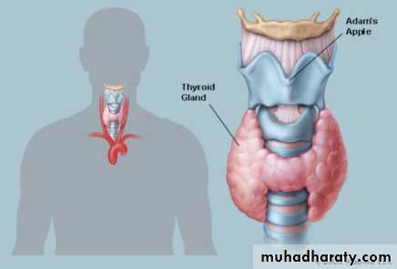
Thyroid Gland
Its largest endocrine gland in body about 25 gr
The thyroid gland butterfly in shape consists of right and left lat. lobesconnected by a narrow isthmus
at mid ant. Triangle . pyramidal lobe , Its often present, and it projects upward from the isthmus, usually to the left of the midline.
A fibrous or muscular(levator glandulae thyroideae) band frequently connects the pyramidal lobe to the hyoid bone
Capsules :-
1- true capsule adherent to gland
2- false capsule derived from the pretracheal layer of deep fascia.
The sheath attaches the gland to the larynx and the trachea. So thyroid move with deglutition
Each thyroid lobe
is pear shaped,
apex ( upper. Pole ) being directed upward as far as the oblique line on the lamina of the thyroid cartilage;
base ( lower pole) lies below at the level of the fourth or fifth tracheal ring.
The isthmus
extends across the midline in front of the second, third, and fourth tracheal rings
Blood Supply
The arteries to the thyroid gland are the superior thyroid artery, the inferior thyroid artery, and sometimes the thyroidea ima. The arteries anastomose profusely with one another over the surface of the gland.1-The superior thyroid artery, a branch of the external carotid artery, descends to the upper pole of each lobe, accompanied by the external laryngeal nerve
2-The inferior thyroid artery, a branch of the thyrocervical trunk, ascends behind the gland to the level of the cricoid cartilage. It then turns medially and downward to reach the posterior border of the gland.
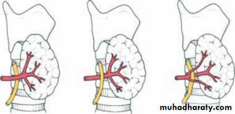
The recurrent laryngeal nerve crosses either in front of or behind the artery, or it may pass between its branches.
3-The thyroidea ima, if present, may arise from the brachiocephalic artery or the arch of the aorta. It ascends in front of the trachea to the isthmus
The veins from the thyroid gland
the superior thyroid, which drains into the internal jugular vein;middle thyroid, which drains into the internal jugular vein;
The inferior thyroid veins of the two sides anastomose with one another as they descend in front of the trachea. They drain into the left brachiocephalic vein in the thorax
Lymph Drainage
The lymph from the thyroid gland drains mainly laterally into the deep cervical lymph nodes.
A few lymph vessels descend to the paratracheal nodes.
Nerve Supply
Superior, middle, and inferior cervical sympathetic ganglia
Functions of the Thyroid Gland
Thyroid gl under effect of thyroid stimulating hormone (TSH) which secreted from pituitary gland , thyroid gl has 2 secretory cells :-
1-Thyroid cells produce thyroid hormones( thyroxine )
Tri iodothyronine(T3) more active increase the metabolic activity of most cells in the body.
Tetra iodothyronine (T4) less active form
2- parafollicular cells produce thyrocalcitonin, which lowers the level of blood calcium
goiter is enlargement of the thyroid Gland can causes
Dysphagia , dyspnea due to presser effect of gland on underling structur
toxic goiter due to increase of thyroid hormones
Development of the Thyroid Gland
The thyroid gland begins to develop during the third week as an entodermal thickening in the midline of the floor of the pharynx as a duct called thyroglossal
duct its distal end becomes bilobed to form primitive thyroid
The thyroid gland migrates inferiorly in the neck and passes either anterior to, posterior to, or through the developing body of the hyoid bone
Developmental abnormalities
1-Ectopic Thyroid Tissue& Lingual thyroid
descent of the thyroid may be arrested at any point between the base of the tongue and the trachea
2- thyroglossal Cysts may occur at any point along the thyroglossal tract
Lingual thyroid
thyroglossal Cysts
Parathyroid Glands
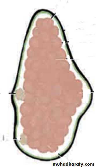
The parathyroid glands are four in number
Lies at post. border of the thyroid within its fascial capsule .Oval shape measuring about 6 mm long in their greatest diameter.
two superior parathyroid glands are the more constant
in position and lie at the level of the middle of the
posterior border of the thyroid gland.
The two inferior parathyroid glands usually lie close
to the inferior poles of the thyroid gland. They may
lie within the fascial sheath, embedded in the thyroid
substance, or outside the fascial sheath.
Sometimes they are found some distance may even
in the superior mediastinum in the thorax.
Blood Supply
The arterial supply to the parathyroid glands is from the superior and inferior thyroid arteries. The venous drainage is into the superior, middle, and inferior thyroid veins.Lymph Drainage
Deep cervical and paratracheal lymph nodes
Nerve Supply
Superior or middle cervical sympathetic ganglia
Functions of the Parathyroid Glands
The chief cells produce the parathyroid hormone, which :-
A) increase serum calcium :-
1- stimulates osteoclastic activity in bones, thus mobilizing
the bone calcium to the blood. osteomalecia
2-stimulates the absorption of calcium from the small intestine
3- the reabsorption of calcium in the proximal convoluted-
tubules of the kidney. renal stone
B) Decrease serum phosphate
in the proximal convoluted tubules of the kidney.
The secretion of the parathyroid hormone is controlled by the calcium levels in the blood