Neurophysiology
Lecture 3
Dr. Noor

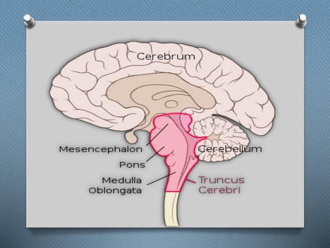
Brain stem

Brain stem
O
The brainstem is the posterior part of
the
, adjoining and structurally
continuous with the
. In
the
the brainstem
includes:
O
O
the
, and
O
the

The brain stem is a tube-shaped mass of
nervous tissue a little over 3 inches (8
cm) long. It is located at the base of the
brain, superior to the spinal cord and
inferior to the cerebrum

• Superiorly
continuous with
forebrain.
• Inferiorly
continuous with spinal
cord .
• Posteriorly
pons and medulla is
separated by forth venricle.

The medulla is the inferior-most region of the
brain stem that connects the brain to the spinal
cord. It is a tube very similar structurally to the
spinal cord, but is wider and contains several
masses of gray matter internally.
Superior to the medulla is the pons, which is
larger and structurally more complex than the
medulla.

Function
There are three main functions of the
brainstem:
1. The brainstem plays a role in conduction.
That is, all information relayed from the
body to the cerebrum and cerebellum and
vice versa must traverse the brainstem.

O
2. The cranial nerves III-XII emerge from
the brainstem. These cranial nerves supply
the face, head, and viscera. (The first two
pairs of cranial nerves arise from the
cerebrum).
O

O
3. The brainstem has integrative functions
being involved in cardiovascular system
control, respiratory control, pain
sensitivity control, alertness, awareness,
and consciousness. Thus, brainstem
damage is a very serious and often life-
threatening problem

basal ganglia
O
The basal ganglia (or basal nuclei) is a
group of subcortical
, of varied origin,
inthe
of
including
which are situated at the base of
the
.

The basal ganglia, like the cerebellum,
constitute another accessory motor system
that functions usually not by itself but in
close association with the cerebral cortex and
corticospinal motor control system.

In fact, the basal ganglia receive most of
their input signals from the cerebral cortex
and also return almost all their output
signals back to the cortex.
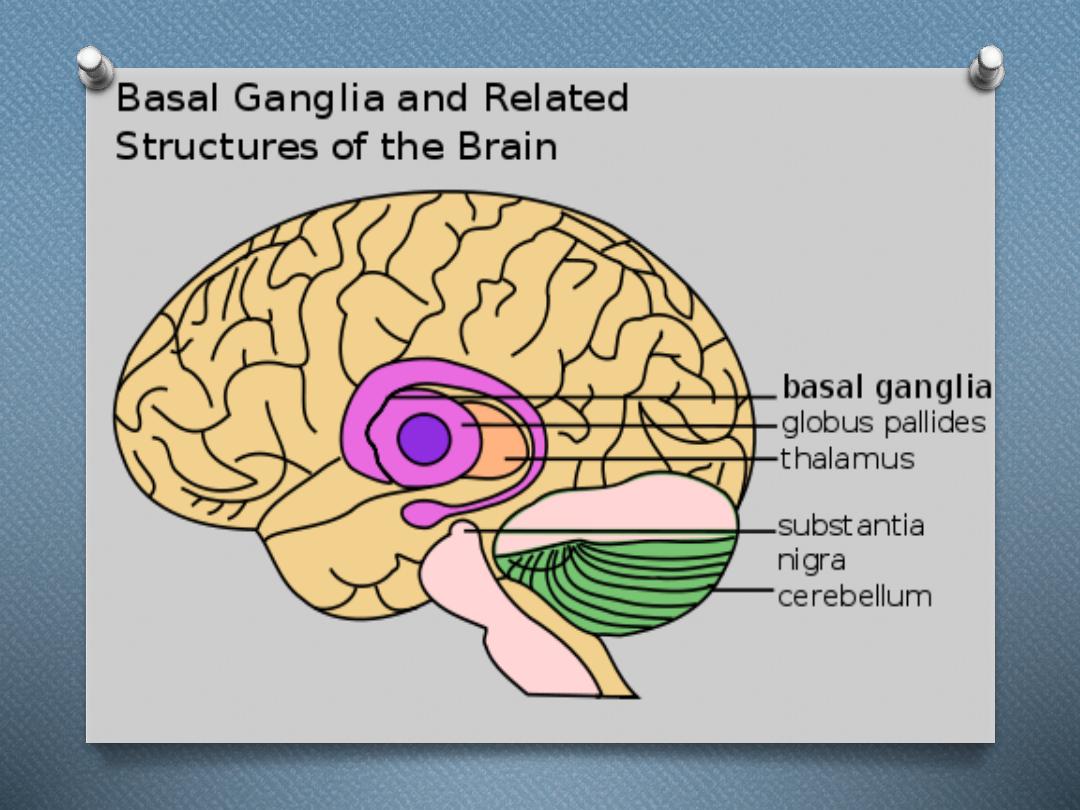

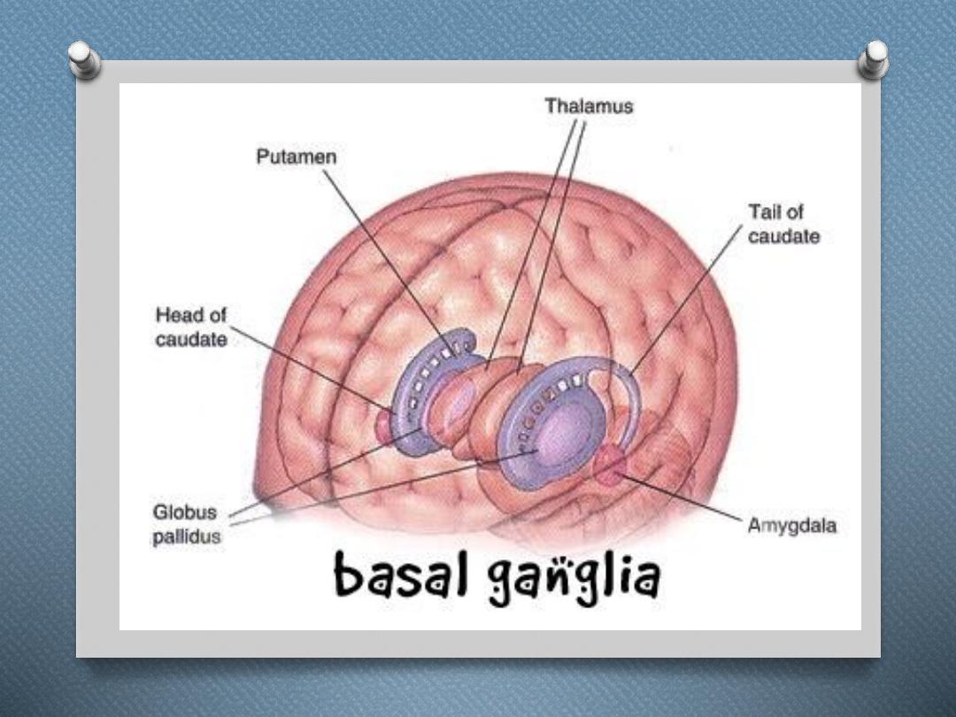
Main components of basal
ganglia
O
Basal ganglia consist of :
O
The caudate nucleus,
O
putamen,
O
globus pallidus,
O
substantia nigra, and
O
subthalamic nucleus.

They are located mainly lateral to and
surrounding the thalamus, occupying a
large portion of the interior regions of both
cerebral hemispheres.

Almost all motor and sensory nerve fibers
connecting the cerebral cortex and spinal cord pass
through the space that lies between the major masses
of the basal ganglia, the caudate nucleus and the
putamen. This space is called the internal capsule of
the brain. It is important for our current discussion
because of the intimate association between the
basal ganglia and the corticospinal system for motor
control

FUNCTION OF THE BASAL GANGLIA
IN EXECUTING PATTERNS OF MOTOR
ACTIVITY
O
One of the principal roles of the basal
ganglia in motor control is to function
in association with the corticospinal
system to control complex patterns of
motor activity.

An example is the writing of letters of the
alphabet. When the basal ganglia sustain
serious damage, the cortical system of motor
control can no longer provide these patterns.
Instead, one’s writing becomes crude, as if
one were learning how to write for the first
time.

Other patterns that require the basal ganglia
are cutting paper with scissors, hammering
nails, shooting a basketball through a hoop,
passing a football, throwing a baseball, most
aspects of vocalization, controlled
movements of the eyes, and virtually any
other of our skilled movements, most of them
performed subconsciously

ROLE OF THE BASAL GANGLIA FOR
COGNITIVE CONTROL OF
SEQUENCES OF MOTOR PATTERNS
O
The term cognition means the thinking
processes of the brain, using both sensory
input to the brain plus information
already stored in memory.

O
Most of our motor actions occur as a
consequence of thoughts generated in the
mind, a process called cognitive control of
motor activity. The caudate nucleus plays
a major role in this cognitive control of
motor activity.

O
Instead, the returning signals go to the
accessory motor regions in the premotor and
supplementary motor areas that are
concerned with putting together sequential
patterns of movement lasting 5 or more
seconds instead of exciting individual
muscle movements.

FUNCTION OF THE BASAL GANGLIA
TO CHANGE THE TIMING AND TO
SCALE THE INTENSITY OF
MOVEMENTS
O
Two important capabilities of the brain in
controlling movement are to (1) determine
how rapidly the movement is to be
performed and (2) control how large the
movement will be.

For instance, a person may write the letter
“a” slowly or rapidly. Also, he or she may
write a small “a” on a piece of paper or a
large “A” on a chalkboard. Regardless of the
choice, the proportional characteristics of the
letter remain nearly the same.

BASAL GANGLIA FUNCTION
O
The basal ganglia and related nuclei are
characterized as one of three types of
nuclei. Input nuclei receive signals from
various sources in the brain. Output
nuclei send signals from the basal ganglia to
the
. .

Intrinsic nuclei relay nerve signals and
information between the input nuclei and
output nuclei The basal ganglia

O
After the information has been processed,
it is passed along to intrinsic nuclei and
sent to output nuclei. From the output
nuclei, the information is sent to the
thalamus. The thalamus passes the
information on to the cerebral cortex.

CORPUS STRATIUM
O
The corpus stratium is the largest group of
basal ganglia nuclei.
O
It consists of the caudate nucleus, putamen
and the globus pallidus. The caudate
nucleus, putamen are input nuclei, while the
globus pallidus is considered output nuclei.
The corpus stratium uses and stores the
neurotransmitter dopamine and is involved
in the reward circuit of the brain

O
Caudate Nucleus - these C-shaped paired
nuclei (one in each hemisphere) are located
primarily in the
region of the
brain. The caudate nucleus is involved in
motor processing and planning.

O
It is also involved in memory storage
(unconscious and long-term),
associative and procedural learning,
inhibitory control, decision making,
and planning
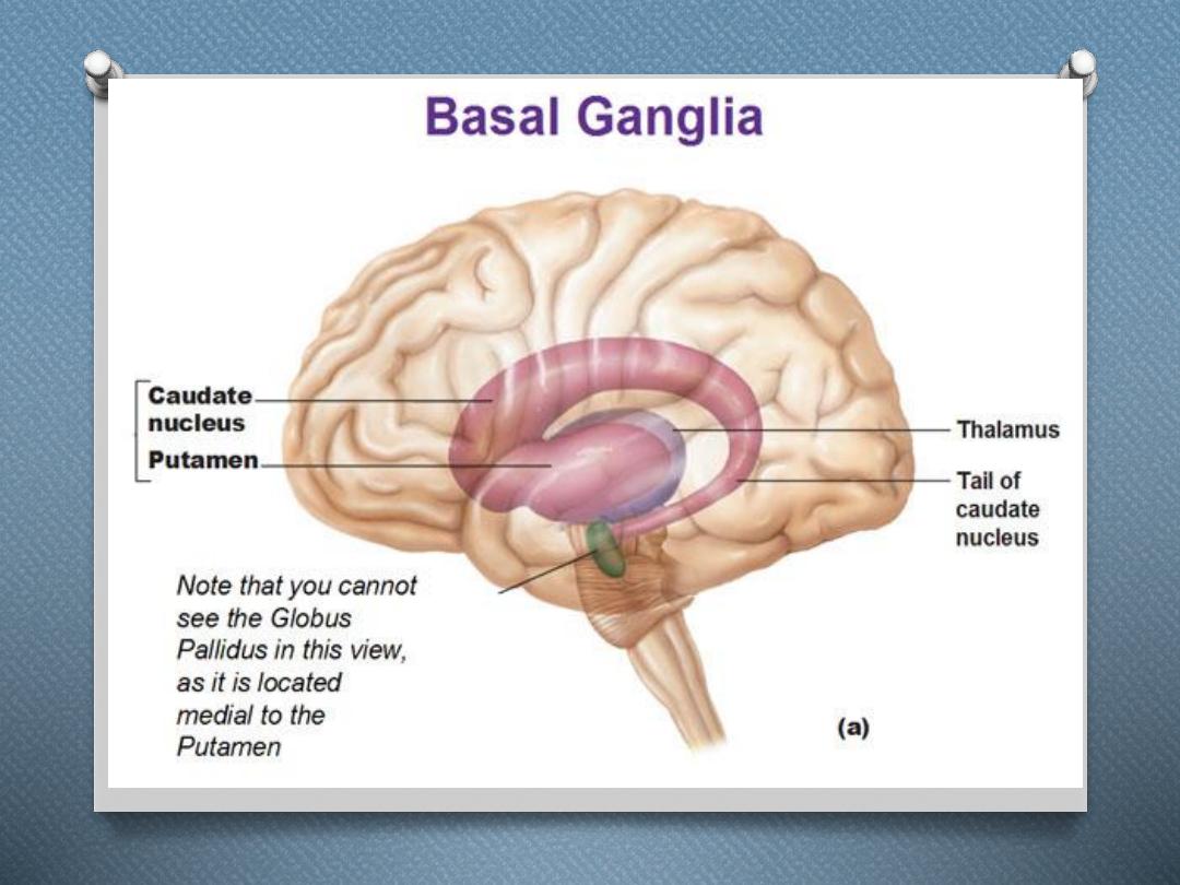

Putamen
O
- these large rounded nuclei (one in each
hemisphere) are located in the
and
along with the caudate nucleus form
the dorsal stratium.

O
The putamen is connected to the caudate
nucleus at the head region of the caudate.
The putamen is involved in voluntary
and involuntary motor control.

Globus Pallidus
O
- these paired nuclei (one in each
hemisphere) are located near the caudate
nucleus and putamen. The globus
pallidus is divided into internal and
external segments and acts as one of the
major output nuclei of the basal ganglia.

.
It sends information from basal ganglia
nuclei to the
. The internal segments
of the pallidus send the majority of output to
the thalamus via the neurotransmitter
gamma-aminobutyric acid (GABA). GABA
has an inhibitory effect on motor function.
The globus pallidus is involved in the
regulation of voluntary movement.

Subthalamic Nucleus
O
- these small paired nuclei located just
below the thalamus. Subthalamic nuclei
receive excitatory inputs from the cerebral
cortex and have excitatory connections to
the globus pallidus and substantia nigra

Subthalamic nuclei have both input and
output connections to the caudate nucleus,
putamen, and substantia nigra. The
subthalamic nucleus plays a major role in
voluntary and involuntary movement.

Substantia Nigra
O
- this large mass of nuclei is located in
the
and is also a component of
the
. The substantia nigra serves
numerous functions including controlling
voluntary movement, regulating mood,
learning, and activity related to the brain's
reward circuit


The basal ganglia include all of the
following except the
a. globus pallidus.
b. putamen.
c. caudate nucleus.
d. thalamus..
e. subthalamic nucleus
f. substantia nigra

The thalamus receives major and direct
input from which basal gangiia structure?
a. internal segment of globus pallidus.
b. putamen.
c. compact part of substantia nigra.
d. nucleus accumbens.