
1
بسم هللا الرحمن الرحيم
Lecture -5- Medical Physiology
2
nd
stage Dr. Noor Jawad
The cardiac cycle
Objective:
1. Definition of cardiac cycle?
2. Phases of cardiac cycle?
The cardiac cycle is described as the repetitive electrical and mechanical
events that occur with each beat of the heart. Each cardiac cycle has two
phases: diastole, the time during which cardiac muscle relaxes and
ventricles fill with blood, and systole, the time during which the cardiac
muscle contracts and blood ejected by ventricles. The duration of cardiac
cycle at heart rate of 75 beat/min is about 0. 8 second, 0.5 second for
diastole and 0. 0.3 second for systole. The long duration of diatsole has
two important physiologic and clinical implications.
most of ventricular muscle perfusion occur during diastole.
ventricular filling occur during diastole.
.
Phases of cardiac cycle
The main phases of cardiac cycle are:
1. Filling Phase
2.Atrial Systole
3. Isovolumetric Contraction Phase
4. Rapid Ejection Phase
2
5. Reduced Ejection Phase
6. Isovolumetric Relaxation Phase
1. Filling Phase
ventricular pressure decreases below atrial, the A-V valves open and
filling occurs rapidly (rapid filling phase), the elastic recoil of the
ventricle may aid in drawing blood into the ventricle .As the ventricle
fills intraventricular pressure increases slowing the rate of
filling(reduced filling phase) or diastasis. At large volumes, and with
ventricles with low compliance, the rapid reduction of filling at the end
of this phase may produce the third heart sound. About 70% of
ventricular filling occur in this phase.
2. Atrial Systole
The sinoatrial pacemaker complex initiates excitation, which spreads
across the atria and is recognised in the ECG as the P-wave atrial
contraction follows increasing the pressure within both atria this increase
in the RA pressure gives the a-wave of the Jugular Venous pulse, or
CVP trace. Atrial systole, force a small extra amount of blood into the
ventricles. atrial contraction is normally not necessary for ventricular
filling,. NB: With reduced ventricular compliance, atrial contraction may
give rise to a fourth heart sound.
3
3. Isovolumetric Contraction Phase
Excitation of the ventricles occurs as the wave of excitation passes
through the AV node, bundle of His and Purkinje system. Ventricular
contraction coinciding with the peak of the R-wave of the ECG, in
response to contraction of the muscle, the ventricular pressure rises very
rapidly this is not true isometric contraction, as some fibers lengthen and
others shorten as the ventricle changes shape. the increasing
intraventricular pressure lead to closure of the mitral and tricuspid
valves, producing the first heart sound. the rapid rise in ventricular
pressure is transmitted across the semilunar valves and appears as a
small rise in the aortic pressure trace.
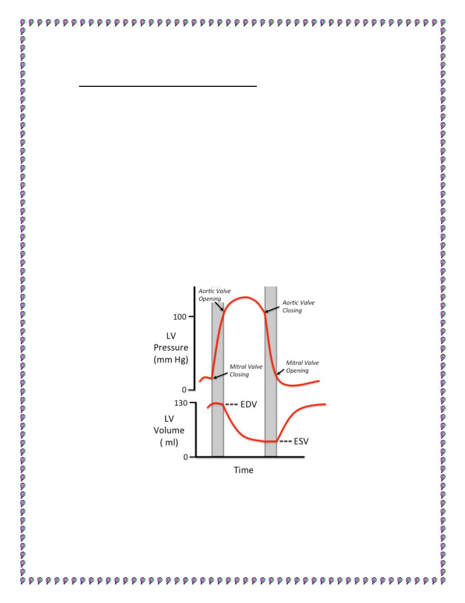
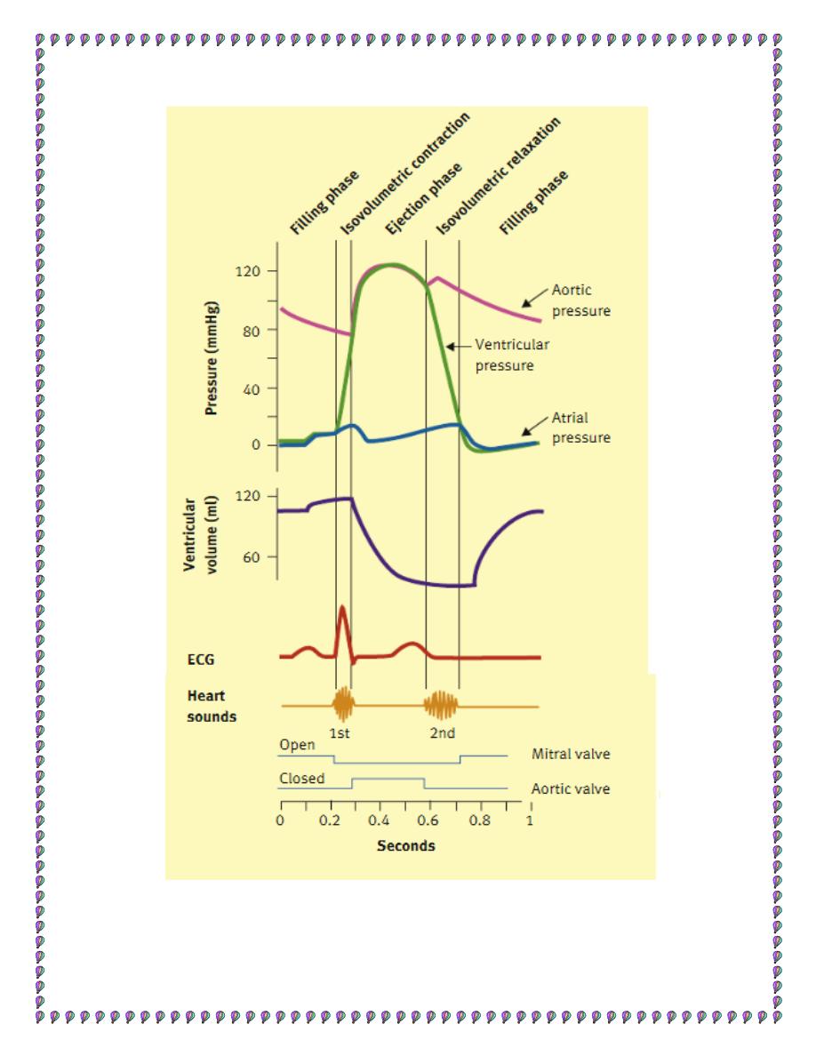
figure1: ventricular pressure and volume curve.
4
4. Rapid Ejection Phase
The intraventricular pressure rises rapidly until it exceeds the pressure in
the great arteries (aorta and pulmonary), leading to opening of the
outflow (Aortic/Pulmonary) valves. There is then a rapid ejection period,
where both intraventricular and arterial pressure rise to a maximum.
Because of the high ventricular pressure, unless the papillary muscles
perfectly compensate, there ,is a tendency for the A-V valves to bulge
into the atria producing the av-wave of the JVP. Immediately after this
the atrial pressure fall due to the descent of the base of the heart and
stretching of the atria, producing the x descent of the JVP. This reduced
atrial pressure aids the return of blood from the periphery.
5. Reduced Ejection Phase
both the contractile forces and the pressure within the ventricles are
decreasing during this phase and are less than those within the aorta by
several mmHg .flow continues due to the momentum of the bolus of
blood into the aorta. This phase coinciding with the Repolarisation of
the myocardium occurs during ( T-wave of the ECG). The ventricular
pressure is decreasing very rapidly and there is reversal of flow toward
the heart, closing the valves and generating the second second heart
sound. The sudden increase in back-resistance to flow increases aortic
pressure giving rise to the incisura, or dicrotic notch in the aortic
pressure trace .
5
6. Isovolumetric Relaxation Phase
both valve sets are closed, so pressure drops very rapidly as there is "no"
change in volume as this is occurring atrial pressure is increasing to its
maximum, due to the:
1. movement of blood from the periphery
2. movement of the base of the heart back to its resting position ® v-
wave of the JVP
6
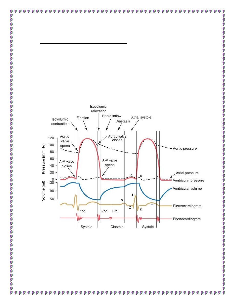
Figure2: cardiac cycle
7
Synchrony of Contraction
1.LV is first to start contraction and the last to start to fill. RV
contraction lags due to the anatomy of the conducting system by ~ 15
ms. The pulmonary valve opens first, and pulmonary flow begins around
10 ms before aortic due to the lower pressure in the pulmonary circuit
2. The isovolumetric period for the LV ~ 40 ms and RV ~ 15 ms
3. Because of higher systemic pressure LV outflow ends first with the
total LVET being shorter that the RVET
4. the mitral valve opens after the tricuspid due to the greater time for
ventricular pressure to drop below atrial pressure.
………………………………………………………………………….
Thank you
References : Guyton and Hall textbook of medical physiology,
thirteen edition