
*
Cardiovascular
system
lecture one
Dr. Noor J. Al maliky

The cardiovascular system (CVS) consists of
• the heart,
• blood vessels
• blood.( approximately 5 liters of blood that the
blood vessels transport).

Function
The four major functions of the cardiovascular
system are
1. To transport nutrients, gases and waste products
around the body
2. To protect the body from infection and blood loss
3. To help the body maintain a constant body
temperature (‘thermoregulation’)
4. To help maintain fluid balance within the body.

An Overview of the Cardiovascular System
The cardiovascular system perform its impressive
work, depending on the heart
together with a network of blood vessels. The
network can be subdivided into two circuits:
• the pulmonary circuit, which carries carbon
dioxide-rich blood from the heart to the gas
exchange surfaces of the lungs and returns
oxygen-rich blood to the heart

• the systemic circuit, which transports
oxygen-rich blood from the heart to the
rest of the body’s cells, returning carbon
dioxide-rich blood back to the heart.

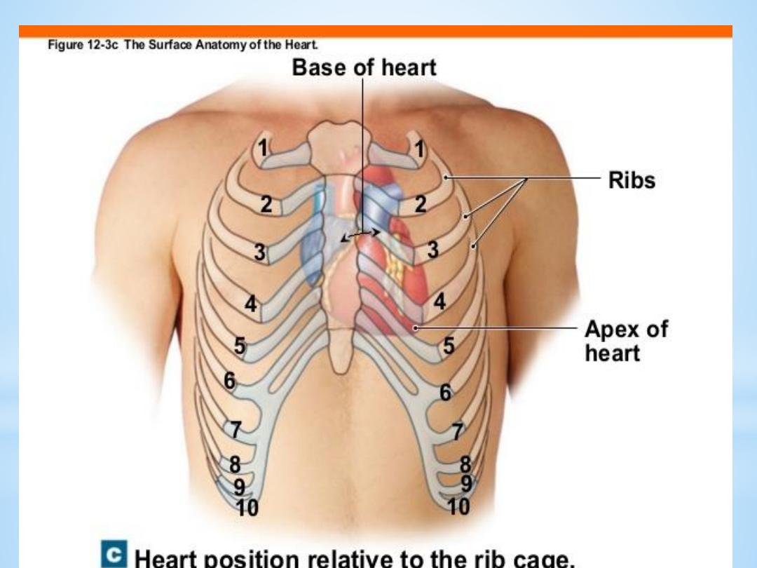
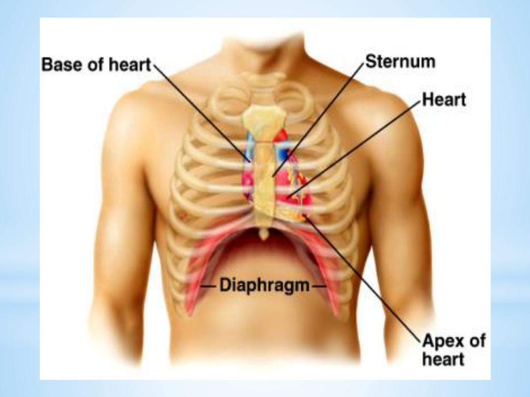
The heart
The heart, is hollow cone-shaped four-chambered
muscular pump approximately the size of a fist. The
heart rests on the diaphragm between the lungs in
the mediastinal space of the intrathoracic cavity in a
loose-fitting sac called the pericardium. It is
suspended by the great vessels, with its broader side
(i.e., base) facing upward and its tip (i.e., apex)
pointing downward, forward, and to the left

. It is located posterior to the sternum and
anterior to the vertebral column. The heart is
positioned obliquely, so that the right side of the
heart is almost fully in front of the left side of the
heart, with only a small portion of the lateral left
ventricle on the frontal plane of the heart.

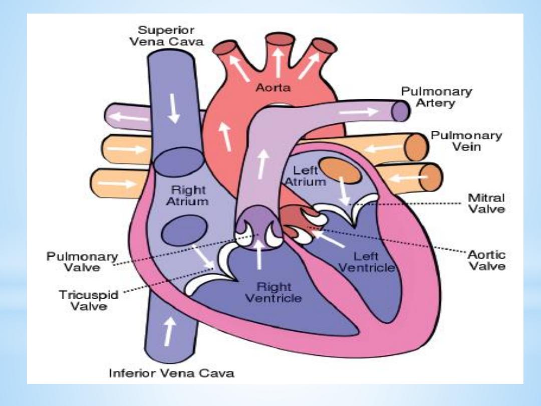
The heart as a pump
The heart is separated by septum into right and left
pump:
The right pump consist of right atrium and right
ventricle, the right atrium receives blood from the
systemic circuit, and the right ventricle discharges
blood into the pulmonary circuit
.

The left pump consist of left atrium and left
ventricle, the left atrium collects blood from the
pulmonary circuit, and the left ventricle ejects
blood into the systemic circuit.
The two are connected in series, when circuits are
connected in series, flow must be equal in the two
circuits.

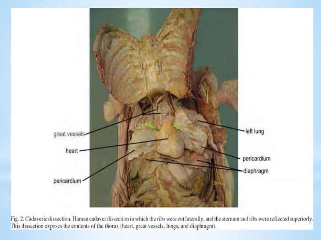
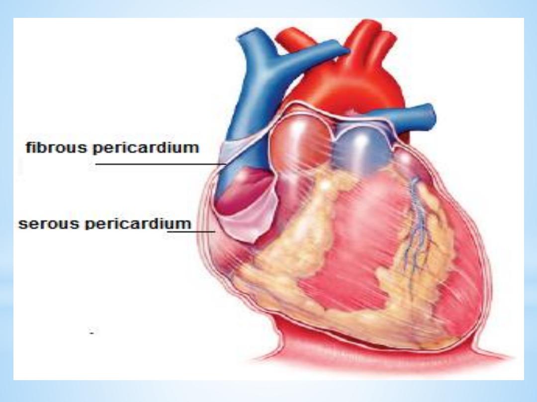
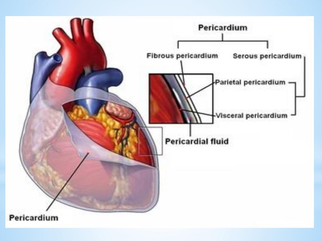
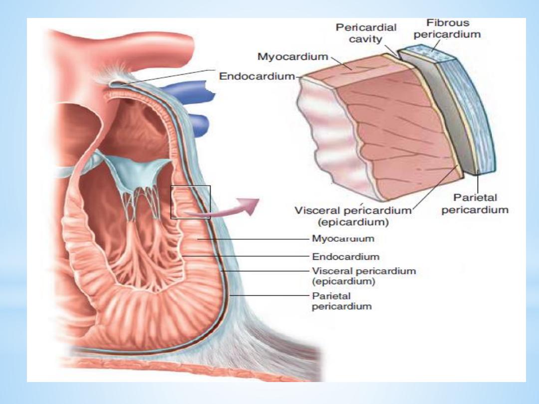
Pericardium
The pericardium forms a fibrous covering around
the heart, holding it in a fixed position in the
thorax and providing physical protection and a
barrier to infection.

The pericardium consists of:
1. a tough outer fibrous layer and a thin inner
serous layer. The outer fibrous layer is attached
to the great vessels that enter and leave the
heart, the sternum, and the diaphragm. The
fibrous pericardium is highly resistant to
distention; it prevents acute dilatation of the
heart chambers and exerts a restraining effect on
the left ventricle.

2.The inner serous layer consists of:
• a visceral layer, also known as the epicardium,
covers the entire heart and great vessels
• the parietal layer that lines the fibrous
pericardium.
Between the visceral and parietal layers is the
pericardial cavity, a potential space that contains 30
to 50 mL of serous fluid. This fluid acts as a
lubricant to minimize friction as the heart contracts
and relaxes.

Clinical points
Pericarditis is inflammation of the pericardium,
usually caused by a viral infection. Although this
disease can cause sharp, piercing chest pain, it is
usually self-limiting and ordinarily does not lead to
further problems.

Pericardial effusion is a collection of fluid around
the heart in the pericardial sac. If the fluid amount
is great enough, it can reduce the heart’s ability to
expand and receive blood, reducing its efficiency.
This condition is known as cardiac tamponade

The wall of heart
The wall of heart consist of
1.the inner surface is lined with endocardium,
which consists of smooth endothelial cells
supported by a thin layer of connective tissue. The
endothelial lining of the endocardium is continuous
with the lining of the blood vessels that enter and
leave the heart.

2.The myocardium, consists largely of cardiac
muscle tissue. The muscle fibers of the
myocardium are branched and tightly joined to
one another. The ventricular muscle is organised
into figure of eight bands that squeeze the
ventricular chamber forcefully in a way most
effective for ejection through the outflow valve.
The apex of the heart contracts first and relaxes
last to prevent back flow .

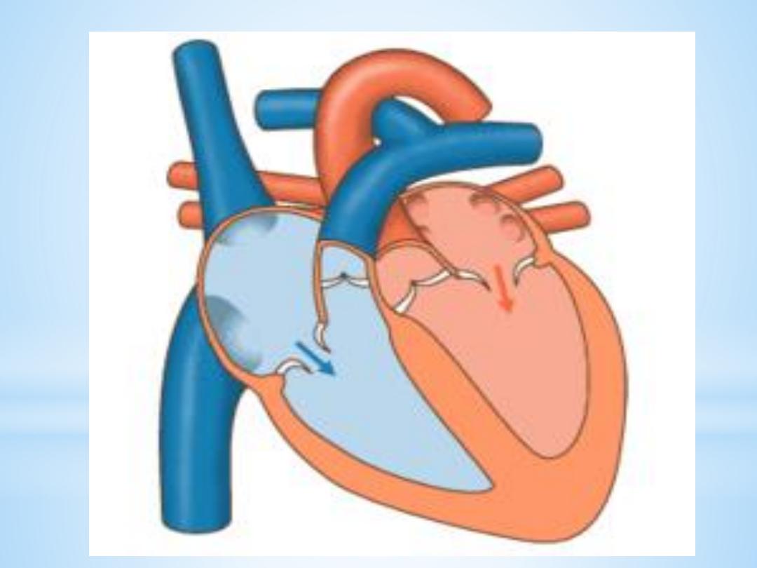
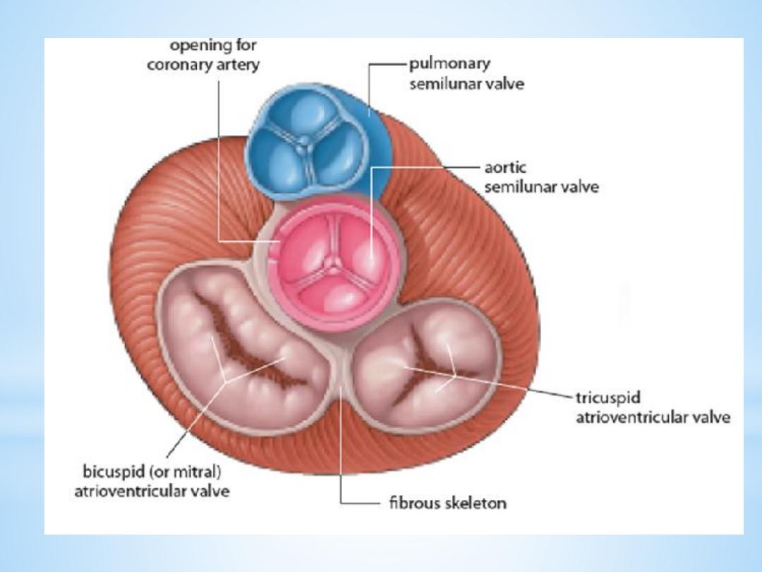
Heart valves
For the heart to function effectively, blood flow
must occur in a one-way direction, moving
forward through the chambers of the right heart
to the lungs and then through the chambers of the
left heart to the systemic circulation.

This unidirectional flow is provided by the heart’s
valves:
1.The atrioventricular (AV) valves control the flow
of blood between the atria and the ventricles . The
thin edges of the AV valves form cusps, two on the
left side of the heart (i.e., bicuspid or mitral valve)
and three on the right side (i.e., tricuspid valve).

The AV valves are supported by the
papillary muscles, which project from
the wall of the ventricles, and the
chordae tendineae, which attach to the
valve.
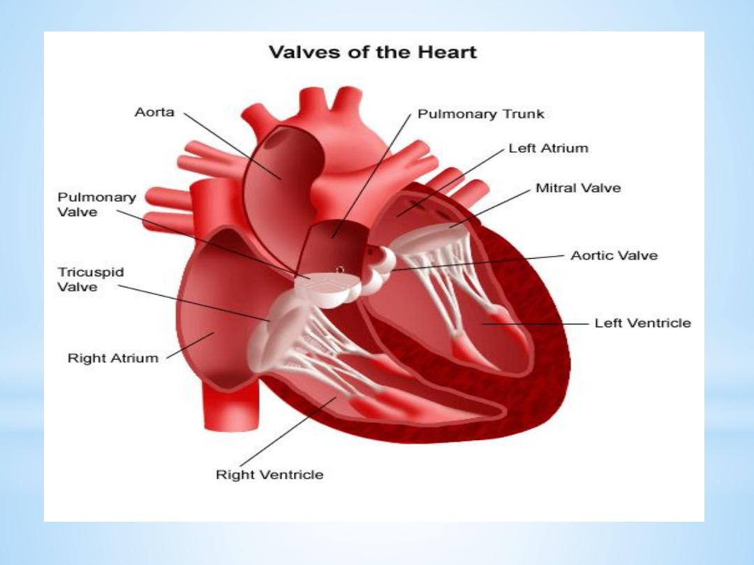
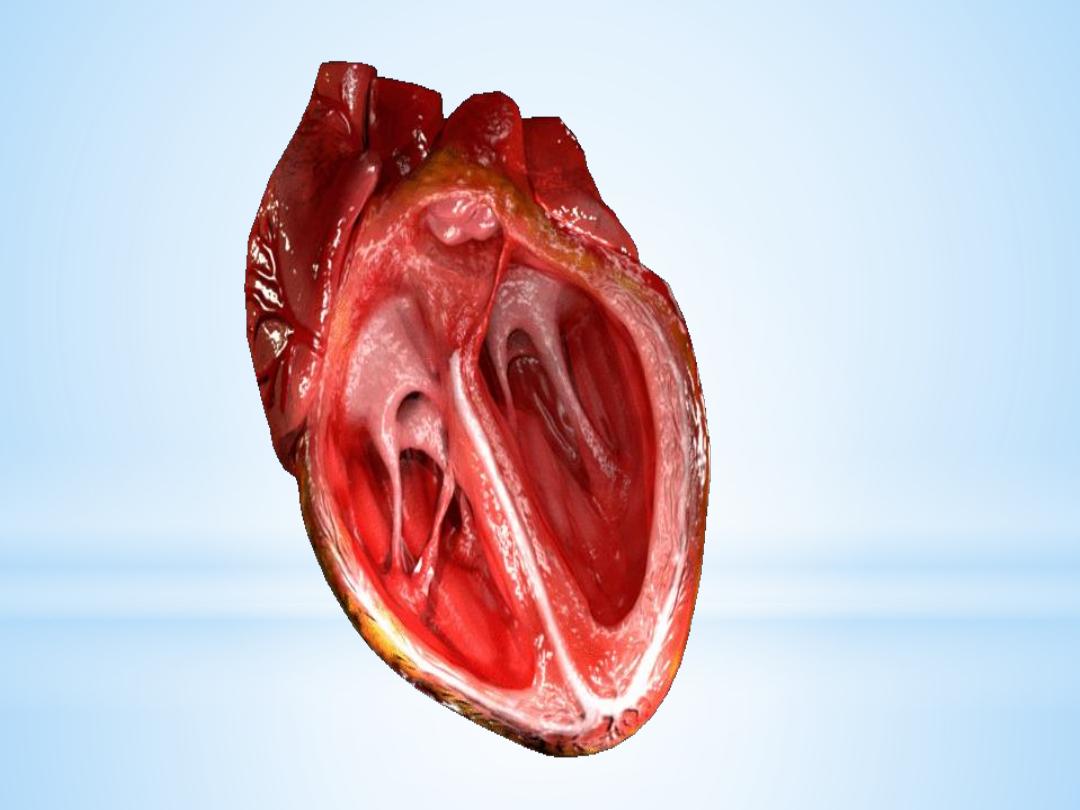

2.The aortic and pulmonic valves control the
movement of blood out of the ventricles. Because of
their half moon shape, they often are referred to as
the semilunar valves. The semilunar valves have
three little teacup-shaped leaflets. These cuplike
structures collect the retrograde, or backward, flow
of blood that occurs toward the end of systole,
enhancing closure.

Intrinsic Control of Heart beat
The rhythmical contraction of the heart is due to the
intrinsic conduction system of the heart, which
consist of:
1. SA (sinoatrial) node
The sinoatrial (SA) node is the normal pacemaker of
the heart and the origin of each normal heartbeat.

The SA node is a collection of specialized
myocytes near the site where the superior
vena cava enters in the wall of the right
atrium.The depolarization begins in the
sinoatrial node (SA node), spread rapidly
throughout the atria via gap junctions between
adjacent myocytes
.

2.Atrioventricular Node(A-V node)
The atrioventricular (AV) node is the only
electrical communication between the atria and the
ventricles. It is characterized by very slow
electrical conduction, ensuring that atrial
contraction is completed before the ventricles are
activated. The AV node is continuous with the
atrioventricular bundle (bundle of His).
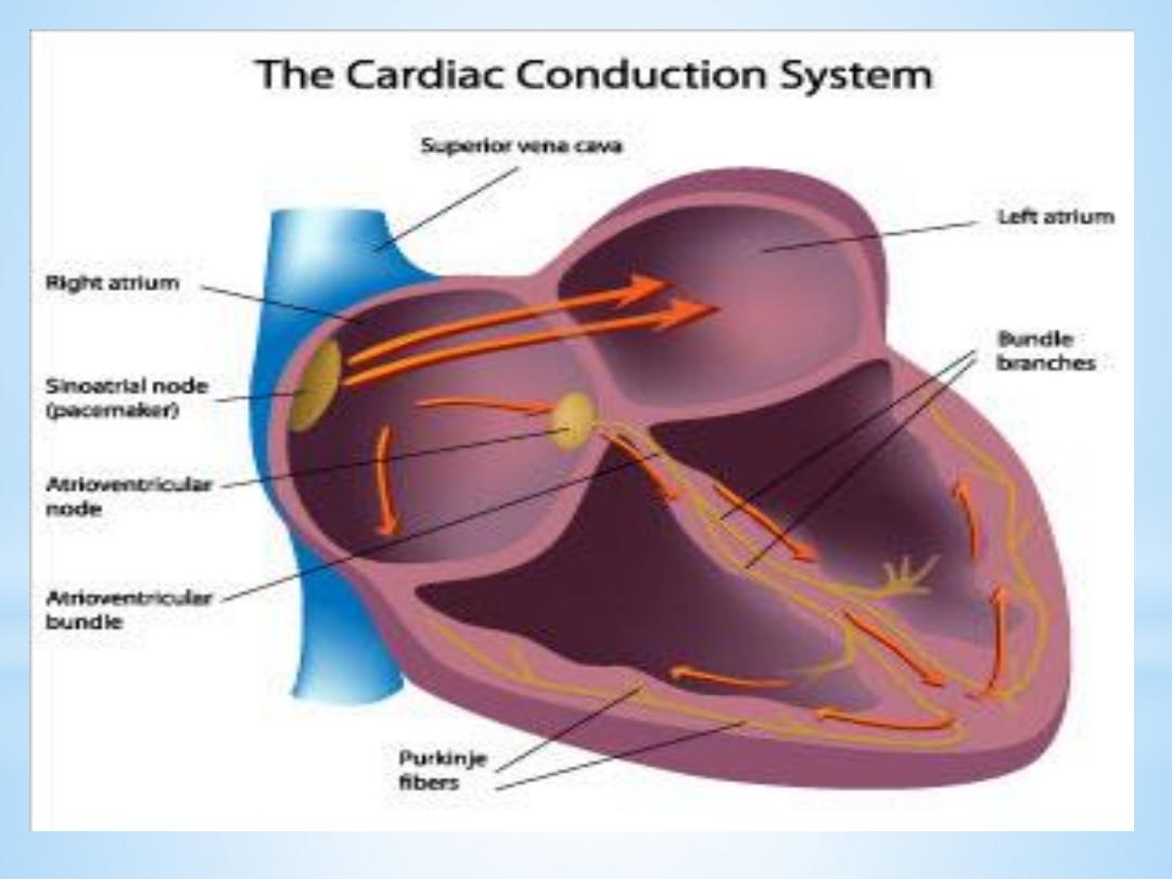

3.Atrioventricular bundle (bundle of His)
The AV bundle carry signals from atrium to the
ventricles, in the ventricles the AV bundle divide into
right and left bundle branch, these branches then
divide into an extensive network of Purkinje fibers.
4. Purkinje fibers
Specialized conducting fibers that transmit
electrical signals very rapidly to all parts of the
ventricular myocardium.

Extrinsic Innervations of the Heart
The excitatory and conductive system of the heart
receive innervations from both division of
autonomic nervous system. Although the basic heart
rate is set by the intrinsic conduction system, fibers
of the autonomic nervous system can modify the
heart beat :

1. The sympathetic nervous system (the
“accelerator”) increases both the rate and
the force of heartbeat.
2.parasympathetic slows the heart rate and
force of contraction.