
Investigations of the
Cardiovascular system
1

At the end of this lecture, you
should be able to appreciate
• The usefulness of each investigation in
diagnosing cardiac disease
• Each type of investigation clarifies and detects
a certain aspect of cardiac pathology
• Non-invasive investigations are increasingly
replacing the old, invasive techniques.
• Rapidly evolving methods of investigation
because of the advances achieved in
technology

Investigations of the CVS
• BNP, Troponin
• Electrocardiography
• Radiology
• Echocardiography
• CT imaging
• MRI
• Cardiac catheterization
• Radionuclide imaging
3

BNP, Pro-BNP
• Peptide released from the atria in response to
stretch
• Very sensitive for the diagnosis of congestive
heart failure
• Levels fall with improvement of heart failure
on treatment, rise with worsening
4

Troponin
• Protein contained within cardiac muscle
• Released when cardiac muscle is injured e.g.
ischemia or inflammation
• Very useful in the diagnosis and follow up of
patients with acute coronary syndrome
5

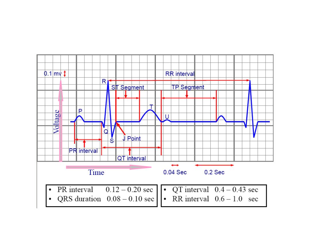
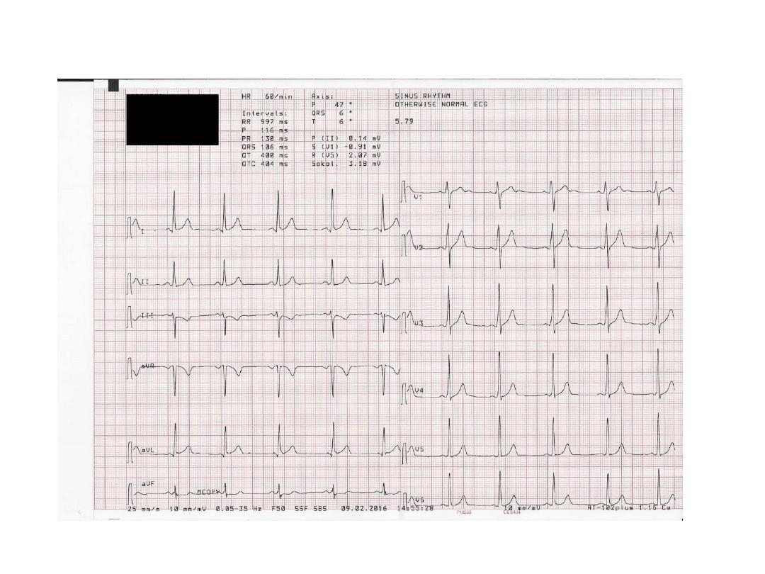
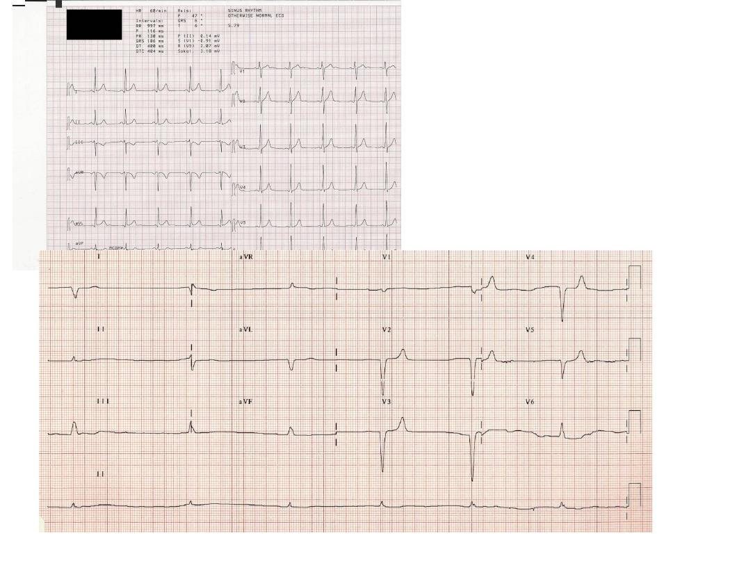
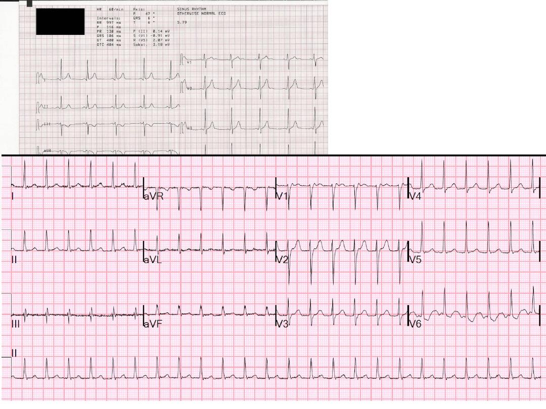
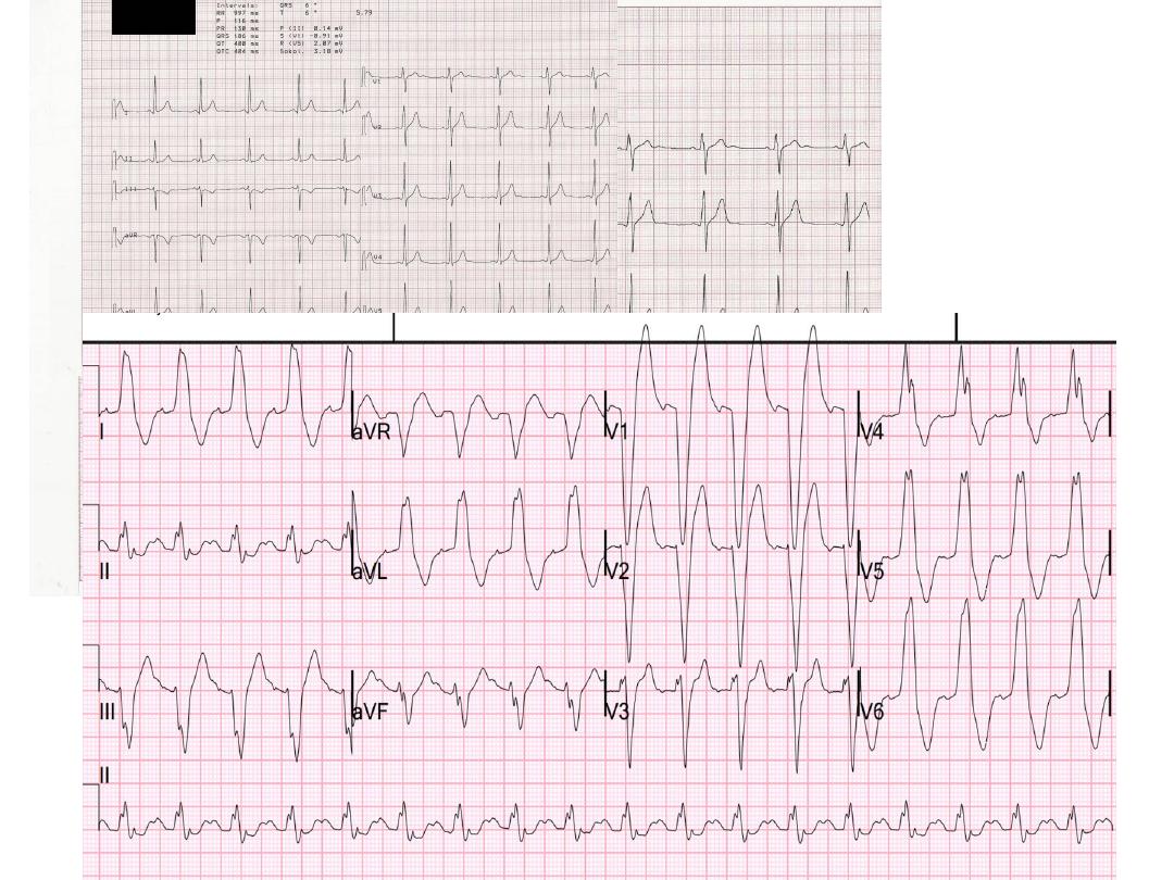
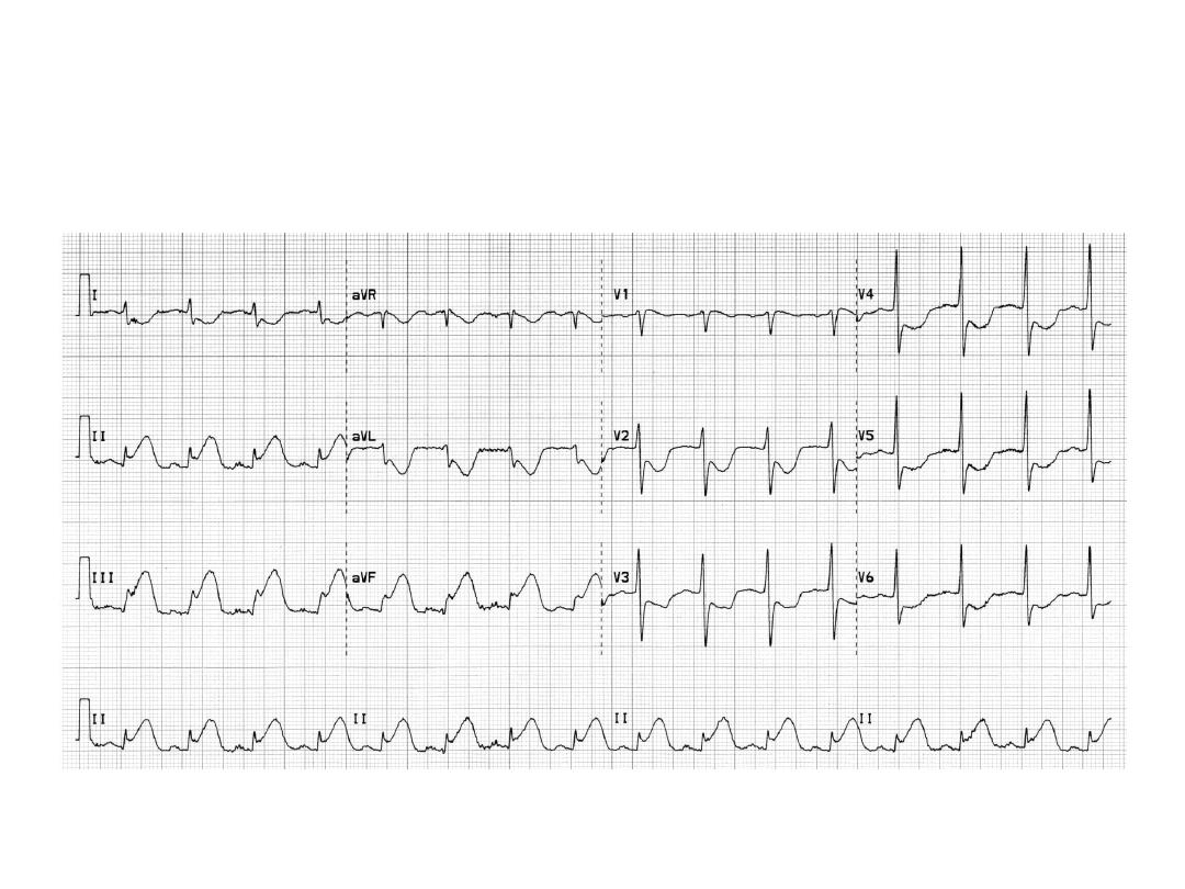
Electrocardiography (ECG)
uses:
• To determine heart rhythm
• Status of the conducting system
• To diagnose myocardial ischemia or infarction
• Chamber enlargement and hypertrophy
• Effects of drugs & metabolic disorders
(electrolyte imbalance, acidosis, etc.)
6
7
8
9
10
11
12

Exercise ECG
• In patients with angina, the resting ECG may
be normal
• The principle of the test is to stress the heart
and observe for ECG changes of ischemia
• ECG and BP are continuously recorded while
the patient is exercising on a bicycle or a
treadmill
13

Ambulatory ECG Monitoring (Holter)
• Continuous recording of ECG over 24 hours or
more
• Used to detect transient episodes of ischemia
or arrhythmia which can rarely be captured
during routine, ordinary ECG recording
14

Imaging
The principle of imaging is to reconstruct a
three-dimensional structure out of a group of
two dimensional images:
• Silhouette imaging: various structures are
overlapped over each other e.g. CXR,
angiography, nuclear imaging
• Tomographic imaging: a group of sections
through the structure to be examined e.g. echo,
CT, MRI
15

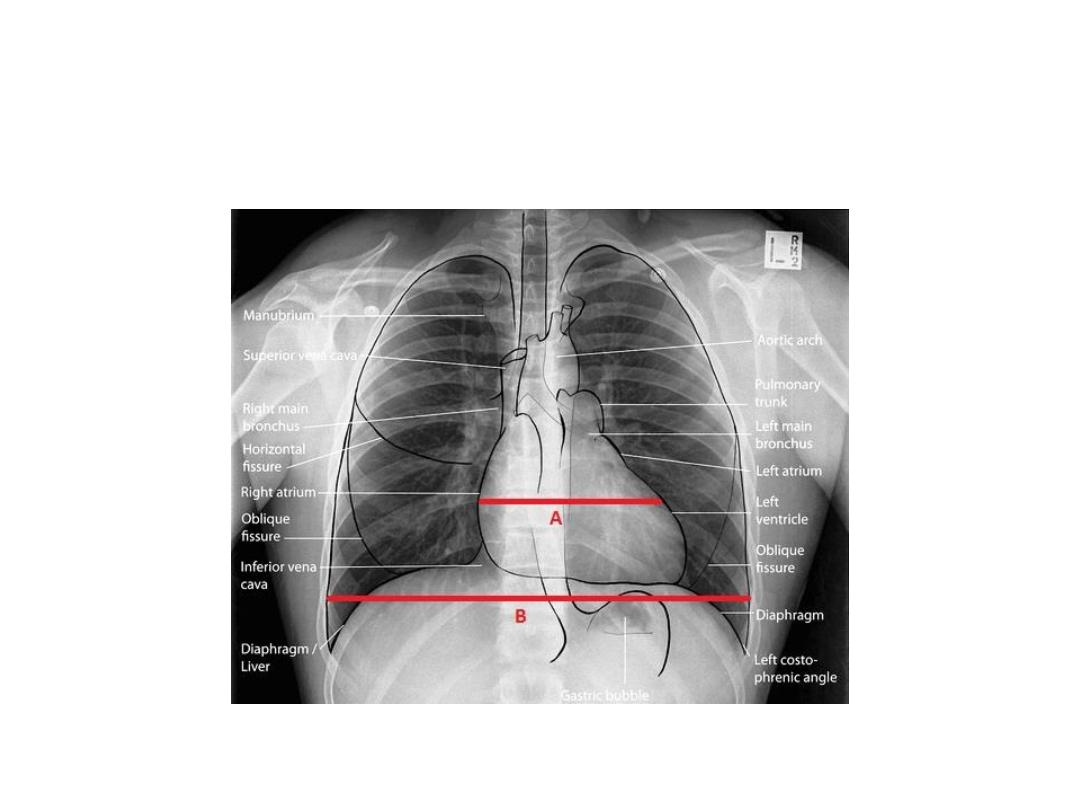
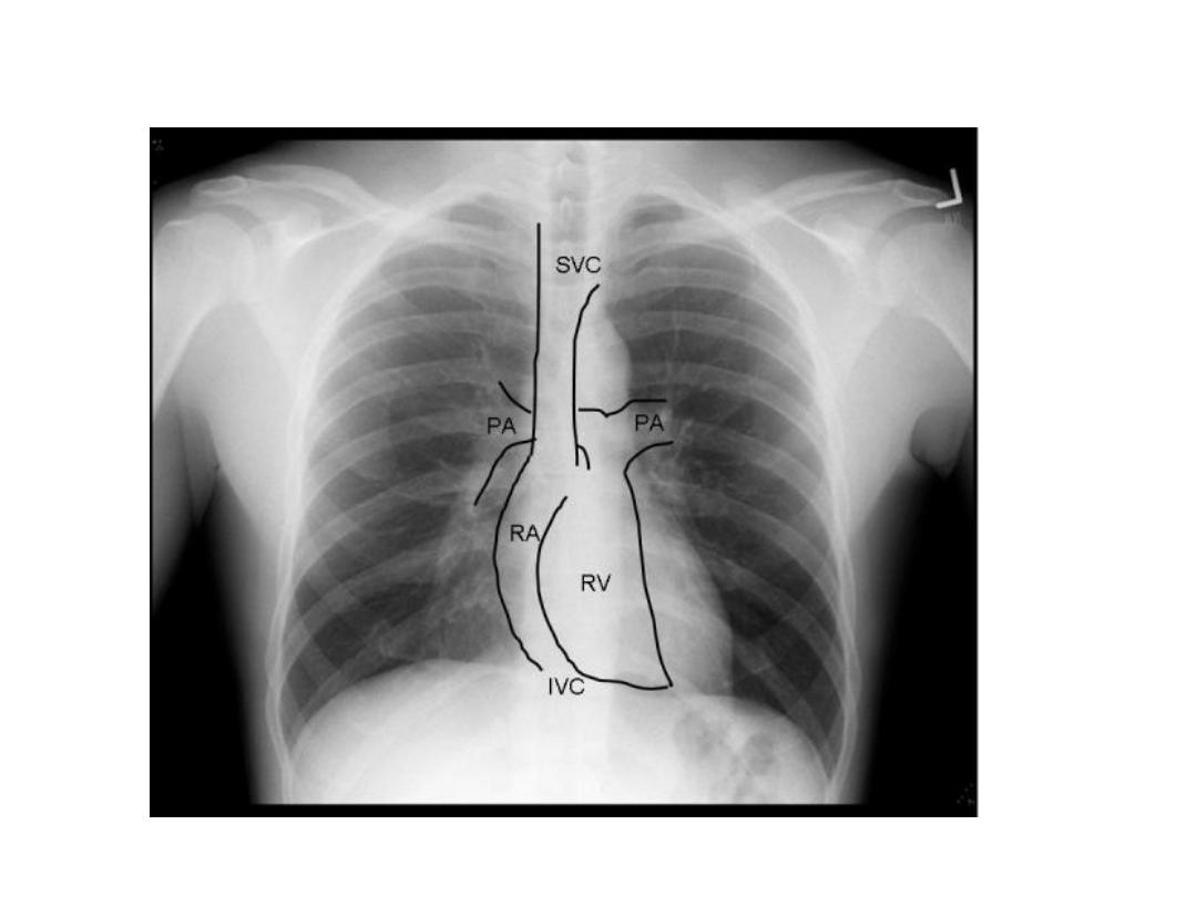
Radiology of the Heart
Chest X-ray: Postero-anterior view (PA view):
• Size of the heart
• Shape of the heart
• Specific chamber enlargement
• Status of the pulmonary circulation
16
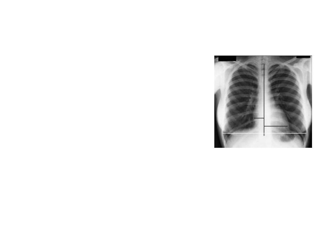
Radiology of the Heart
Cardiac size:
• Cardio-thoracic ratio (CTR):
– Normally < 0.5
• Enlargement of the heart
(cardiomegaly):
– LV dilatation and dysfunction
– Pericardial effusion
17
18
19
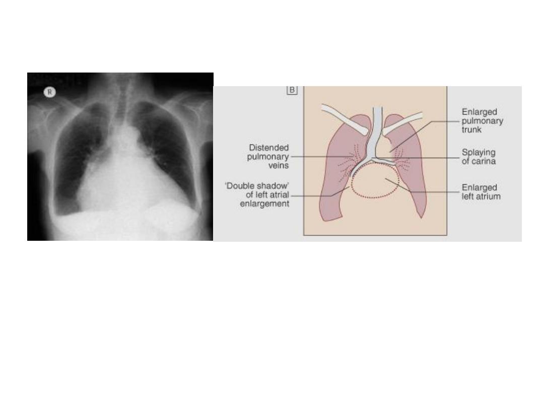
Radiology of the Heart
Left atrial enlargement:
• Straight heart border (LA appendage)
• Widening of the carinal angle
• Double contour of the right heart border
20
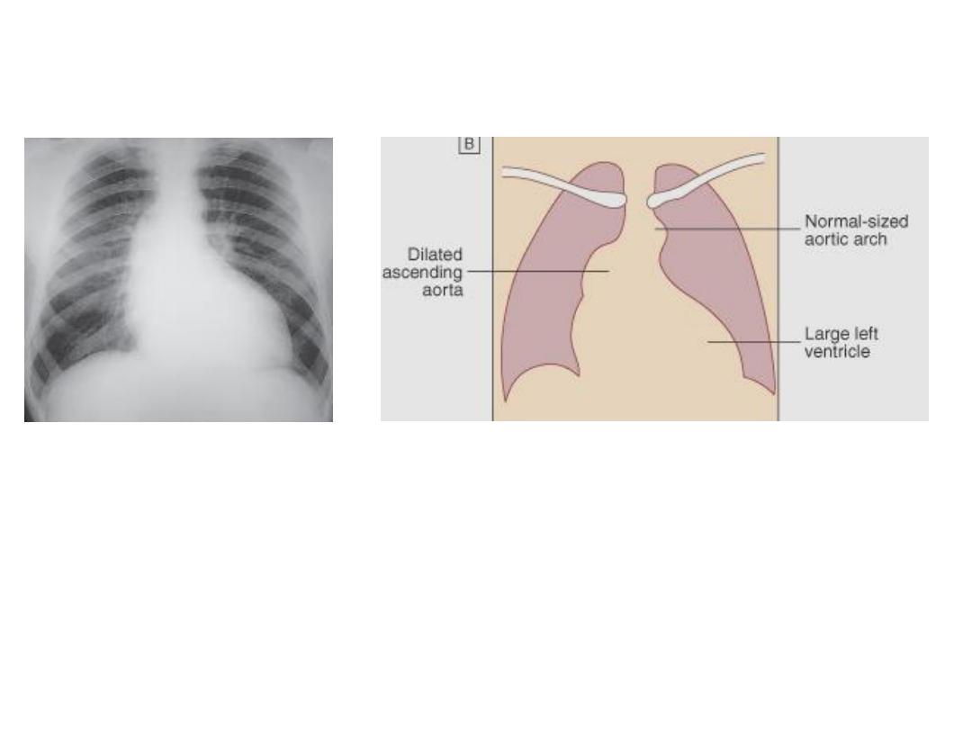
Radiology of the Heart
LV enlargement:
• Enlarged cardiac silhouette
• Prominent left heart border
21

Radiology of the Heart
RV enlargement:
• Cardiomegaly
• Straightening of the left heart border
• Apex displaced upwards
Right atrial enlargement:
• Prominence of the right border of the heart
22

Radiology of the Heart
Lung fields:
• Congestion & edema in patients with left heart
failure
• Increased blood flow (prominent arteries and
veins) in shunt lesions
• Oligemic lungs in pulmonary stenosis
• Pleural effusions in advanced heart failure
23
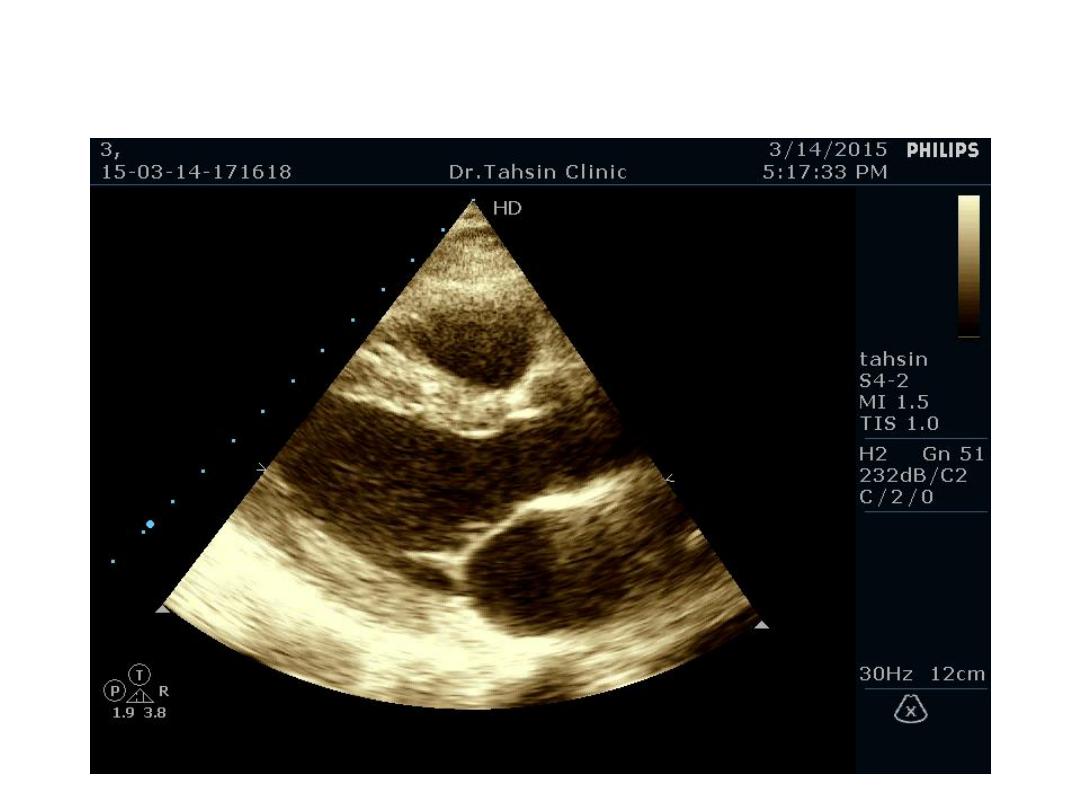
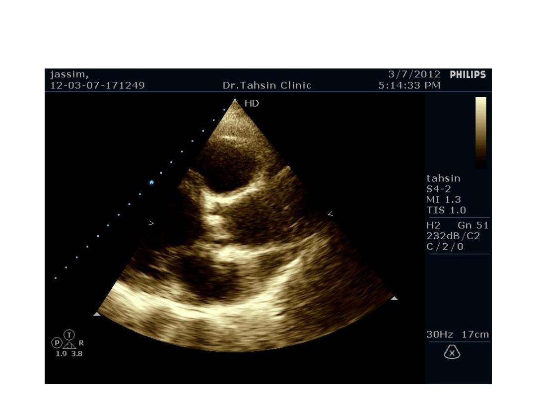
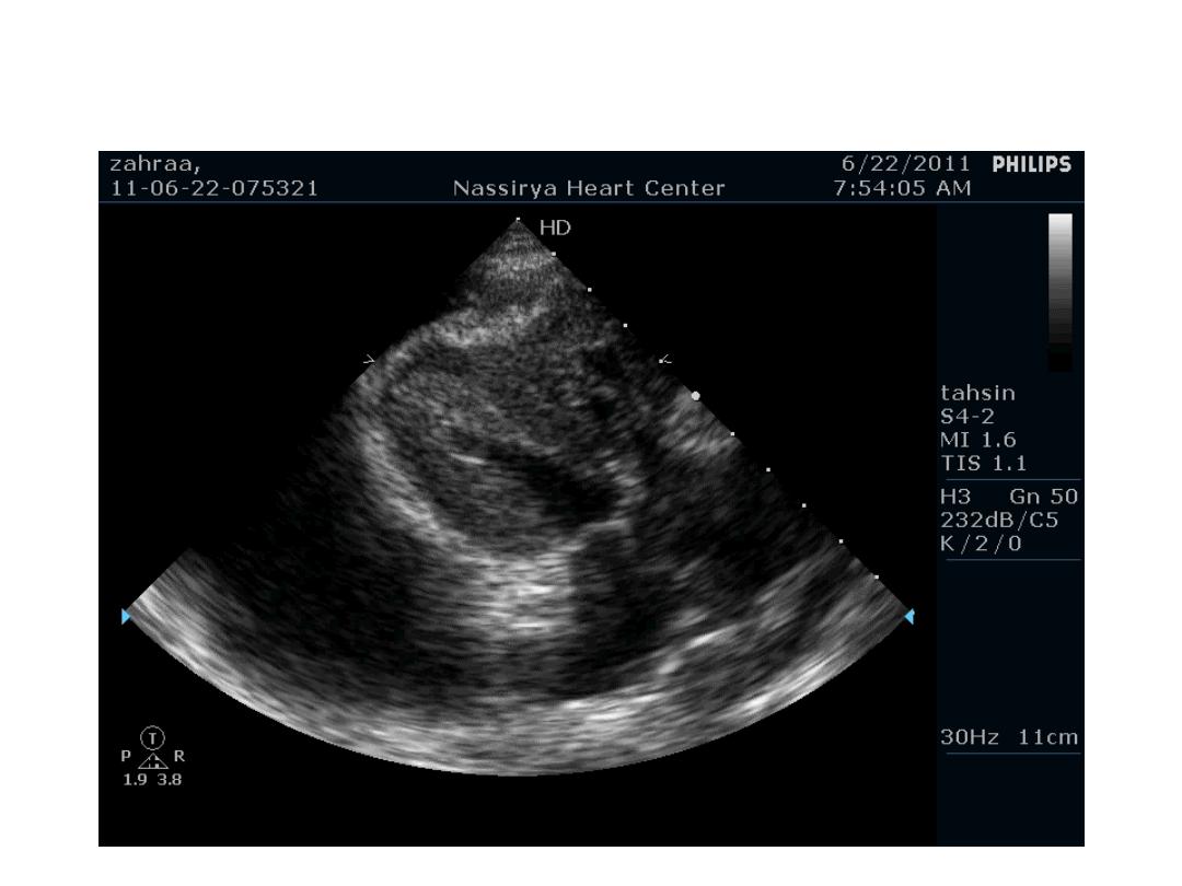
Two Dimensional Echocardiography
• Ultrasound beam passing through
the heart generates cross
sectional images or “slices” of
the heart
• Various structures can be seen in
real time
24
25

Two Dimensional Echocardiography
indications
• Assessment of LV function
• Diagnosis & quantitation of severity of
valvular lesions
• Identification of vegetations
• Identifying the source of systemic embolism
• Detection of pericardial effusion
26
27
28
29

30

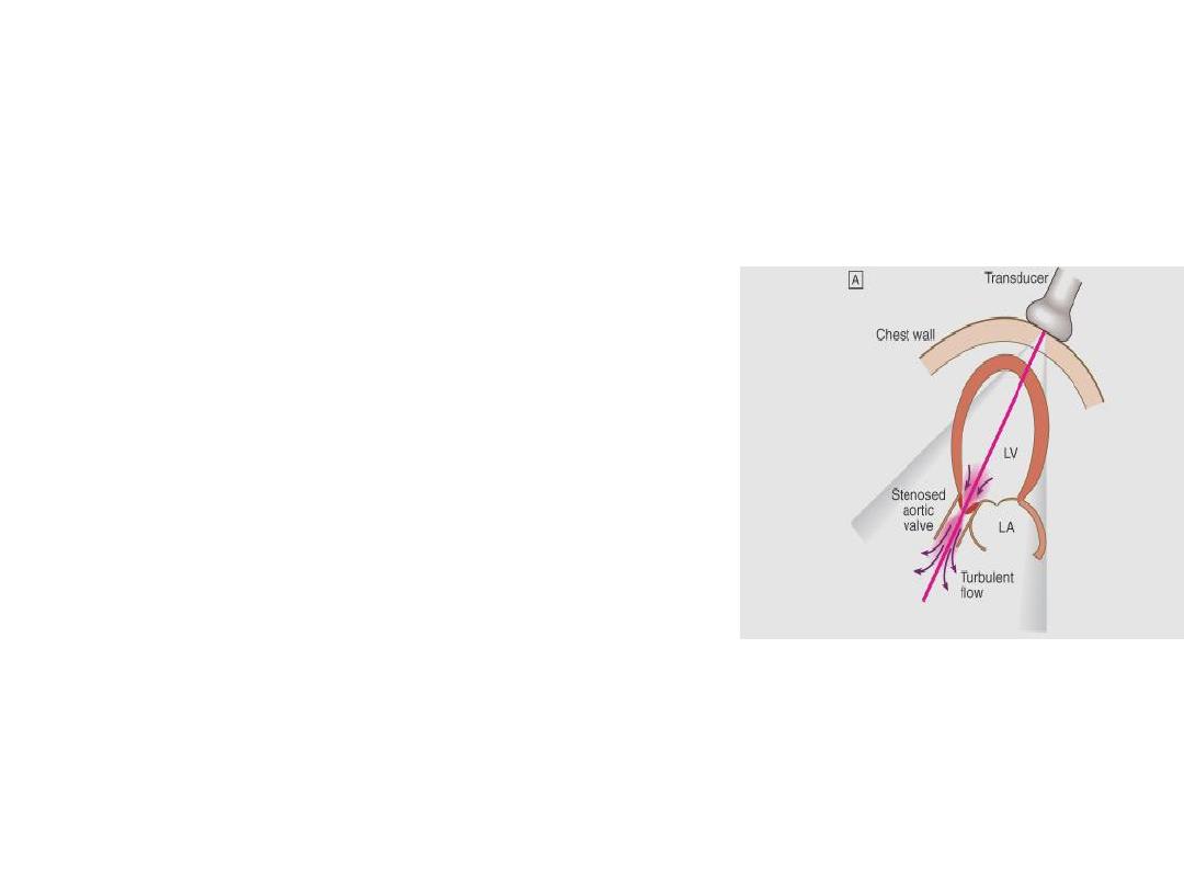
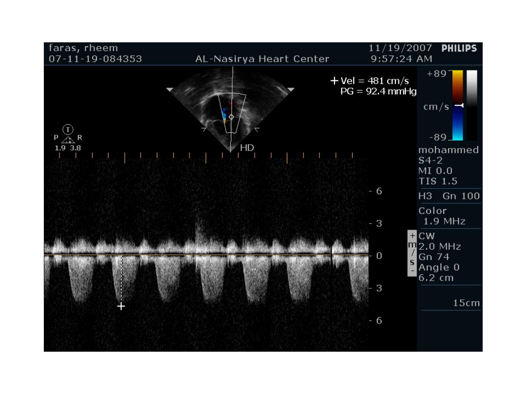
Doppler Echocardiography
• Sound waves reflected from moving RBCs
undergo frequency shift
• The faster the blood velocity , the greater the
frequency shift
• The direction of moving blood determines
whether the reflected signal is positive or
negative
31
32
33

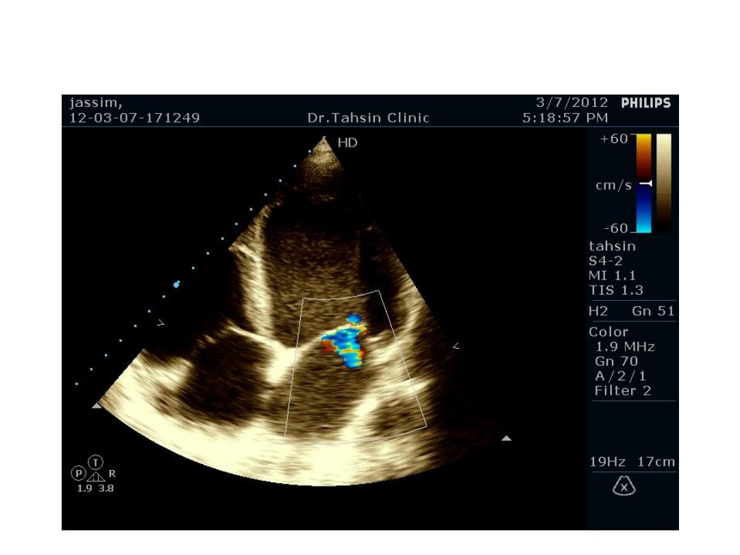
Doppler Echocardiography
• The derived signal can be plotted graphically
against time
• Or, color can be assigned for the reflected
signal and superimposed over the 2D image
(color flow mapping)
34
3-Dimentional Echocardiography
35
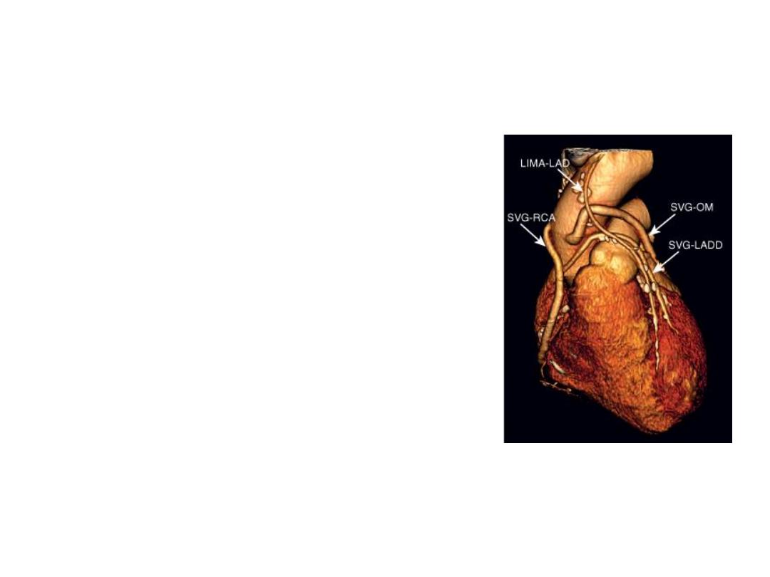
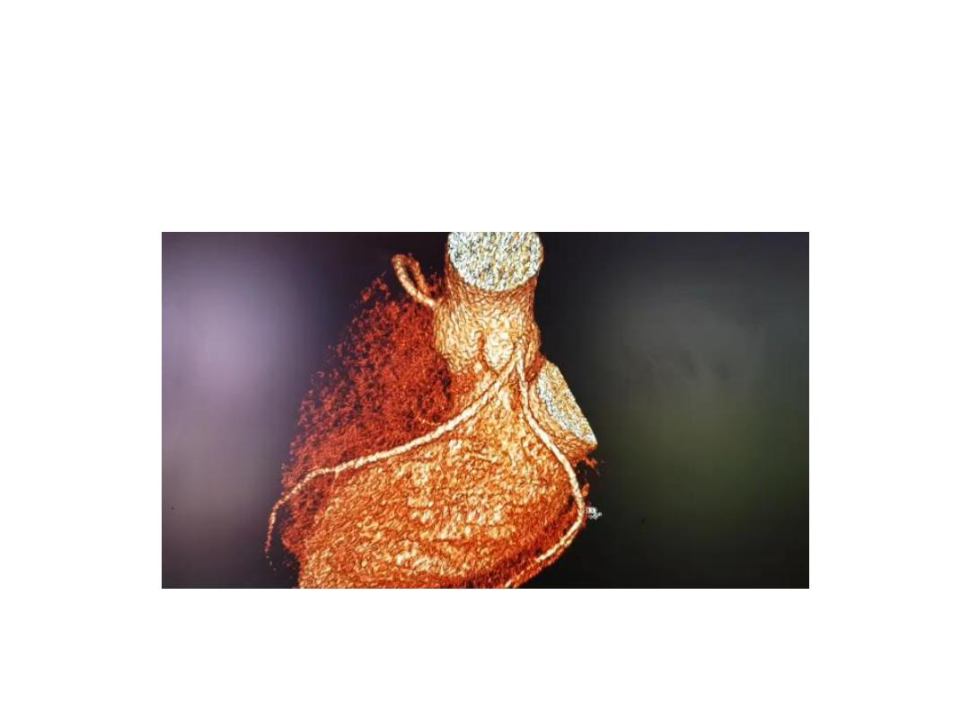
Other non-invasive imaging:
CT and MRI
• Chambers of the heart
• The great vessels
• The pericardium
• Diseases of the aorta
• The pulmonary arteries
• Non-invasive imaging of the
coronary arteries
36
37

Invasive investigation: cardiac
catherization
• A small tube is passed into the heart via a
peripheral artery or vein under fluoroscopic
guidance
• Pressure can be measured, flow volumes
calculated, radiographic dyes can be infected
to outlime the specific chamber or vessel
(angiography)
38