Early development of human
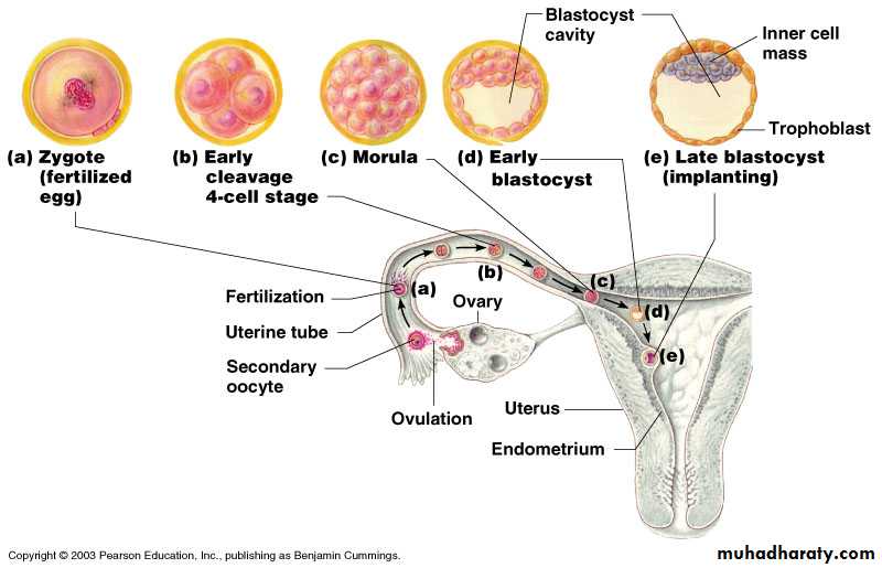
Early human development may be summarized by the following four phases: (1) fertilization; (2) implantation; (3) gastrulation; and (4) embryogenesis.Fertilization: This stage takes place during the first week of human embryology. Fertilization occurs when the cell membrane of a sperm fuses with the cell membrane of the oocyte (egg), injecting its nucleus. The egg then undergoes its second division of meiosis, and the resulting haploid nucleus fuses with the haploid sperm nucleus to re-form the diploid number of chromosomes. These events occur in the oviduct, or fallopian tube. The fertilized egg, now termed a "zygote," continues to move down the oviduct to the uterus, where it lodges in the wall. Cleavage defined as the mitotic division of cells in the early embryo

Morula, a solid ball of 32 cells that resembles a raspberry, moves downward toward the uterus
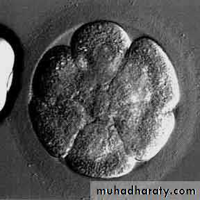
Blastula, a ball of more than 64 cells surrounding a fluid-filled cavity (the blastocele)
Blastula is produced by the repeated mitotic division of a zygote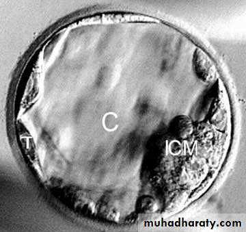
Implantation: The zygote implanted in the uterine wall, the cells that will form the embryo proper divide and organize themselves into a bilaminar (two-layered) disc. This disc is surrounded by an outer ring of cells, the trophoblast, which does not contribute to the new organism's tissues. After implantation, trophoblast cells multiply rapidly and invade the endometrium (uterine wall). Together, the trophoblast and endometrium form the placenta, through which all the nutrition for the developing embryo will pass. This rich mass of tissue is filled with blood vessels, allowing rapid exchange of nutrients and waste. Another group of cells separates from the developing embryo near this time, and these cells also do not form part of the new organism. Instead, they develop into the amnion, the membrane that will surround the fetus to form the embryonic sac. This fluid-filled sac helps to cushion the fetus during later development. This phase begins during the second week of development.
Gastrulation. During the third week, the embryo undergoes the process of gastrulation, forming a trilaminar (three-layered) disc. Gastrulation establishes the three germ layers—the endoderm, ectoderm, and centrally placed mesoderm—all of which will give rise to the various organ systems. Mesoderm also combines with trophoblast tissue to form the umbilical cord, which transports nutrients and wastes between the fetal circulation and the placenta.
Ectoderm forms the outer layer. Ectoderm forms skin, hair, sweat glands, epithelium, brain and nervous system.
Endoderm forms the inner layer. The endoderm forms digestive, respiratory systems, liver, pancreas, gall bladder, and endocrine glands such as thyroid and parathyroid glands.
Mesoderm forms the middle layer. The mesoderm forms body muscles, cartilage, bone, blood, reproductive system organs and kidneys
Embryogenesis: At this point, the developing human enters the actual embryonic phase, which lasts from the third week through the eighth week after conception. The organ systems differentiate at greatly varying rates during this phase. For example, the circulatory system is largely functional at the end of this period, whereas the nervous system is still engaged in massive cell division and only beginning to establish functional connections. Most embryological malformations occur during this embryonic phase.
The remainder of human development, from weeks nine to thirty-eight, is called the fetal period, the time during which the embryo first acquires human appearance.
The Fetal Period
The fetal period is characterized by two processes. The first is rapid growth (increase in size and cell number) and the second is continued tissue and organ differentiation (specialization of cells to perform distinct functions).
Tissue and organ development: Early in fetal development the head dominates the body, constituting half its length. The face is broad and flat, eyes are still wide apart, and ears are low. The intestines temporarily protrude through the abdominal wall until the tenth week, and the external genitalia appear similar between the sexes. By the end of the fourth month, the rest of the body has caught up to the head and the limbs have grown to give the fetus proportions more nearly like those of a newborn. The fifth month is marked by the first fetal movements perceived by the mother, known as quickening. The skin of the fetus secretes a lipid -rich covering substance. It also exhibits a temporary covering of fine hair.
By the sixth month, the fetus acquires the capacity for independent existence because the lungs have finally matured to the point where the fetus can breathe. During the seventh month the nervous system develops many basic reflex responses, including the constriction of the pupils in response to light. Other reflexes controlling breathing, swallowing, and general movement can be detected much earlier, around the middle of the third month, although the effective coordination of such movements requires several more months in uter.
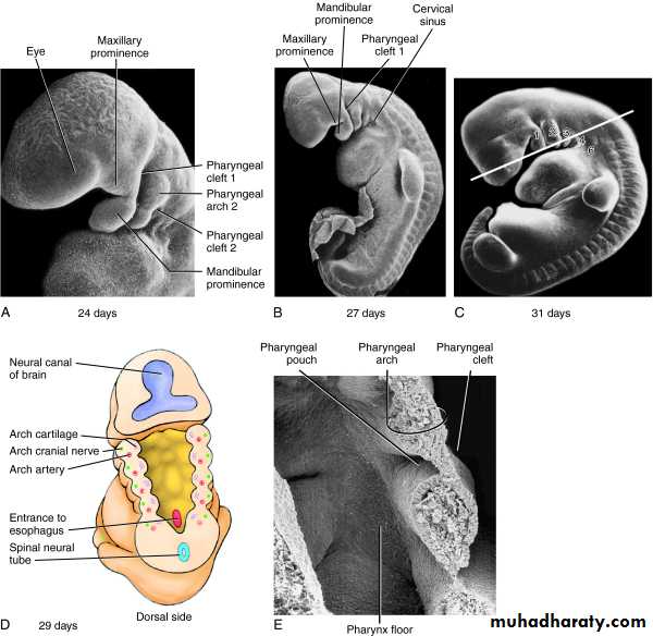
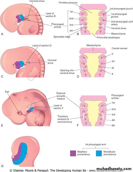
The branchial apparatus
The branchial (=Gr gill) apparatus comprises an early structure during embryologic development. It is associated with the formation of the head and neck. It consists of the branchial arches, the pharyngeal pouches, the branchial clefts or grooves and the branchial membranes. A four-week-old embryo features four visible branchial arches separated by branchial grooves. They are numbered craniocaudally. A fifth and a 6th are also present but are very small. A primitive mouth appears as a small depression referred to as stomatodeum (stomodeum). The oropharyngeal (buccopharyngeal) membrane separates the stomatodeum from the primitive pharynx (cranial part of the foregut). This membrane ruptures at 24 to 26 days and communication with the amniotic cavity is established.
Adult Derivative
ArchNerve
1
(Mandibular Arch; Merckel’s Cartilage)
CN V2CN V3
Mesoderm: Muscles of mastication (temporal, masseter, medial & lateral pterygoids), mylohyoid, anterior belly of digastric, tensor veli palatine, tensor tympani
Neural Crest: Maxilla, mandible, incus, malleus, zygomatic bone, squamous temporal bone, palatine bone, vomer, sphenomandibular ligament
1st Aortic Arch: Maxillary artery, external carotid artery (?)
2
(Hyoid Arch; Reichert’s Cartilage)
CN VII
Mesoderm: Muscels of facial expression (buccinator, auricularis, frontalis, platysma, orbiuclaris oris & obicularis oculi), posterior belly of digastric, stylohyoid, stapedius
Neural Crest: Stapes, styloid process, stylohyoid ligament, lesser horn and upper body of hyoid bone
2nd Aortic Arch: Stapedius artery
3
(Glossal Pharyngeal Arch)
CN IX
Mesoderm: Stylopharyngeus, common carotid arteries, internal carotid arteries
Neural Crest: Greater horn and lower body of hyoid bone
3rd Aortic Arch: Common carotid artery, internal carotid artery
4
CN X (superior laryngeal)
Mesoderm: Muscles of soft palate (except tensor veli palatine), muscels of the pharynx (except stylopharyngeus), cricothyroid, cricopharyngeus, laryngeal cartilages, rt. subclavian artery, arch of aorta
Neural crest: none
4th Aortic Arch: Arch of aorta (left side), right subclavian (right side)
5
n/a
Rudimentary:
Develops in fish not humans (branchia = gill)
Pharyngeal apparatus used to be called “branchial” apparatus
6
CN X (recurrent laryngeal
Mesoderm: Intrinsic muscles of larynx (except cricothyroid), upper muscles of the esophagus, laryngeal cartilages, pulmonary arteries, ductus arteriosus
Neural Crest: none
6th Aortic Arch: Left pulmonary artery (left side), ductus arteriosus (left side), right pulmonary artery (right side)
Pouch
1
Epithelial lining of auditory tube and middle ear cavity
2Epithelial lining of palantine tonsil crypts
3Inferior parathyroid gland, thymus
4Superior parathyroid gland, ultimobranchial body♦
Groove1
Epithelial lining of the external auditory meatus
2, 3, 4Obliterated
Membrane1