


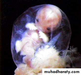
Human Development
week 10
Explain the stages of development starting with fertilization and ending with the neonatal period.Discuss the major events of the first, second, and third trimesters of development.
I addition we will discuss the formation process maxilla, mandible and cranium
Prenatal Development
Embryonic developmentfertilization - 8 weeks
Fetal development9 weeks - birth
Postnatal Development
time period from fertilization to birth = gestation= pregnancy

Stages of development
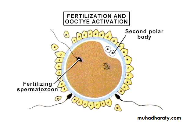
Fertilization:it is the process of fusion of mail and female germs to form zygote. Typically this cell contains 46 chromosomes and gametes contain 23 chromosomes
Taking place in ??
Viability of gametes:
Oocyte 12-24 h
Sperm 12-48 h
Single sperm fuses with oocyte
Cleavage
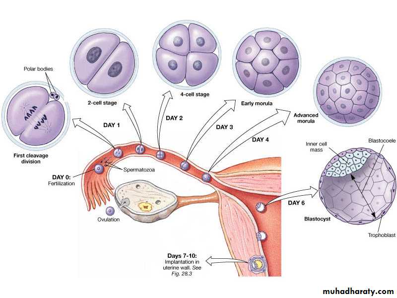
Blastocyst
with blastocoele cavity 64 cellsMorula
solid ball of cells 16 cells
Early division of zygote into multiple cells without increase in size. Cleavage is the mitotic division of cells in the early embryo which occurs in sequence of 2.4.8.16. 32,64 and so on
Zygote
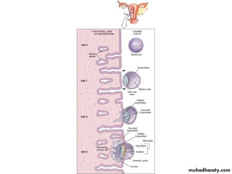
• Implantation Embedding of blastocyst into uterine lining begins at day 7 end at 9th dayBall of mass cell called blastocyst. Out layer form trophoblast and inner layer form embryo.Trophoblast converted to chorion and finally establish to form placenta
Formation of Placenta
Development of placenta from edges of blastocystPlacenta = organ that allow the embryo/fetus to exchange nutrients and waste.
Nutrient and gas exchange happens without actual blood exchange
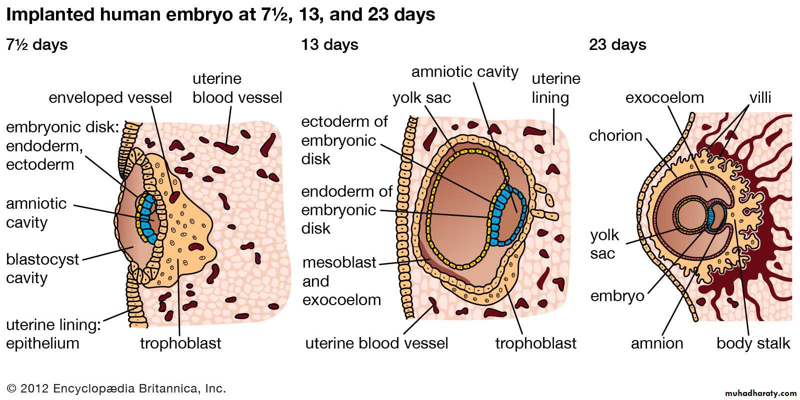
Gastrulation: 3 Germ Layers Formed
Ectoderm (forms from epiblast)Nervous system
Epidermis, epithelium lining skin mucosa of lips, nasal cavity, floor of mouth palate, exocrine sweat gland parotid gland. Enamel of teeth
Endoderm (forms from hypoblast)
Mucosae (eg: GI-tract
Associated glands
Mesoderm
Dentin, heart blood vessels etc.
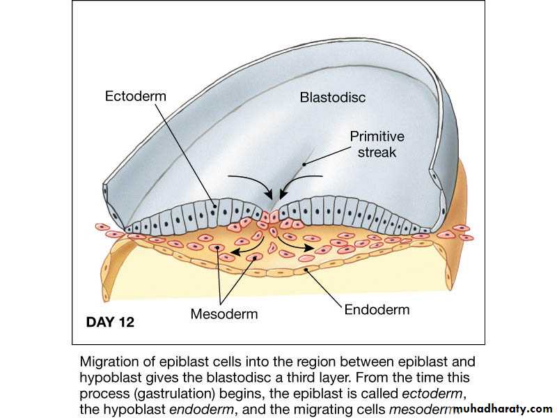
day 12:
Embryogenesis
Following gastrulation, formation of viable embryoHead fold and tail fold develop
Critical period organogenes
Teratogens, Teratology = ?
Rubella and syphilis
X-rays
FAS and smoking
Fetal development from 9th -38th weeks
Characteristics1. rapid growth
2.tissue differentiations
Fetal development
Tissue and organ developmentFace
Eyes
External genitalia
Limbs
Lung development
Nervous system
Pharyngeal Apparatus
The head and neck region of four week human embryo somewhat resemble these regions of a fish embryo of comparable stageThis explains the former use of designation branchial apparatus
Branchial is derived from the Greek word branchia or gill
Pharyngeal Apparatus
Pharyngeal apparatus consists of:Pharyngeal arches
Pharyngeal pouchesPharyngeal grooves/clefts
Pharyngeal membrane
Pharyngeal Arches
Pharyngeal arches begin to develop early in the fourth week as neural crest cells migrate into the head and neck regionThe first pair of pharyngeal arches (primordium of jaws) appears as a surface elevations lateral to the developing pharynx
Soon other arches appear as obliquely disposed, rounded ridges on each side of the future head and neck regions
Each pharyngeal arch consists of a core of mesenchyme
Is covered externally by ectoderm and internally by endodermIn the third week the original mesenchyme is derived from mesoderm
During the fourth week most of the mesenchyme is derived from neural crest cells that migrate into the pharyngeal arches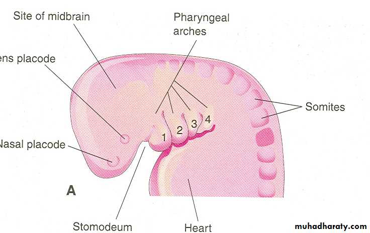
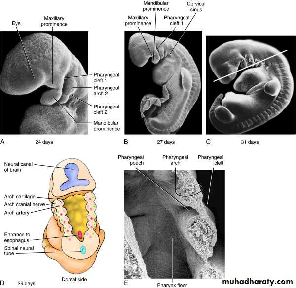
Pharyngeal Arches
By the end of the fourth week, four pairs of pharyngeal arches are visible externallyThe fifth arch is rudimentary and are not visible on the surface of the embryo
The pharyngeal arches are separated from each other by fissures called pharyngeal groovesArches
Each arch containsCartilage
Cranial nerve
Artery
Muscle component
All neural crest origin
6 arches, only 5 form structures in humans
1, 2, 3, 4, and 65th fails to develop
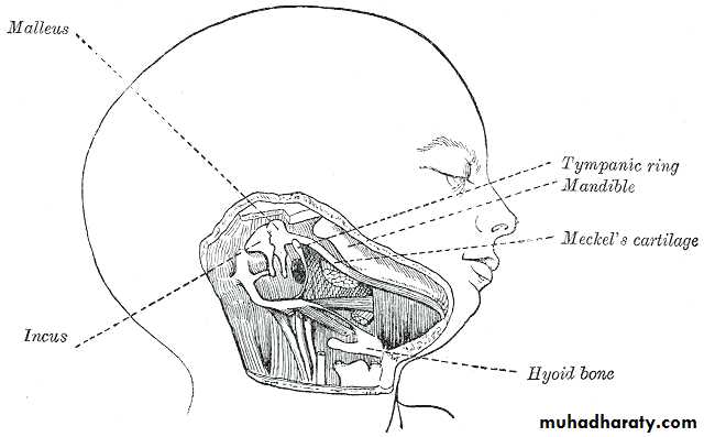
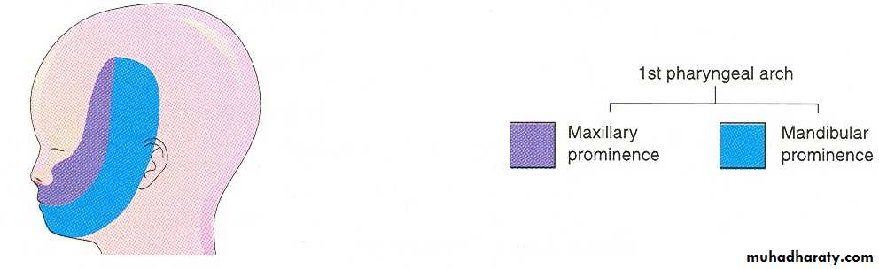
1st Arch “Mandibular Arch”
Skeletal componentsMeckel’s cartilage
Framework for mandible
Malleus head and neck
Incus body and short process
Muscles
Muscles of mastication
Anterior digastric
Mylohyoid
Tensor tympani
Tensor veli palatini
Nerve
CN V (Trigeminal)
Artery
Maxillary; external carotid
2nd Arch “Hyoid Arch”
Skeletal componentsReichert’s cartilage
Stapes
Malleus manubrium
Incus long process
Styloid process
Hyoid bone (lesser horn and upper body)
Muscles
Facial expression, buccinator, platysma, stapedius, stylohyoid, posterior digastricNerve
CN VII (Facial)
Artery
Stapedial3rd Arch
Skeletal componentsHyoid (greater horn and lower body)
Muscles
Stylopharyngeus
Nerve
CN IX (Glossopharyngeal)
Artery
Common/Internal carotid
4th Arch
Skeletal componentsThyroid, epiglottic, cuneiform cartilages
Muscles
Cricothyroid, inferior constrictors
Nerve
Superior laryngeal
Artery
Subclavian, aortic arch
6th Arch
Skeletal componentsCricoid, arytenoids, corniculate
Muscles
All intrinsic muscles of larynx (except cricothyroid)
Nerve
Recurrent laryngeal
Artery
Pulmonary artery
Branchial Clefts and Pouches
4 clefts and 4 pouches5th and 6th contribute to the 4th
Clefts provide “covering” to structures of the corresponding arch and pouch
Pouches
1st PouchEustachian tube, middle ear, mastoid, inner layer of tympanic membrane
2nd Pouch
Tonsils, root of tongue, foramen cecum, pharynx(part)3rd Pouch – ventral and dorsal wings
Ventral wing – ThymusDorsal wing – inferior parathyroid glands
Pouches
4th PouchSuperior parathyroid glands
Parafollicular C-cells of thyroid gland
5th Pouch
Contributes to Parafollicular C-cells
6th Pouch
Contributes to laryngeal musculature and cartilage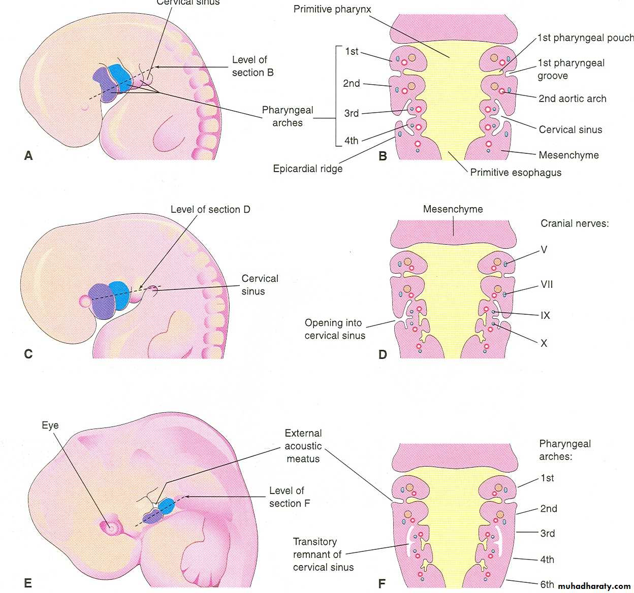
Fate of Pharyngeal Arches
A typical pharyngeal arch contains:An aortic arch, an artery that arises from the truncus arteriosus of the primordial heart
A cartilaginous rod that forms the skeleton of the archA muscular component that differentiates into muscles in the head and neck