
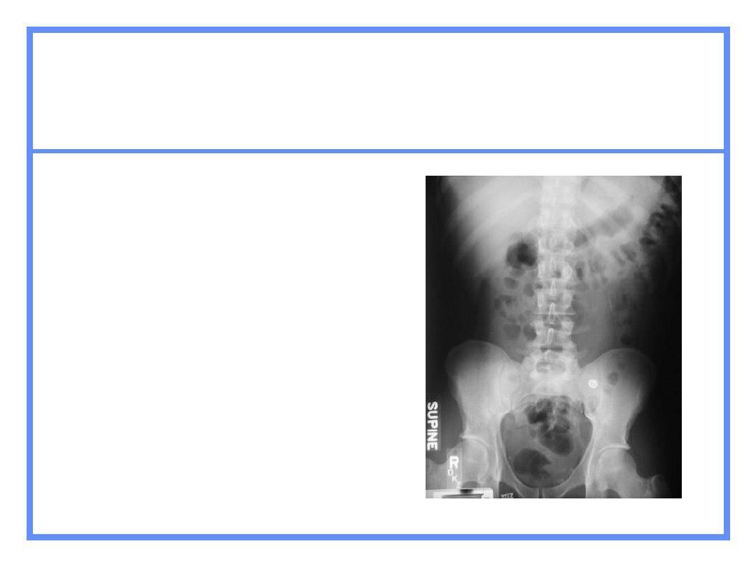
The plain x-ray abdomen
The standard plain films of the abdomen are
1- supine A.P for abdominal soft tissue shadow and
abnormal calcification
2-erect A.P.to detect fluids levels in case of intestinal
obstruction
3- lateral decubitus view (A.P. view taken and the patient
lying on his side)taken in patient unable to sit or stand
What to Examine
Gas pattern
Extraluminal air
Soft tissue masses
Calcifications
What to look
1
-
gas and fluid levels
fluid levels become abnormal when :three or more small –bowel fluids
levels longer than 2.5 cm
2-look for any gas outside the lumen of the bowel
3-identify the liver :seen as ahomogenous opacity in the right upper
quadrant usually extending in to the left upper quadrant
4-identify the borders of the spleen ,kidney,bladder and psoas muscles
5-look for any soft tissue masses in the abdomen and pelvis
6-if there is any calcification try to localize it
7- look for vertebrae if any abnormality

Normal Gas Pattern
Stomach
Always
Small Bowel
Two or three loops of non-distended bowel
Normal diameter = 2.5 cm = 1 US quarter
Large Bowel
In rectum or sigmoid
– almost always
Gas in
stomach
Gas in a few
loops of
small bowel
Gas in
rectum or
sigmoid
Normal Gas Pattern

Normal Fluid Levels
Stomach
Always (except supine film)
Small Bowel
Two or three levels
possible
Large Bowel
None normally
Erect Abdomen
Always
air/fluid level
in stomach
A few
air/fluid
levels in
small bowel
Large vs. Small Bowel
Large Bowel
Peripheral
Haustral markings don't
extend from wall to wall
Small Bowel
Central
Valvulae extend across lumen
Maximum diameter of 2"

Complete Abdomen
Obstruction Series
Supine
Prone or lateral rectum
Erect or left decubitus
Chest - erect or supine
Complete Abdomen
Supine
Looking for
Scout film for gas
pattern
Calcifications
Soft tissue
masses
Substitute
– none
Complete Abdomen
Erect
Looking for
Free air
Air-fluid levels
Substitute
– left
lateral decubitus
Complete Abdomen
Erect Chest
Looking for
Free air
Pneumonia at bases
Pleural effusions
Substitute
– supine
chest

•Normal Anatomy
•Types of Projection
•Assessing the Film
•Technical Qualities
•Gas containing structures
•Solid Organs
•Bones
•Soft Tissues
•Presenting the film

Abdominal x ray
• The abdominal x-ray (AXR) has a much more limited
value in diagnosis than a chest x-ray.
• The radiation exposure of an AXR compared to a CXR is
also considerably higher. One AXR is equivalent to 35
CXRs.
• The AXR is of most use in the patient with an acute
abdomen. It may guide further imaging (Other Imaging
Modalities Lecture)
• As with a CXR, an appreciation of normal structures is
vital

abdominal x ray projections
• Supine 99%
• Erect
• Lateral decubitus
.
• Knowledge of the anatomy of the abdomen allows
localization of the abnormalities observed on the AXR.
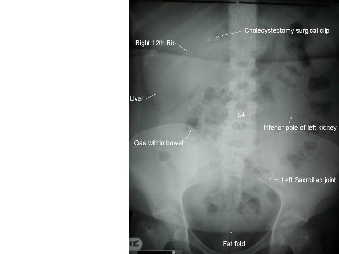
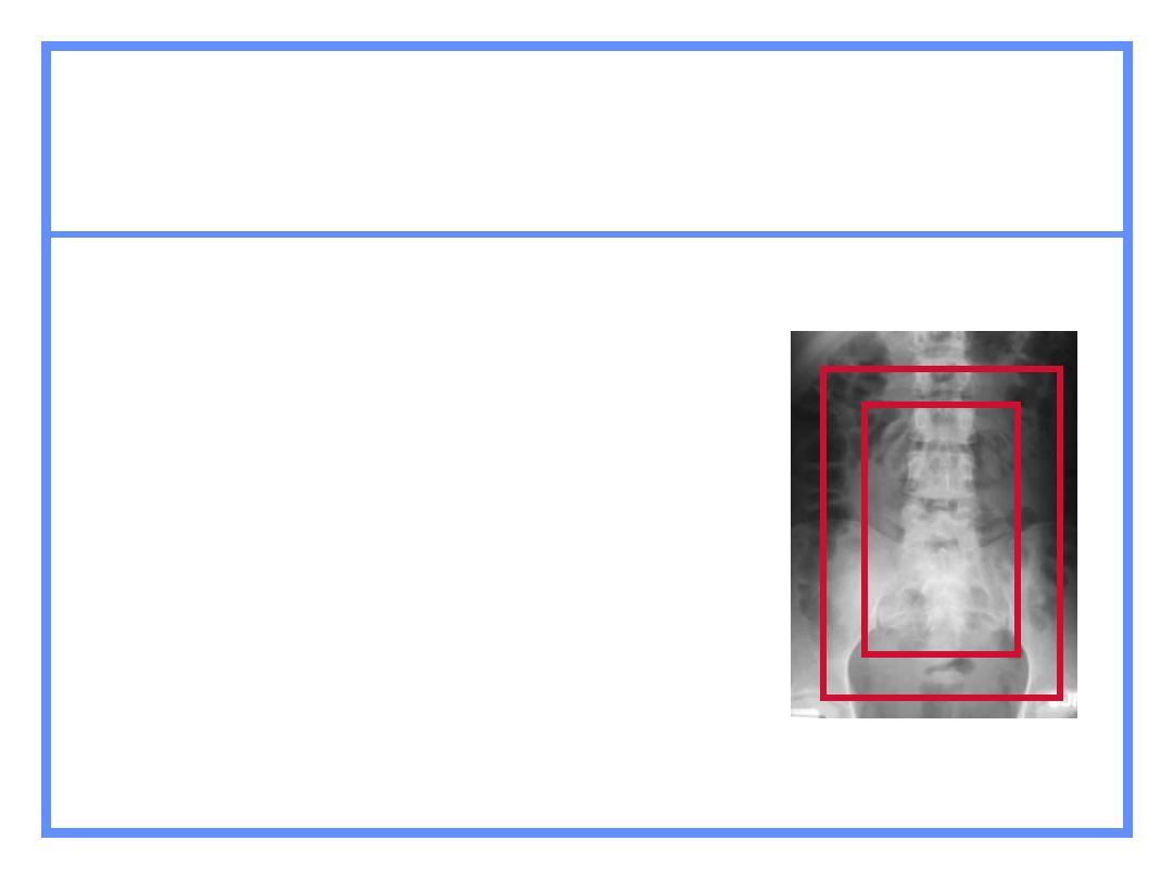
Anatomy
on the
Abdominal
X-Ray:
Abdominal X-Rays:
AXR-3
AXR-4
Abdominal X-Rays:
AXR-1
AXR-2

Film Specifics and Technical Factors
the nitial assessment of an AXR is the same as
for a CXR:
• Film Specifics:
• Name of Patient
• Age & Date of Birth
• Location of Patient
• Date Taken
• Film Number (if applicable)
• Film Technical factors:
• Type of projection (Supine is standard)
• Markings of any special techniques used

• BLACK BITS’
• Intra-luminal gas can be normal.
• Extra-luminal gas is abnormal.
• However, intra-luminal gas can be abnormal if
it is in the wrong place or if too much is seen

• BLACK BITS’ (Continued) -
Intra-luminal gas:
• The maximum normal diameter of the large
bowel is 55mm.
• Small bowel should be no more than 35mm in
diameter.
• The natural presence of gas within the bowel
allows assessment of caliber - although the
amount varies between individuals.
• The caecum is not said to be dilated unless
wider than 80mm.
• Large and small bowel may be distinguished by
looking at bowel wall markings, as shown in the
box below.

•
Intra-luminal gas (continued):
• It is usual to see small volumes of gas
throughout the GI tract and the absence in one
region may in itself represent pathology.
• For example, if gas is seen to the level of the
splenic flexure and nothing is seen beyond this,
a site of the obstruction at this site
– a ‘cut off’
point is noted.
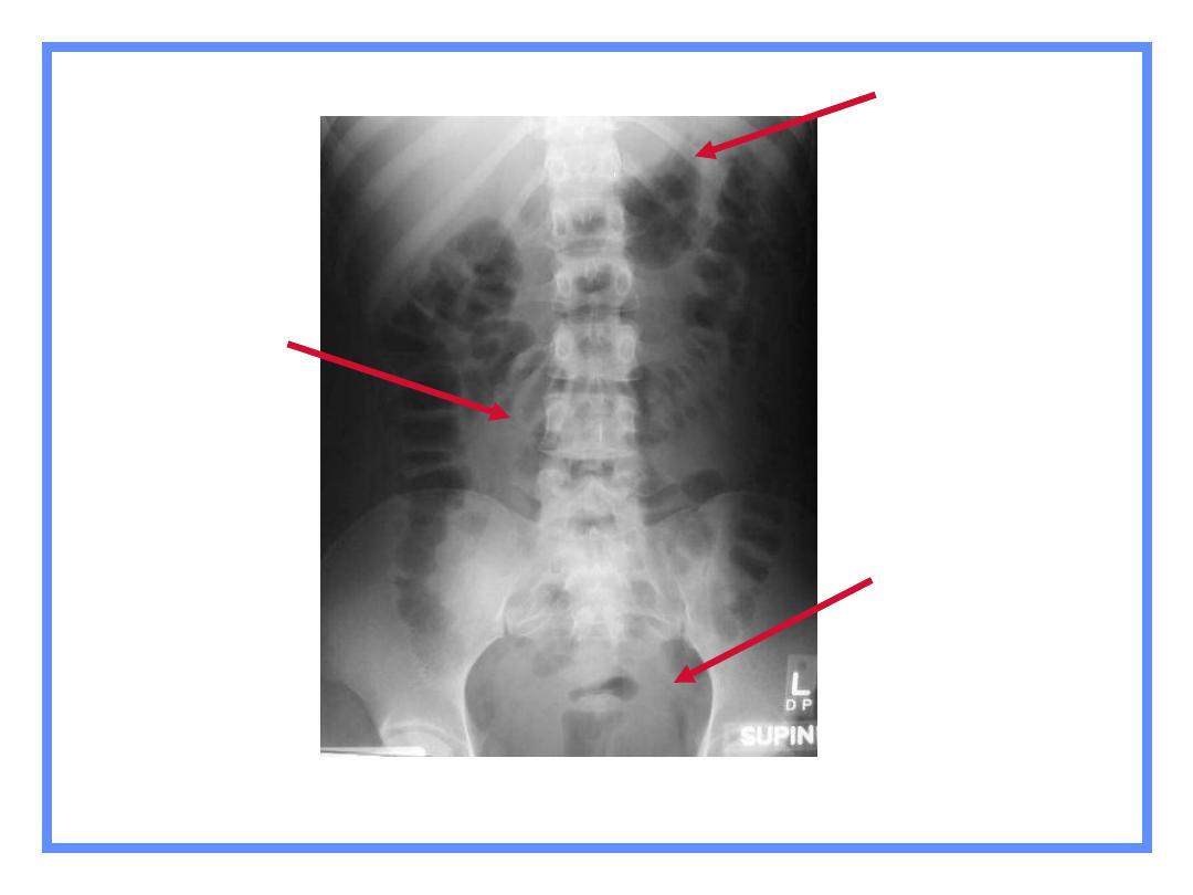
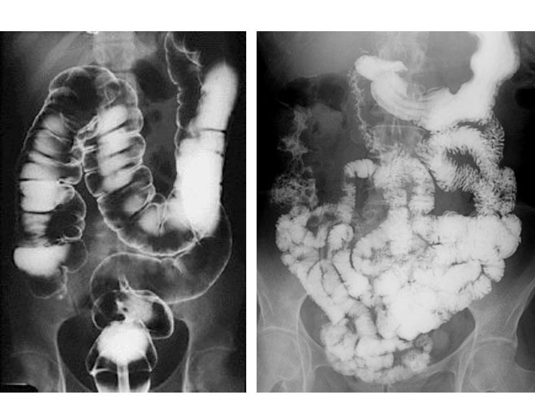
Assess the Film in Detail:
luminal Gas:
-
Intra
Low Small Bowel
Obstruction
Small Bowel obstruction.
Assess the Film in Detail:
If bowel obstruction is
observed try to look for
the cause. For example
a hernia as the cause of
obstruction.
Hernia.
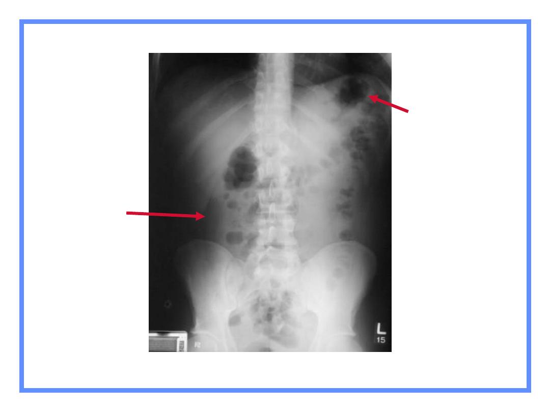
Assess the Film in Detail:
luminal Gas:
-
Extra
When an bowel is
obstructed, or any other gas
containing structure
perforates, its contained gas
becomes extra-luminal.
Extra-luminal gas is never
normal, but may be seen
following intra-abdominal
surgery or endoscopic
retrograde cholangio-
pancreatography (ERCP).
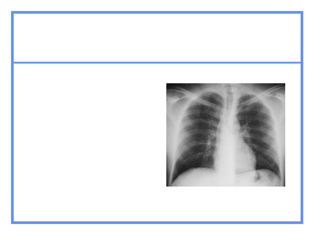
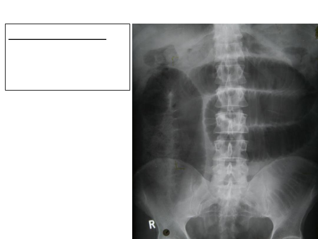
Extra-luminal gas seen on erect
CXR.

•
Causes of Extra-luminal gas
:
• Post Abdominal Surgery/ERCP
• Perforation of viscus (eg. bowel, stomach)
• Gallstone ileus
• Cholangitis (infection with gas forming
organisms)
• Abscess
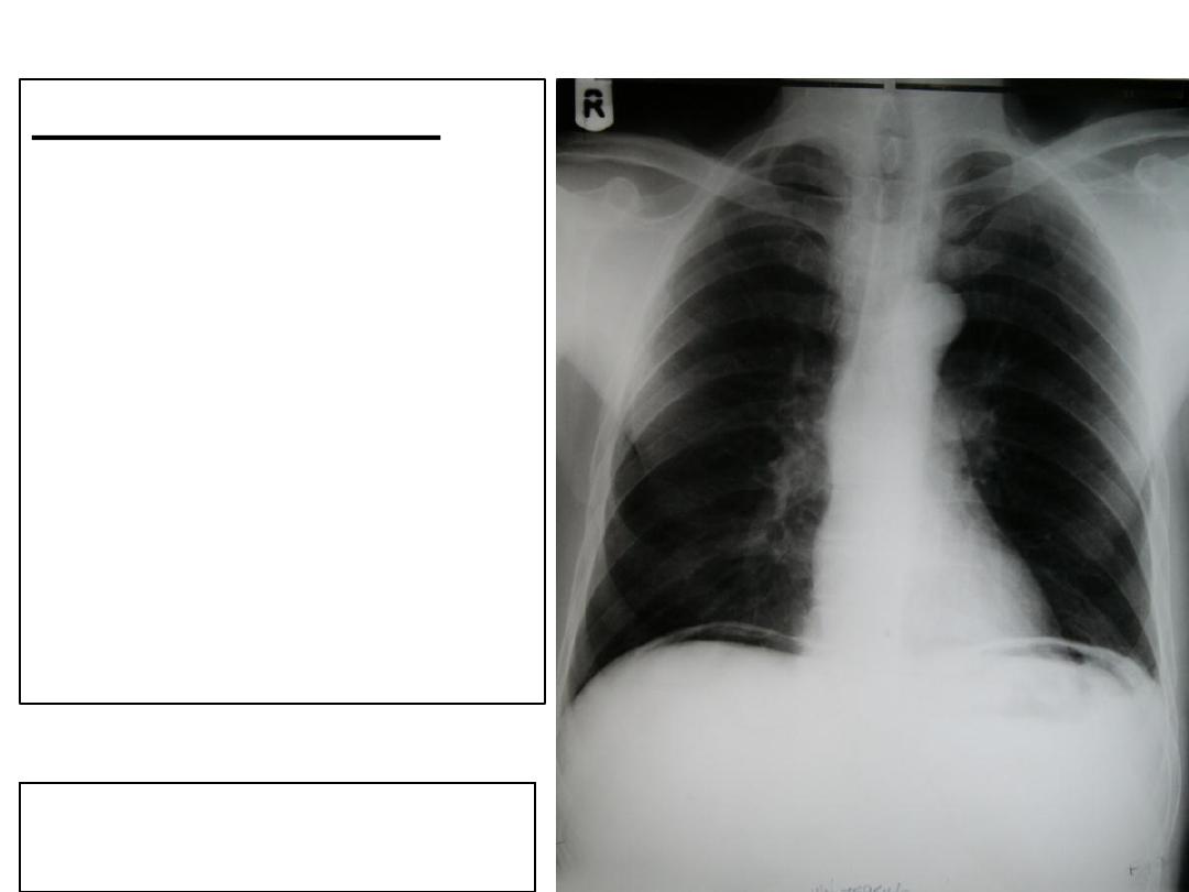
• An erect CXR (not AXR) is the best
projection to diagnose a pneumoperitoneum
(gas in the peritoneal cavity).
Assess the Film in Detail:
Pancreatic Calcification
Gallstones

• ‘GREY BITS’ = Soft Tissues
• Soft tissues represent most of the contents of
the abdomen and feature heavily in the AXR.
However, these tissues are poorly seen when
compared to other imaging techniques such as
ultrasound or CT.
• The kidneys, spleen, liver and bladder (if filled)
can be seen in addition to psoas muscle
shadows and abdominal fat. Rarely would
action be taken on the basis of this imaging
alone.
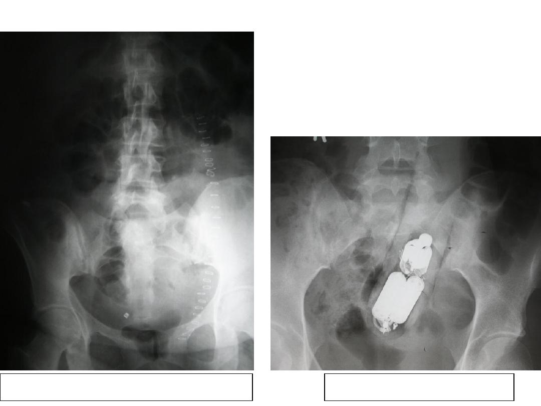
Assess the Film in Detail:
Sterilisation and Surgical Clips
Foreign body per rectum
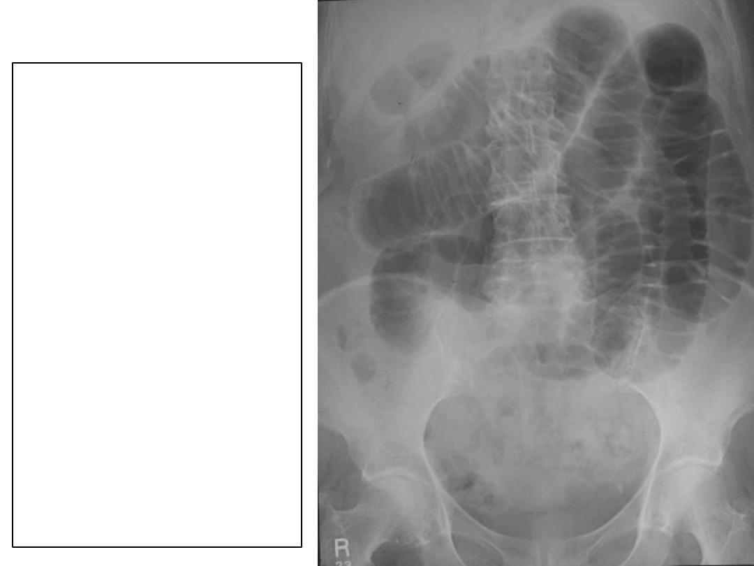
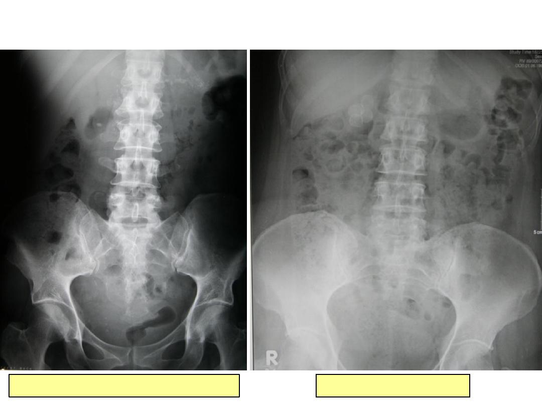
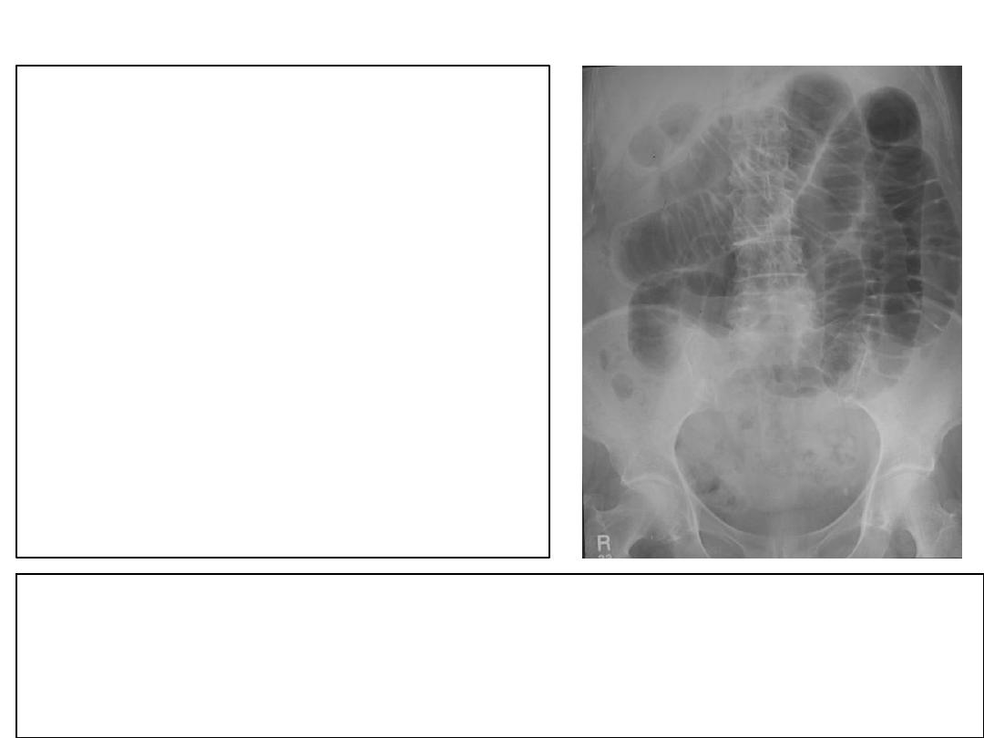
Case
1
:
This
60
year-old
women presented to
the surgical ward
with a distended
abdomen and
vomiting
.
Present this x-ray
Give a diagnosis and
potential causes
Case
1: Answer
Radiology Report
:
Plain abdominal radiograph
.
Multiple dilated loops of small bowel
within the central abdomen. Gas is not
seen in the large bowel. No evidence of
hernia or gallstone to suggest potential
cause of the dilated loops
.
These findings are in keep with a low
small bowel obstruction
.
I would like to know if the patient has a
history of abdominal surgery as the
commonest cause is surgical admissions
.
The three commonest causes of small bowel obstruction are:
•Surgical adhesions
•Herniae
•Intraluminal mass eg, small bowel lymphoma or gallstone (in gallstone ileus)
Case
2
:
This 71 year-old
gentleman visits his GP
complaining of blood in
his urine. He has had a
number of UTI’s in
recent years.
Present this x-ray
Give a diagnosis and
potential causes
Case
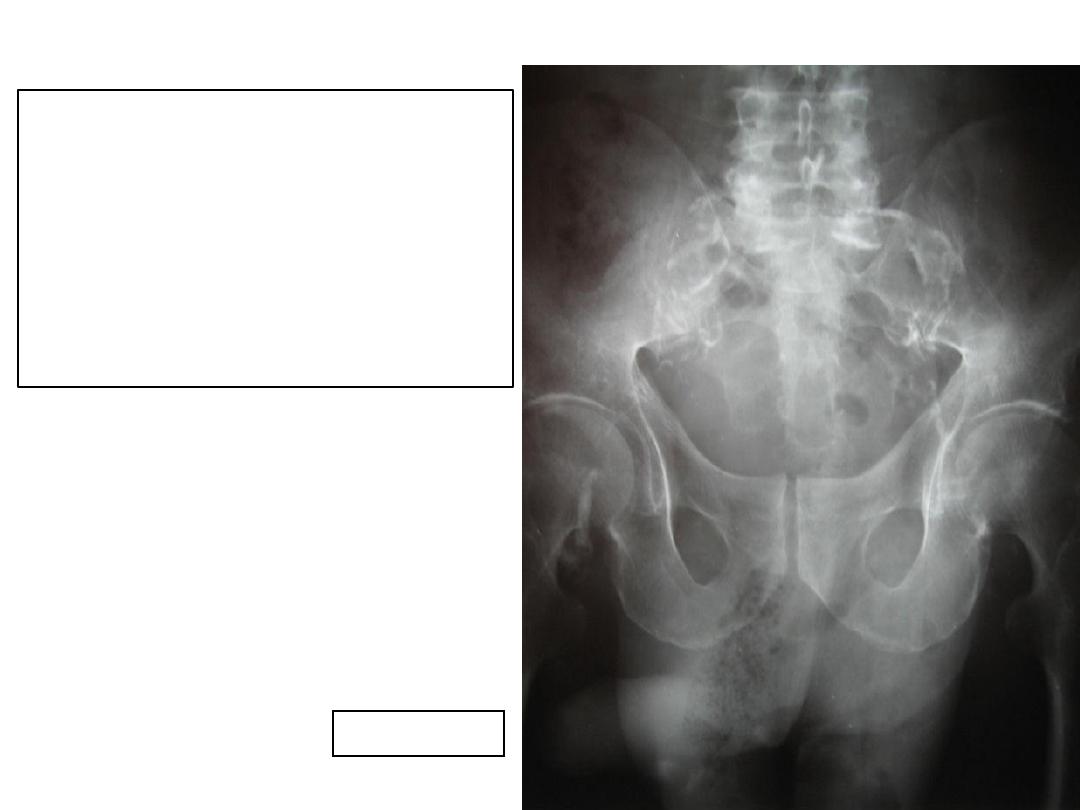
2: Answer
Radiology Report
:
Plain abdominal radiograph
.
Two rounded radio-opacities measuring
4cm within the pelvis. Both opacities are
smooth in outline, laminated in nature,
have the same density as bone and
project over the bladder. No other renal
tract calcification
.
Does the patient have a history of
neurogenic bladder
?
Given the size of these stones and history
of UTI’s these are bladder calculi.
Bladder calculi are more common in those with a history of:
•UTI’s
•A neurogenic bladder
•Bladder diverticulum
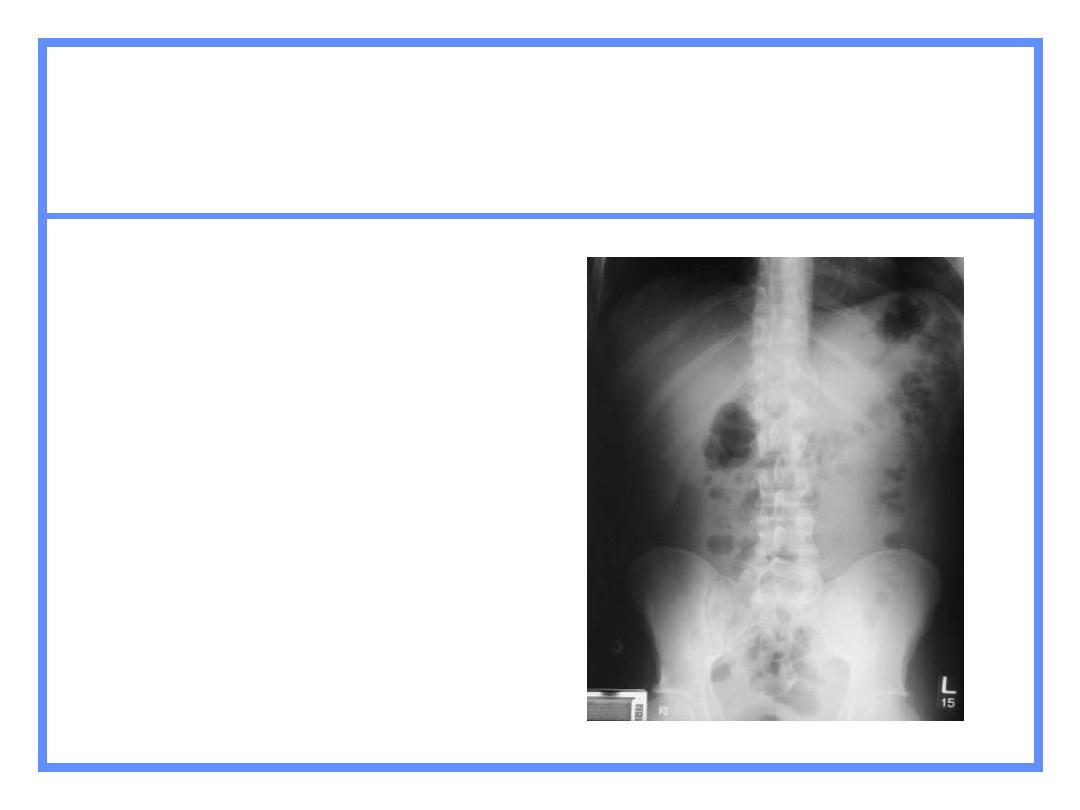
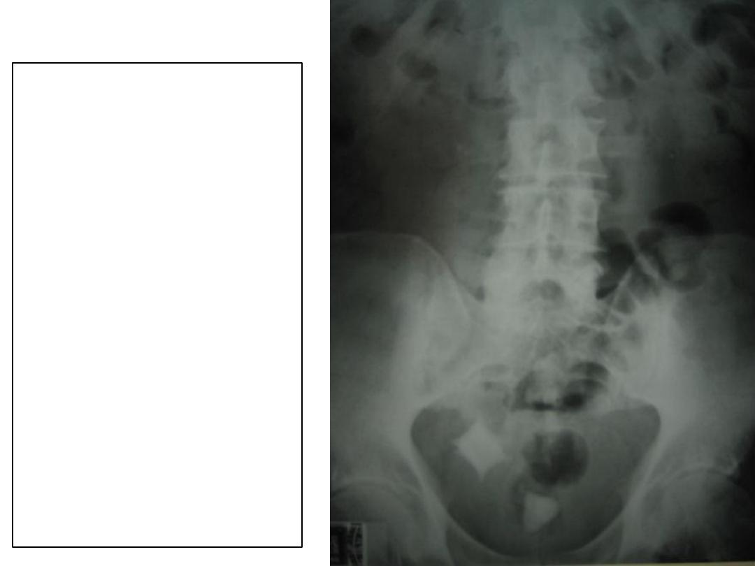
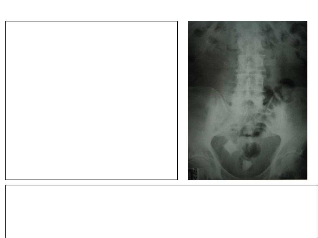
Case
3
:
This patient was
admitted with poor renal
function.
Present this x-ray
Give a diagnosis and
potential causes
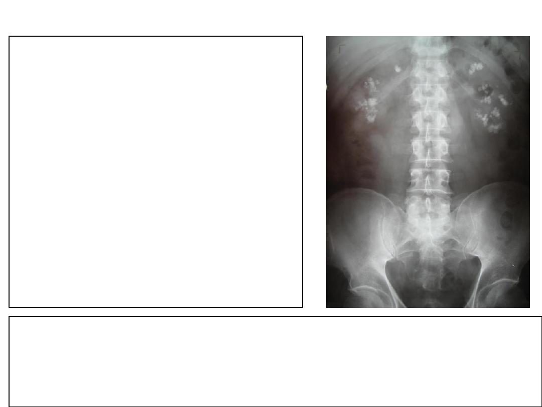
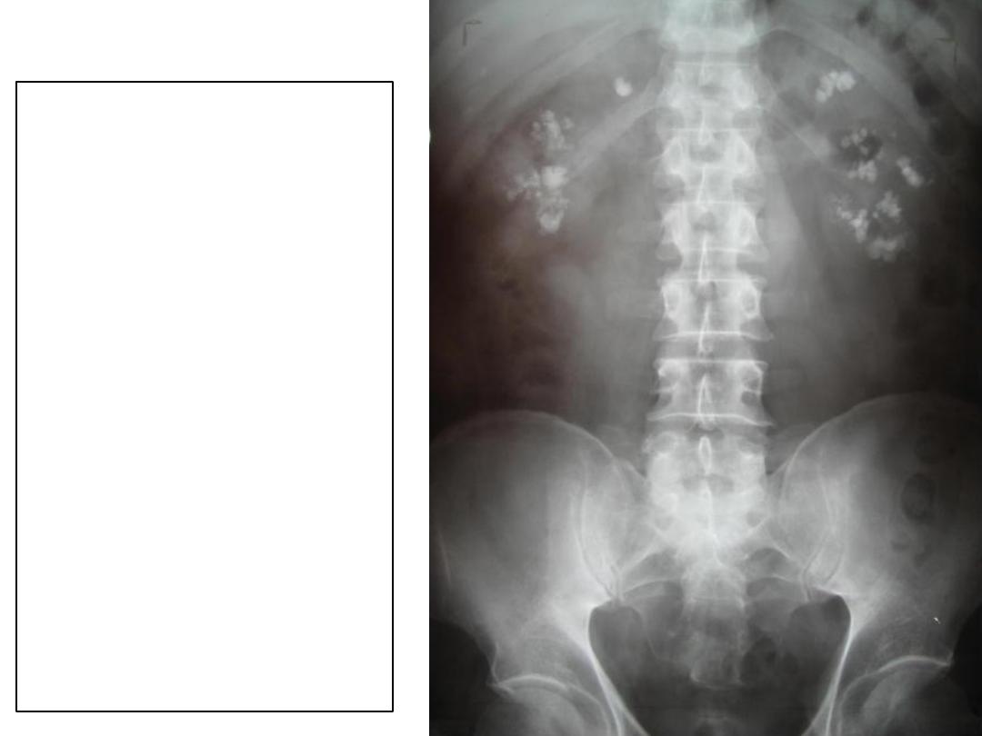
Case
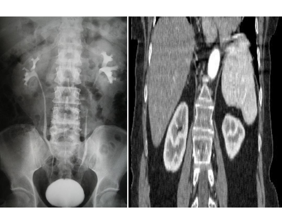
3: Answer
Radiology Report
:
Plain abdominal radiograph
Multiple areas of punctuate calcification
project over the renal outlines bilaterally
.
The calcification is within the medulla of
the renal parenchyma. The bones are
normal in appearance
.
These findings are consistent with
nephrocalcinosis
Causes of Nephrocalcinosis include:
•Hyperparathyroidism
•Medullary sponge kidney