Dental fluorosis
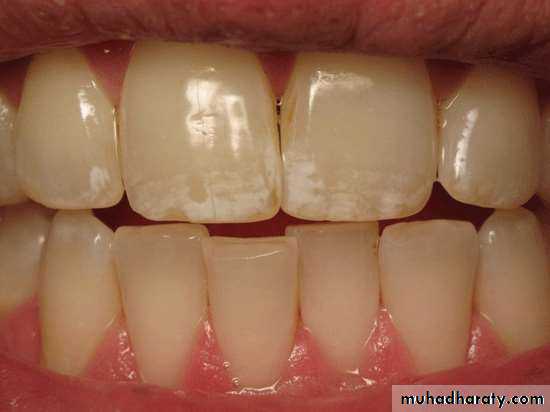
Dental fluorosis
is a defect of the teeth marked by increased porosity of the enamel ( a condition known as “Hypomineralization“ or enamel mottling )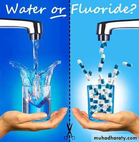

Causes
This defect of tooth enamel caused by too much fluoride intake during the first 8 years of life. the damage to the enamel is permanent. The fluorosis of primary tooth enamel is very rare.
Causes of fluorosis include:
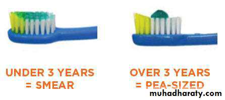
• Excess fluoridation of drinking water.• Inappropriate use of fluoride containing dental products such as Toothpastes and mouth rinses.
• Consumption of processed food made with fluoridated water.
• taking a higher-than-prescribed amount of a fluoride supplement during early childhood ( As use overuse of Fluoride tablets(.
• taking a fluoride supplement when fluoridated drinking water or fluoride fortified fruit juices and soft drink already provide the right amount

HISTORY
1888 : “KUHNS” described teeth of persons inareas of Mexico that were opaque, discolored and
disfigured. (Kuhns1888; Moller 1982).
1901 Dr. Fredrick Mckay of Colorado USA discovered permanent stains on teeth of his patients which were referred as Colorado stains.
Mckay named then “mottled enamel”.
An Assitant surgeon of U.S marine hospital service reported similar condition in Italians
emigrating from USA from Naples named it denti di chiaie. ( Eager 1901).
1916 Mckay and Black published a series of articles in dental cosmos.

Mechanism of action of fluoride:
, an increase in fluoride intake results in an increase in degree and extent of porosity of the enamel ; the enamel changes described may be a result of a fluoride damage of secretory ameloblasts.This can either be due to a fluoride-induced change in composition of enamel matrix, or be a result of a disturbance of the cellular processes during enamel maturation.
Optimal Level of Fluoride and Fluorosis:
1 ppm → minimal threshold of fluorosis .When fluoride concentration is more than 1 ppm undesirable mottling began to be seen in about 10% of the population.Variables Affecting Prevalence and Severity of Dental Fluorosis:
Fluoride concentration in drinking water.Total amount of fluoride ingested
which include amount of fluoride in water, food, drugs, dentifrices
Temperature
water requirement increase in hot temperature which increase possibility of fluorosis
Duration of exposure
fluorosis increased with the longer time teeth are exposed to fluoride.
Malnutrition
in some studies showed that malnourished children develop more dental fluorosis than well-nourishedDental fluorosis index
Dean‘s Index – 1934
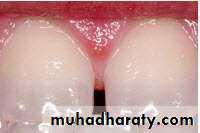
Dean’s 0 Normal: The enamel surface is smooth, glossy and usually a pale creamy-white color.
Dean’s 1 Questionable: The enamel shows slight aberrations from the translucency of normal enamel, which may range from a few white flecks to occasional spots.
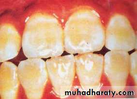
Dean’s 2 Very mild: small opaque white area scattered irregularly over the tooth, but not involving more than 25% of the tooth surface, for posterior teeth it is an area of 1 – 2 mm white opaque area at the tips of the cusps of molars and premolars.
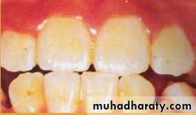
Dean’s 3 Mild: we have white opaque area in the enamel more extensive than for code 2 but involving not more than 50% of the tooth surface.
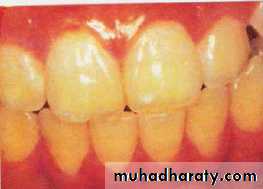
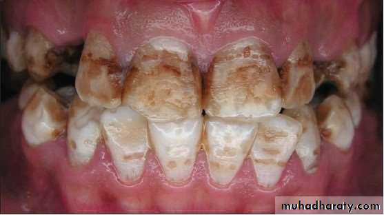
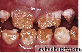
Dean’s 4 Moderate: all enamel surface are effected, surfaces are subjected to attrition, it showed there is a marked wear brown stain.
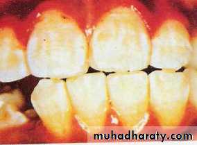
Dean’s 5 Severe: when the all enamel surface are badly affected and the hypoplasia is so marked that the general form of the tooth may be affected, There are pitted or worn areas and brown stains are widespread; the teeth often have a corroded appearance.
Code 6 : All 4 anterior teeth absent.
Enamel Mottling by AL – Alousi et al . (1975)
Selection of teeth and scoringLabial surface of permanent incisor teeth . Calculation of the prevalence of different types of opacities in both individual and teeth :
Diagnosis and criteria:
:Type A : White area less than 2 mm in diameter .
Type B : White area of , or greater than 2 mm in diameter .
Type C : Colourd (brown) area less than 2 mm in diameter, irrespective of there being white area.
Type D : Colourd (brown) area of, or greater than 2 mm in diameter, irrespective of there being any white area.
Type E : Horizontal white lines , irrespective of there being any white non-linear lines.
Type F : Colourd (brown) or White area or lines associated with pits or hypoplastic area.