
1
Epistaxis
;(
Bleeding from the nose)
Etiology of epistaxis
I. Idiopathic
.
: Spontaneous bleeds without any proven precipitant or causal factor. It is
the common causes 70-80% of all cases of epistaxis, common in children and adolescents
II. Local causes :
1.Traumatic. *Nose picking.
*Blow to the nose.
*Foreign body in the nose.
* Fracture nasal bones and anterior skull base, and fracture
sinuses.
*Iatrogenic .Nasal surgery especially follows turbinectomy., Endoscopic
sinus surgery(Anterior ethmoidal a., internal carotid artery.)
2. Inflammatory. Rhinitis and sinusitis ,either acute or chronic, specific(Diphtheria, TB )
or non-specific infection.
3. Neoplastic: Benign or malignant tumor in the
*nose. like haemangioma of the septum.
*sinuses. like angioma of sinus,Sequmous cell Ca.
*nasopharynx. Angiofibroma,sequamous cell ca.
4.Septal causes. deviation, and perforation.
5.Environmental. Over exposure to air condition and heat, smoke and industrial fumes
results in dryness and crustation. Dry and cold air during the autumn and winter months
makes epistaxis more common in these seasons
III. Systemic causes:
1. Raised blood pressure. temporary or permanent.
A. Raised arterial pressure. Hypertension common in adult and elderly,.
B. Raised venous pressure. In congestive heart failure,emphysema wheeping cough,
pneumonia, associated with venous bleeding.
2. Blood dyscreasia and diseases of blood vessels. Like Leukemia, Haemophilia, Von
wilbrand disease,purpura.etc .
3.Hepatic failure.(hypoprothrombinemia)
4.Renal failure.(platelete dysfunction)
2
5.Drugs taken. Like anticoagulants (heparin,warfarin), antiplatlet aggregation like
aspirin and NSAID.
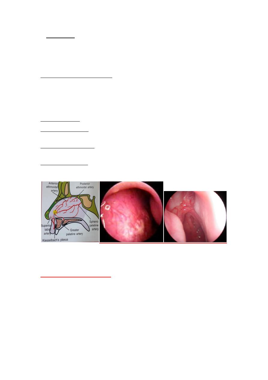
Site of bleeding :
1. Little's area(Kiesselbach's plexus).The commonest site of bleeding (90%)located in the
antero-inferior part of the nasal septum, when anastomosis of poorly supported blood
vessels which are;
1.Anterior ethmoidal a. 2. long sphenopalatine a.
3.greater palatine a. 4. Superior labial a.
2. Wood ruff's area. Venous plexus in the posterior end of inferior turbinate.
3. Retrocolumellar vein.lies immediately behind the columella in the superficial areas, and
is a common causes of venous epistaxis in children.
4. Above middle turbinate from anterior and posterior ethmoidal arteries, usually in case
of hypertension.
5. From middle meatus (rare) from maxillary and ethmoidal sinuses.
Anterior septal vessels. Posterior dilated vessels.
Clinical feature of epistaxis
:
Epistaxis is a common ENT emergency.
Bleeding varies in degree from trivial to lethal.
Usually occur from anterior naries, may flow back in to the pharynx and in the opposite
nostril. Occationally inhaled and may be suspected haemoptysis,or swallowed and get
haematemesis,malaena in sever bleeding.
*Anemia in recurrent sever bleeding.
*It is of two types 1. Anterior epistaxis .2. posterior epistaxis

3
Type of epistaxis
Anterior epistaxis
Posterior epistaxis
Incidence
More common(90%)
Less common(10%)
Age
Younger patients <18 years
Older patients>40 years
Site
Common is little's area.
Common isWoodruff's area
Common cause
Idiopathic
Hypertension
Localization
Easy
Difficult
Management
Easy to manage
More troublesome
Treatment
*Cautery.*If fail nasal packing
,merocele,nasal ballon.
*Endoscopic diathermy.
*if fail posterior pack ,or
nasal ballon with anterior and
posterior component..
Management:
In acute active epistaxis priority is given to control bleeding and deal with hypovolemia
and blood loss.
I. Brief history.
Looking for* severity (amount of blood loss) and
*for predisposing factors.
*Duration.(short in venous bleeding, prolonged in arterial bleeding).
*Frequency. Recurrent in angiofibroma,osler's disease,
*Trauma (Facial traum a, nasal surgery)
*Medical history. hypertension.
*Drug history. aspirin, warfaren.
*Family history.haemophilia
*Nasal symptoms. Obstruction,rhinorrhea.
II. Examination:
1. *
General Assessment
for hypovolemic shock : Weak rapid pulse , hypotension,
cold extremities, irritability, decreased urine output.
4
*Hypertension.
*pallor.
*Fever.
*in sever bleeding insert I.V line, take blood sample for blood group and Rh,Hb%,give fluid,
do cross matching and blood transfusion when needed.
2. Local examination to identify the bleeding site.
* Anterior rhinoscope.
*Posterior rhinoscope.
*Endoscopic examination.
III. Arrest bleeding.
1.Firs aid measures.
*Pressure:Pinching the ala nasi to compress the vessels of anterior part of the septum
which common site of epistaxis by thumb and index finger for 5-10 minutes.
*Position: The patient sit up, Lean forward slightly with the flexion of the head tilted to
prevent blood get to post nasal space, and breathing quietly through the mouth.
*Ice or cold packing to the bridge of the nose causes reflex vasoconstriction.
*Local vasoconstriction (. Oxymetazoline drops. Phenyl phrine and ephedrine are more
cardiotoxic)
5
Correct nose pinching Correct position Incorrect nose pinching
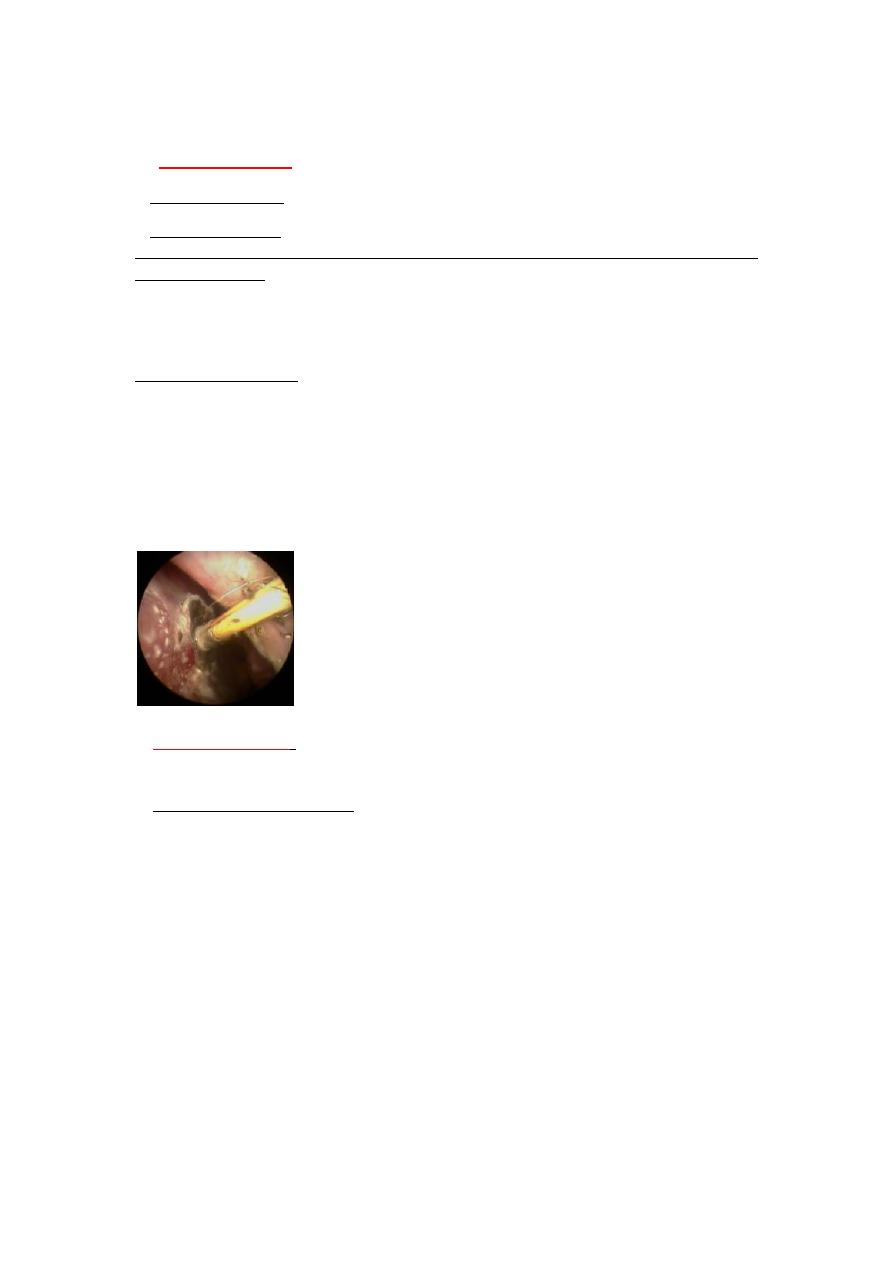
2. Cauterization
.
when identify the bleeder, the bleeding arrested by
a.Chemical cautery using caustic agents (silver nitrate sticks,Trichloracetic acid) .
b.Electrical cautery (Gelvanic cautery, Bipolar diathermy )
c.Endoscopic guidance using hot wire cautery,or modern single fiber bipolar electrodes. for
posterior bleeding.
*Post cautery give lubricants ,antibiotics, and sedation.
*Avoid bilateral septal cautery results in septal perforation
Post cautery instruction:
*No manipulation,*No nose blowing,*Open mouth when sneezing,*No straining, lifting
or strenuous activity for one week,*No smoking or alcohol for one week,*No hot drink or
food for one week,*Elevate the head of the bed for one week,*No aspirin, warfaren for
one week,*Cold mist humidifier at the bed time,*lubricant drops (normal saline)for one
week,
Silver nitrate sticks
3.Nasal packing
:
When failure of cautery or not see the bleeding site.it is of two
type
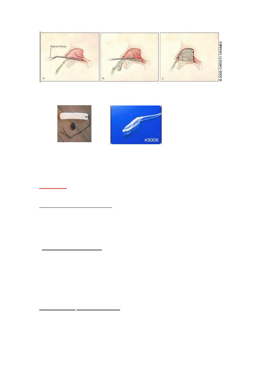
A; Anterior nasal packing.
Using ribbon gauze(half inch)lubricated with petroleum gelly or Bismuth Iodoform
Paraffin Past(BIPP).
*Under local anesthesia ,done in layers without traumatizing the nasal mucosa using
Tilly's forceps, inserted along the floor of the nose then build mup in successive loop from
floor upward till every part of nasal cavity finally fitted. Usually done bilaterally to increase
pressure on nasal septum ,with anti biotic cover.
Pack usually left for 24-72 hours, if bleeding restart needs further evaluation and
reinserted or do posterior packing
Complications; sinusitis,septal perforation, hypoxia.
***Modern variation using special tampons like Merocel nasal packing ,gelfoam , and
nasal balloon.
6
Anterior nasal packing.
Nasal tampon(merocel),
Nasal balloon,for anterior epistaxis
Merocel ;
Is compressed dehydrated sponge, which can inserted in the nasal cavity
then rehydrated by blood, expanding to 3 times it is normal size filling nasal cavity.
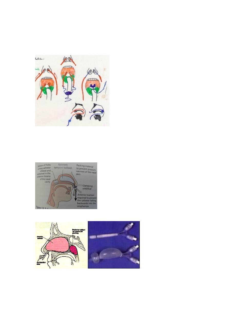
B.Posterior nasal packing
:
failure of anterior nasal packing ,or bleeding arise
from back(ex. sphenopalatine a. in postnasal space).
*Can do it under local anesthesia ,but general anesthesia is preferable. Using gauze packs
inserted transorally and positioned by means when of tapes pass from the posterior
choana to anterior naries bilaterally
.
Foley urethral catheter
(size 12-14)inserted along the floor of the nasal cavity until
the nasopharynx is reached, then the Foley catheter is inflated up to 15 ml of water, pulled
forward to engage in the posterior choana and anterior nasal packing inserted. The Foley
catheter secured anteriorly avoiding pressure over the columella.
3. Nasal balloon .used the anterior and posterior balloon –integral airway
*Pack should be left in position for minimum of 48hours.
Complications of posterior pack :
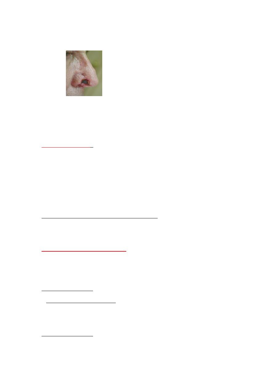
1*Necrosis of septum and columella,and alar nasi ,2*Sinusitis,3*otitis media,4*hypoxia ,so
the patient must admitted in the hospital.
7
Antibiotics, and analgesia are necessary.
**Never pack the nose of any unconscious person when skull fracture
or cribriform plate injury is suspected
*posterior nasal packing
Foly's catheter as the balloon fit the
nasaopharynx.
for temponade of Nasal balloon used bleeding
Inflate the anterior and posterior component may be seen.
The anterior and posterior balloon –integral airway
8
Alar
necrosis
4. Arterial ligation
:
Indications: Intractable bleeding cannot be located or controlled by the methods described
above.
1*Ligation of external carotid a.
2*Ligation of internal maxillary a.
3*Ligation of anterior/posterior ethmoidal a.
4*Endonasal sphenopalatine a. ligation.
**.Selective angiography and Embolization
for Intractable bleeding from surgically inaccessible sites, *patient
not fit for surgery. Only able to embolize external carotid & branches
5:Search for causes and treated
Complete blood tests, and serological tests, with imaging studies tailored according to
the history and ENT and systemic examinations
Biopsy for nasal mass
Medical Treatment.
*
systemic inhibitors of fibrinolysis
.
like Tranexamic acid and epsilon aminocaproic acid .
*Control Hypertension.
*Treat renal failure ,hepatic failure,thrombocytopenia.Correcion of coaggulopathy.
Surgical treatment.
*
Septoplasty for access to bleebing point.

9
*Remove of tumor ex. angiofibroma
*Septodrmoplasty for Osler's disease.
* Nd-YAG Laser.
===========================