
5
th
year 2012/2013 by dr. Ammar
Page
1
Anatomy & Physiology of the Ear
The external ear
consists of the pinna and the external auditory canal
.
Pinna
Elastic cartilage surrounded on either side by a layer of skin. There is minimal subcutaneous tissue
between the skin and the perichondrium.
Function :::>>, the pinna acts to funnel sound waves from the outside environment into the ear
canal. The shape of the external ear provides approximately 20 dB of gain to sounds in the middle
frequency range (2–4 kHz).
External Auditory Canal
(EAC)
Approximately 2.5 cm in length.
Outer one third ::::;>cartilaginous[fibrocartilaginous]
Inner two-thirds :::::>bony..
The narrowest part of the external auditory canal (isthmus)is located between the fibrocartilaginous
and the bony canal.
Anterior to the bony canal is the temporomandibular joint. The skin of the ear canal is thicker in the
cartilaginous canal and contains glands that secrete cerumen (ear wax). The skin of the bony ear
canal is very thin and fixed to the periosteum. No cerumen is secreted in the bony ear canal.
Nerves
The great auricular nerve (from nerve roots C2 and C3) provides sensory innervations to the skin
overlying the mastoid process as well as the majority of the pinna. Cranial nerves V (the trigeminal
nerve), VII (the facial nerve), and X (the vagus nerve) innervate the external auditory canal.
Middle Ear ::::consist of
1-Tympanic Membrane

5
th
year 2012/2013 by dr. Ammar
Page
2
2-tympanic cavity and its contents
3-eustachian tube
4-mastoid air cells
The tympanic membrane consists of three layers::::
The outer layer, …… squamous epithelium.
The middle layer ……… fibrous of the tympanic membrane consists of both radial and
circumferential fibers. Which maintain the strength of the tympanic membrane.
The inner layer :::::::::::::: cuboidal mucosal epithelium.
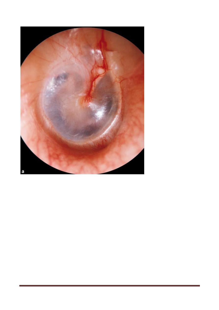
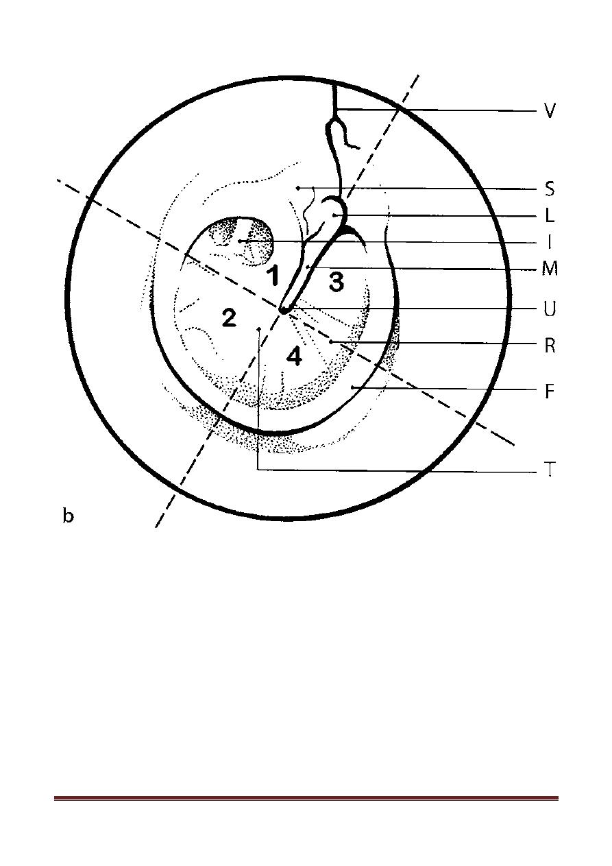
The tympanic membrane has an oval shape and is approximately 8 mm wide by 10 mm high, pale
grey in colour, surrounded by fibrous annulus, which is incomplete superior to the anterior and the
posterior malleal folds.
This section of the tympanic membrane above the anterior and posterior malleal folds is the pars
flaccida, while the section inferior to the folds is the pars tensa. Blood vessels enter the tympanic
membrane through the superior external auditory canal skin (the vascular strip) as well as
circumferentially from around the fibrous annulus.
Landmarks visible at otoscopy:: the 1-lateral or short process of the malleus 2- the handle of the
malleus, 3-the pars flaccida, and the pars tensa with 4- the umbo and the triangular cone of light
(light reflex).
(In the thin, transparent tympanic membrane: the chorda tympani, the long process of the incus and the
head of the stapes)
5
th
year 2012/2013 by dr. Ammar
Page
3
5
th
year 2012/2013 by dr. Ammar
Page
4
Middle Ear Cavity
Boundaries
Laterally------------------------tympanic membrane
medial wall-------------------- promontory of inner ear
Posterior---------------------- mastoid air cells
Anterio-medially------------------Eustachian tube opening
The mastoid air cells is connect with the attic portion of the middle ear cavity through the aditus and
antrum. The middle ear cavity and mastoid air cells are lined with ciliated mucosal epithelium. The
blood supply of the middle ear and mastoid originate from the internal and external carotid arteries.

5
th
year 2012/2013 by dr. Ammar
Page
5
Ossicular Chain
There are three ossicles the malleus, the incus, and the stapes.
The malleus has a long process, a short process, and a head. The malleus is bonded to the tympanic
membrane from the tip of the long process (the umbo) to the short process. The head of the malleus
articulates with the body of the incus in the attic.
The incus has a long process and a short process. The short process is tethered to the posterior wall
of the middle ear cavity for structural support and the long process is connected to the stapes head.
The stapes consists of a footplate and a superstructure. The superstructure includes the anterior and
posterior crus, which are attached at the head. The footplate is the bony covering that sits within the
oval window.
Muscle :::: The stapedius muscle. The tensor tympani muscle is anchored to the malleus.
Eustachian Tube
About 35 mm in adult life
The bony part (11–14 mm) opens into the protympanum (tympanic ostium), and the
fibrocartilaginous part (20–25 mm) opens into the lateral wall of the nasopharynx.
Lined with respiratory ciliated mucosa, the tube connects the nasopharynx with the middle
ear and permits ventilation of the pneumatized temporal bone spaces. It opens on swallowing.
Physiology of the Middle Ear
The middle ear amplifies the airborne sound vibration in two ways. First, the large surface area of
the tympanic membrane, compared to the small surface area of the stapes (14:1), Second, the lever
arm effect of the malleus and incus increase in vibrational amplitude (1.3:1.0). The total middle ear
gain is 18.3:1.0 or 20–35 dB. This gain counteracts the loss that would result from the impedance
mismatch between airborne sound and the fluid vibrations in the cochlea.
Changing the mass and stiffness of the middle ear modulates its frequency response, which can be
observed clinically. For example, the stapedius and tensor tympani muscles contract through a neural
reflex arc mediated by loud sounds (>80 dB). They act to stiffen the ossicular chain and protect the
inner ear from noise damage, particularly at low frequencies.
Inner Ear
The cochlea (the auditory apparatus)
The vestibular apparatus, composed of the utricle, saccule, and semicircular canals. These are
surrounded by the petrous portion of the temporal bone known as the otic capsule.
The Cochlea
5
th
year 2012/2013 by dr. Ammar
Page
6
The cochlea resembles a snail shell and spirals for about 2 3/4 turns around a bony column.
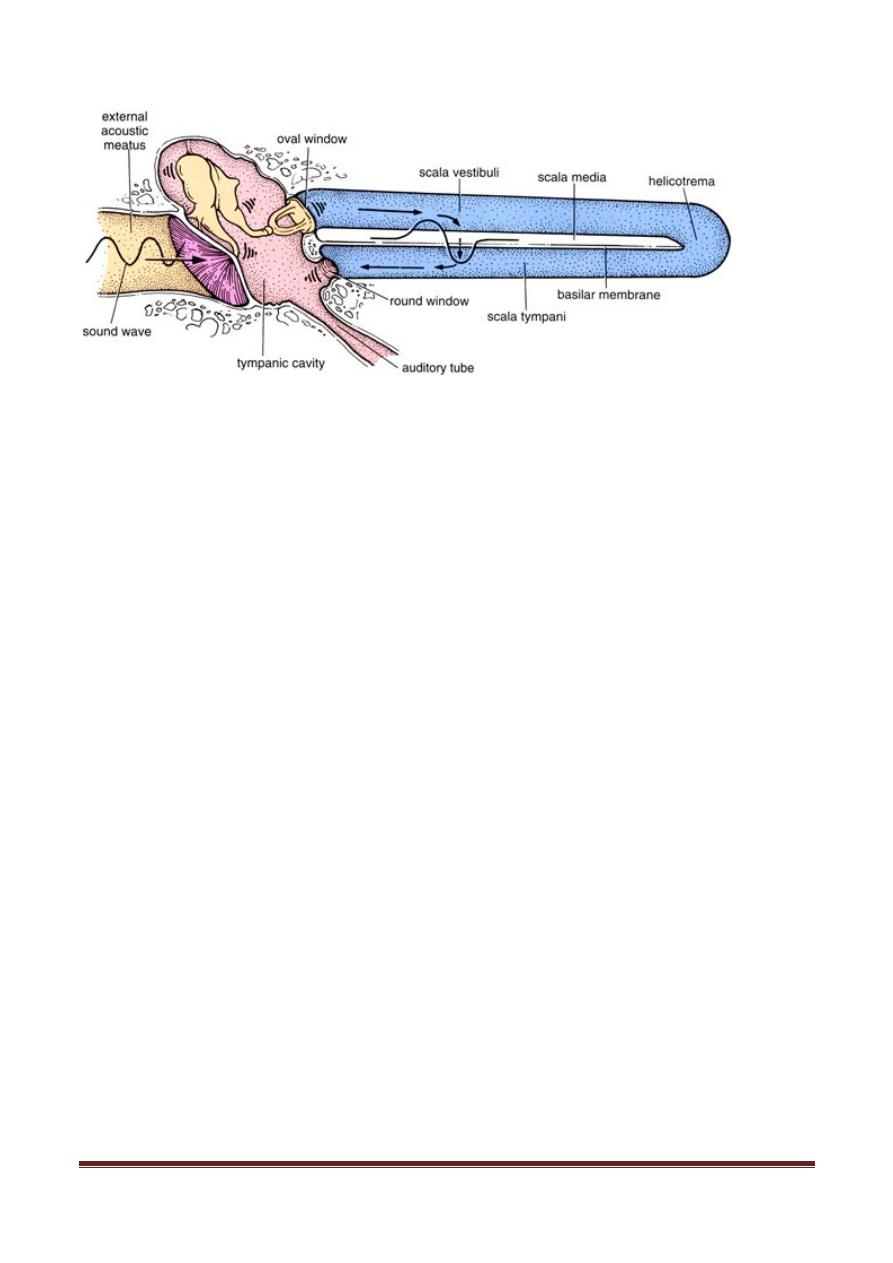
Within the cochlea are three canals:
o
Scala Vestibuli ------proximal end covered by oval window and stapes footplate.
o
Scala Tympani------- proximal end covered by round window
o
Scala Media
5
th
year 2012/2013 by dr. Ammar
Page
7
Fluid Compartments
The inner ear is divided into two fluid-filled chambers, one inside the other. The fluid in the outer or
bony chamber is filled with a sodium salt solution called perilymph that resembles the salt
composition in the blood or the fluids found in the brain. The inner or membranous chamber is filled
with a potassium salt solution called endolymph that resembles the fluid that is normally found
inside the cells of the body. The difference in the chemical composition between perilymph and
endolymph provides the electrochemical energy that powers the activities of the sensory cells.
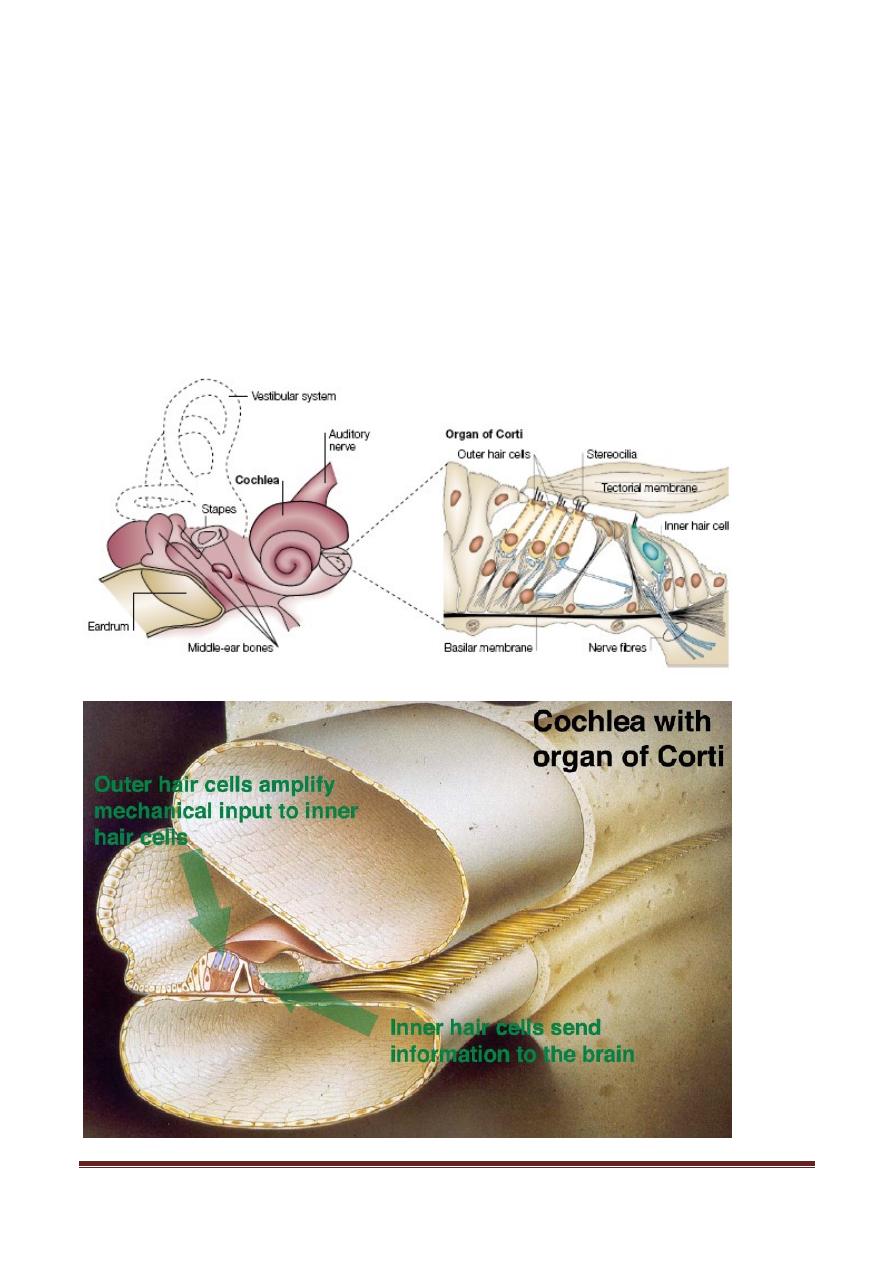
Organ of Corti
It is the key sensory area within the scala media. Here, inner and outer hair cells are rest on basilar
membrane and can be stimulated, via bending of their stereocilia, by sound waves.
Hair cells are specialized mechanoreceptors that convert the mechanical stimuli associated with
hearing and balance into neural information for transmission to the brain. Their name derives from
the fact that they have about 100 stereocilia at their apical end.
Physiology of hearing
Sound waves from the vibrating stapes footplate in the oval window enter the scala vestibuli at the
posterolateral end of the basal turn to circulate through the cochlear fluids. These waves are
transmitted from the vibrating stapes footplate into perilymphatic fluid to displace the basilar
membrane, on which the hair cells rest. The scalae vestibuli and tympani communicate at the
cochlear apex.
Vestibular System
Anatomy & Physiology of the Vestibular Organs
The utricle and saccule make up the portion that perceives linear acceleration and position sense.
They are located just deep and posterior to the end of the basal turn of the cochlea.

5
th
year 2012/2013 by dr. Ammar
Page
8
The semicircular canals—lateral, superior, and posterior—comprise the other portion of the
vestibular system. They are oriented in three planes, each perpendicular to the other. These sense
―rotational‖ head motion, or angular acceleration, interacting with the neck and eyes to maintain
orientation during turning motions
The vestibular nerve, receiving information from the balance organs, has a superior and inferior
division. These divisions converge with the auditory nerve from the cochlea to form cranial nerve
VIII. Cranial nerve VIII is immediately adjacent to the facial nerve (cranial nerve VII) as the two
enter the brainstem via the IAC and its internal auditory meatus.