Infections in gynecology
Dr Hiba Ahmed SuhailM.B. Ch. B./F.I.B.O.G.College of medicineUniversity of MosulOther causes of pelvic (inflammatory disease ( PID
TuberculosisMycobacterium tuberculosis can spread through the genital tract via the blood or lymphatic.
There is mostly tuberculosis elsewhere, usually pulmonary.
Granulomata develop in the Fallopian tubes endometrium and the ovaries
Presentation
Abnormal uterine bleeding is a presenting symptom in 10-40 percent of patients.Infection may remain subclinical, presenting ultimately with:
Amenorrhea, infertility .
PID, with chronic, low-grade pelvic pain.
Examination findings are either:
Normal .
An adnexal mass .
Fixed pelvic organs due to adhesion .
Diagnosis can be confirmed by
Obtaining endometrial tissue by biopsy which containing caseating material .
A Mantoux test should be reactive in a woman with active tuberculosis unless she is immunosuppressed.
A chest X- ray should performed to look for evidence of pulmonary tuberculosis.
Abdominal X-rays After chronic infection, bilateral tubal calcification may be seen
Laparoscopy and laparotomy .
Treatment with anti tuberculosis drugs .

Actinomycosis
This infection is almost seen in women with an intrauterine device (IUD)Causative organism actinomycosis israili.
It can be detected on cervical cytology.
If there is any history of pelvic pain, the IUD should be removed and antibiotic treatment with penicillin initiated.
If un treated actinomycosis can progress to widespread pelvic involvement with an inflammatory mass and fixing of the pelvic organs.
Toxic shock syndrome
Is a rare condition associated with the retention of tampons or foreign bodies .An overgrowth of staphylococci producing a toxin causes systemic shock with fever, diarrhoea, vomiting and an erythematous rash causes with an offensive discharge . There is a 10 per cent mortality rate.
Genital ulcer disease
Differential diagnosis of genital ulcer
Infective
• HSV
• Primary syphilis
• LGV
• Chancroid
• Donovanosis
• Human immunodeficiency virus (HIV)
Non-infective
• Aphthous ulcers
• Trauma
• Skin disease, e.g. lichen sclerosis
• Behget's syndrome
• Other multisystem disorders, e.g. sarcoidosis
• Dermatitis
Malignancy
particularly squamous cell carcinoma.
Herpes simplex virus
An incurable STDIt can be transmitted to neonates during pregnancy.
Traditionally, herpes simplex virus type I (HSV-I)
causes oral lesions (cold sores) and type II (HSV-II)
causes genital herpes. BUT , 50 per cent of genital
Infection is caused by type I (HSV-I)
Patient with one type of HSV infection can develop symptomatic infection from the other type.
Primary herpes
Primary herpes presents up to 3 weeks after infection
There is usually widespread involvement of the external genitalia , the vagina and cervix can also be affected
Painful vesicles which coalesce into multiple
ulcers.
Peri-urethral involvement may cause severe pain, and urinary retention this may also be due to involvement of the sacral nerves.
Recurrent herpes
Following a primary infection, herpes colonizes the
neurons in the dorsal root ganglia, establishing a latent infection.
Secondary herpes May be manifested as:
• Asymptomatic shedding of virus.• Apparently trivial ulcers, resembling small abrasions on the external genitalia .
• Localized clusters of vesicles and ulcers .
• Widespread or chronic ulceration resembling a primary infection can be seen in pregnant women.
• If a woman is immune suppressed, large atypical chronic ulcers may develop.
Diagnosis
Usually the diagnosis made clinically
It confirms by collecting fluid from a vesicle with a small-gauge needle and syringe or by
applying a cotton-tipped swab to ulcers and examine by :
electron microscopy virus is demonstrated.
Culture
Serology antibodies are used to type the virus.
Treatment
Advise to keep the area clean by washing .Use analgesia and Lignocaine gel locally
If retention of the urine develop catheterization of the bladder .
Antiviral treatment stops viral replication
Aciclovir 200 mg five times a day for 5 days
Famciclovir and valaciclovir
With genital herpes develop frequent recurrences (more than six to eight attacks a year) or are incapacitated during attacks then prescribe long-term suppression with aciclovir 400 mg twice a day.

Syphilis
Syphilis is a systemic STI caused by Treponema pallidum.. Following such an infection, the serological tests for syphilis may remain positive for life, causing diagnostic confusion.
Primary syphilis
The first manifestation of venereal syphilis is a painless rubbery consistency ulcer (chancre) at the site of inoculation
A chancre usually arises 3-6weeks after infection and are accompanied by inguinal lymphadenopathy
These are usually single but can be multiple.
The commonest site for a chancre is on the cervix; it may
therefore pass unnoticed and will resolve spontaneously without treatment after a few weeks..
Secondary syphilis
Can arise as the chancre disappears or up to 6 months later.
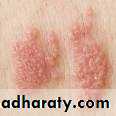
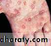
This manifested by a systemic eruption, most often a non-itchy maculopapular rash. It is symmetrical and involves the palms of the hands and soles of the feet.
More florid lesions resembling warts (condylomata lata) are seen particularly peri-anally.
Oral lesion mucous patches and linear (snail track) ulcers are seen on the mucosal surfaces.
Generalized lymphadenopathy.
Other alopecia, arthritis , meningitis and sensorineural deafness .


Following the resolution of secondary syphilis, a period of latency occurs( no manifestations).
Secondary syphilis can relapse for up to 2 years (early latent syphilis).
Primary and secondary syphilis are not life threatening.
Syphilis is also important because of the risk of vertical transmission, causing intrauterine death or a severely affected neonate.
late tertiary syphilis
The importance of the diagnosis rests on the risk of late tertiary syphilis.
Neurosyphilis can be manifest within 5 years of infection in the form stroke, tabes dorsalis, or general paresis.
cardiovascular syphilis as thoracic aortic aneurysm or aortic regurgitation .
The diagnosis of syphilis is made by
Microscopy demonstrating the organism by dark-field microscopy. Dark field illumination. Treponema pallidum can be seen as tightly wound spiral organisms, which move and bendSerological tests for syphilis, including:
Fluorescent treponemal antibody (FTA) test (most sensitive and specific test )
Specific treponemal test such as the Treponema pallidum
Haemagglutination assay (TPHA)
Treponema pallidum particle agglutination (TPPA).
A reaginic or non-specific test such as the VenerealDisease Reference Laboratory (VDRL) test
Rapid plasma reagin (RPR) test
In early primary syphilis, the serological tests may all be negative.
In secondary syphilis, the serological tests are positive with a VDRL titre of usually 1 in 32 or greater.
Following treatment of primary or secondary syphilis) the titre of VDRL should fall twofold every 3 months, becoming negative within 2 years.
Biopsy If there is any doubt, biopsies must be taken. Extensive infiltrate of lymphocytes and plasma cells histologically. Specialized stains (silver) reveal the presence of spirochaetes.

Treatment
Treponema pallidum replicates slowly, with an estimated doubling time of 20 hours.• Procaine penicillin 1.2 MU daily by intramuscular
injection for 12 days.
• Benzathine penicillin 2.4 MU by intramuscular
injection, repeated after 7 days.
• Doxycycline 100 mg two times a day for 14 days.
• Erythromycin 500 mg four times a day for 14 days.
Lymphogranuloma venereum
Lymphogranuloma venereum is caused by specificserovars (Ll-L3) of Chlamydia trachomatis.
It isfound in the Far East, sub-Saharan Africa and SouthAmerica.
In the early stages there is a small superficial ulcer that can slowly increase in size but goes unnoticed.
More obvious are the enlarged nodes, which become compressed by the inguinal ligament leading to the 'groove sign', the nodes can become matted together and discharge pus.
In women, a severe proctocolitis can progress to fistulae and strictures.
The diagnosis can be confirmed serologically by a complement fixation test.
Treatment tetracycline or erythromycin .
Chancroid
Chancroid is an infection caused by Haemophilus ducreyi.The geographical distribution is similar to that of LGV
It starts with small, shallow ulcers, which are usually multiple and painful the edges are irregular may persist for several months.
There is localized lymphadenopathy and the glands suppurate through the skin.
The diagnosis
Culture organism can only be grown on specialized culture medium
Biopsy There is a characteristic appearance on when Ducrey's bacillus seen.
Treatment azithromycin ,ceftriaxone ,erythromycin .
Granuloma inguinale (Donovanosis)
Granuloma inguinale is an infection caused by Klebsiella granulomatis .
It is endemic in India, southern Africa.
It is usually a slowly progressive infection starting with discrete papules on the skin which can enlarge to form 'beefy red' painful ulcers. These spread slowly around the genitalia and perineum.
As they heal, fibrosis can develop, which may lead to lymphoedema and elephantiasis.
Diagnosis is best confirmed by biopsy or crush preparation in which Donovan bodies are visible.
Treatment tetracycline ,erythromycin .
Viral Infection in gynecology
Human papillomavirus(HPV)More than 70 different types of HPV have been found
Certain genital strains preferentially infect the genital mucosa.
These are sexually transmitted.
Infection is often asymptomatically and may be carried for years, probably lifelong.
The virus can infect the perineum, the vagina, cervix
and rectum
Warts are frequently multiple and slowly increase in size
The majority of genital warts are caused by HPV types 6 and 11 - viruses that have little oncogenic potential.
HPV types 16 and 18 may cause flat warts and have been linked with the development of cervical carcinoma. (squamous cell carcinomas)
Smoking and low immunity is an important risk factors.
Treatment
Visible genital warts are usually treated with
physical methods such as cryotherapy.
podophyllin application once or twice a week for up to 6 weeks
Surgical treatment is used for intractable cases (lasers, electrocautery or scissor excision).
In immune compromised Immunebased therapies with interferon topical application imiquimod (a cream that stimulates local cytokine)
If cervical cytology has not been performed within 3 years, it should be done
Woman with warts on the cervix should be referred for colposcopic assessment..
Molluscum contagiosum
This poxvirus produces painless, pearly lesions with a dimple, up to 5 mm in diameter.The fluid from the vesicles is infectious
These are common in childhood and clear after a few months. Adults may acquire infection as STI
The lesions can be mistaken for genital warts.
Infection resolves with cryotherapy or following curettage and the application of phenol.

HIV infection
It is caused by HIV virusIt is a devastating disease because of the stigma of sexual transmission and the risk of vertical transmission to children.
A resurgence in tuberculosis has occurred hand in hand with the AIDS epidemic.
Virology
Human immunodeficiency virus is a retrovirus with
its genetic code in a single strand of RNA.
Reverse transcriptase is carried within the virus to enable
It to replicate within an infected cell.
The aim of therapy is to
• Reduce the level of virus in the plasma to zero with a combination of antiretroviral agents.
• Increase the CD4lymphocyte count (If therapy is effective, the CD4 lymphocyte count rises progressively)
Natural history
Twenty per cent of those infected with HIV experience an acute seroconversion illness a few weeks after acquisitionClinical features include fever, generalized lymphadenopathy, a macular erythematous rash , pharyngitis and conjunctivitis
. A steady decline in immune function over the first few years may manifest as non-life-threatening opportunistic conditions such as
• Recurrent oral and vaginal candidiasis,
• Single dermatome herpes zoster (shingles)
• Frequent and prolonged episodes of oral or genital herpes
• Persistent warts.
• Furry white patches on the sides of the tongue, termed hairy oral leukoplakia (HOL)
• Persistent generalized lymphadenopathy may be present.
Life threatening infections
Pulmonary• Pneumocystis carinii pneumonia
• Tuberculosis - pulmonary or extrapulmonary
Neurological
• Cerebral toxoplasmosis
• Cryptococcal meningitis
• AI DS dementia
Gastrointestinal
• Diarrhoea and wasting syndrome, which may be due to
infection with Cryptosporidium, Microsporidium,
Isospora
• Oesophageal candidiasis
Ophthalmic
• Cytomegalovirus retinitis
Malignancy
• Kaposi's sarcoma (Fig. 15.10)
• Non-Hodgkin's ,lymphoma
Systemic
• Mycobacterium avium intracellulare complex infection
Gynaecological manifestations
Genital warts often persist despite aggressive surgical treatment.( Persistent atypical warty lesions of the skin or vulva should be biopsied).
Chronic HPV infection can result in the development of cervical carcinoma, vulval intraepithelial neoplasia and cervical cytology annually in women with HIV
Other infections(PID) can also be more persistent in HIV-infected individuals
Postpartum endometritis is common .
Eruptions of secondary genital herpes become widespread, severe and persist often presents as deep, painful ulceration
Diagnosis
Human immunodeficiency virus infection is diagnosed by :Finding antibodies to gp-120, during sero conversion, p24 antigen is detectable in the serum before antibodies are produced.
We monitor the disease by measuring the level of CD4 lymphocytes in peripheral blood.
Using PCR technology, we can also measure the concentration of viral RNA in the plasma. A high level (> 100 000 particles/mL) predicts rapid disease development.
Transmission
HIV is principally spread as a STIIntravenous drug use .
Blood transfusion .
Genital infections are risk factors for HIV transmission and acquisition, including genital ulcer disease, Chlamydia and gonorrhoea and BV.
Treatment
With combination antiretrovira[ therapy can improve life expectancy and reduce hospital admissionsThese may include two or more nucleoside Analogue reverse transcriptase inhibitors, such as zidovudine or didinasine,
A non-nucleoside reverse transcriptase inhibitor, such as nevirapine.
Protease inhibitors, such as nelfinavir
This combination is expensive and complex to manage and there is considerable drug toxicity.
Vaginal discharge in children
Vaginal infections are common in childhood and . Streptococcal infections are the commonest cause.
Shigella spp. can cause a haemorrhagic chronic vaginitis, with history of diarrhoea.
Recurrent vaginal infections should lead to suspicion of a foreign body.
An examination under anaesthesia necessary to exclude or remove the cause.
Pinworms (Enterobius vermicularis) are common and migrate from the anus at night, causing intense irritation and inevitable scratching by the child. ( the nocturnal pattern). Selotape test can be performed to look for eggs if the worms have not been witnessed at night.
Treatment by treat the underlying cause
Infestations
Pubic lice and scabies are transmitted by contact. Pubic lice (Phthirus pubis) attach their eggs to the base of pubic hair. . Infected individuals may report small itchy papules, . Lice are treated by the application of topical agents such as malathion, carbaryl or permethrin,treatment should be repeated after 7 days.Scabies (Sarcoptes scabiei) causes an intensely itchy papular rash. , it may be initially confined to the genital area. It responds to applications of malathion or permethrin; however, symptoms may take up to 6 weeks to resolve completely