
1
Operative
Cariology
Dr Maan
Lesion , Etiology , prevention and control
Dental caries is an infectious microbiologic disease of the teeth that
results in localized dissolution and destruction of the calcified tissues.
It is a preventable disease and can be arrested or reversed in its
early stages.
Dental Plaque (Biofilm): A gelatinous mass of bacteria
adhering to the tooth surface .
Carious lesions occur under a mass of bacteria producing
acidic environment to demineralize tooth structure after
metabolization of refined carbohydrates.
Enamel demineralization occur at pH of 5.5 or less while
remineralization occurs as the local pH rises above 5.5 .
Saliva contains high concentrations of calcium and phosphate ions that
serve as a supply for the remineralization process.
Understanding the balance between demineralization and
remineralization is the key for caries management.
Etiology of Dental Caries
Dental caries has a multifactor etiology, However four principle factors
are necessary for the production of a carious lesion
1. Dental Plaque (Bacteria)
Carious lesions only occur under a mass of bacteria capable of producing
a sufficiently acidic environment to demineralize tooth structure .

2
2. Diet
(Substrate such as a fermentable carbohydrate or dietary sugars) which
is necessary for microorganism to as nutrition with subsequent
production of acid as byproduct that would initiate the demineralization.
3. Tooth
(A susceptible tooth) as teeth composed of minerals (calcium and
phosphate) therefore low pH will create environment for
demineralization of the minerals resulting destruction of hard tissue.
4. Time
To produce demineralization process sufficient time at low pH is
required It is difficult to determine the exact time for demineralization
inside patient mouth .
Elimination of one of these factors is required for the prevention of
dental caries.
Over 300 species of bacteria present in the oral cavity, only mutans
streptococci, are caries causing (cariogenic) organisms MS are the
primary causative agents of initial coronal caries because they
(1)adhere to enamel
(2) produce and tolerate acid
(3) Live and grow in a sucrose-rich environment
(4) produce bacteriocins. Substances that kill off competing organisms
Mutans Streptococci and lactobacilli can produce great amounts of acids
(acidogenic), and appear to be the primary organisms associated with
canes in man.
Mutans Streptococci are most strongly associated with the onset of
caries while lactobacilli are associated with active progression of
cavitated lesions.
3
Caries can be classified according to location , extent , and rate.
Location
(1)Pits and fissures of enamel
Which is the most susceptible site because the pits and fissures of newly
erupted teeth are rapidly colonized by bacteria.
Pit-and-fissure caries expands as it penetrates into the enamel_ Thus,
the entry site may appear much smaller than the actual lesion, making
clinical diagnosis difficult.
Detection of the lesion by explorer might be destructive method as the
lesion could be treated conservatively through the remineralization
process, however explorer might breakdown the dental tissue and
create cavity that will act as harbor for dental plaque and debris that
initiate caries progression. In addition the remineralization process will
be more difficult to achieve in cavitated lesion than white spot lesion.
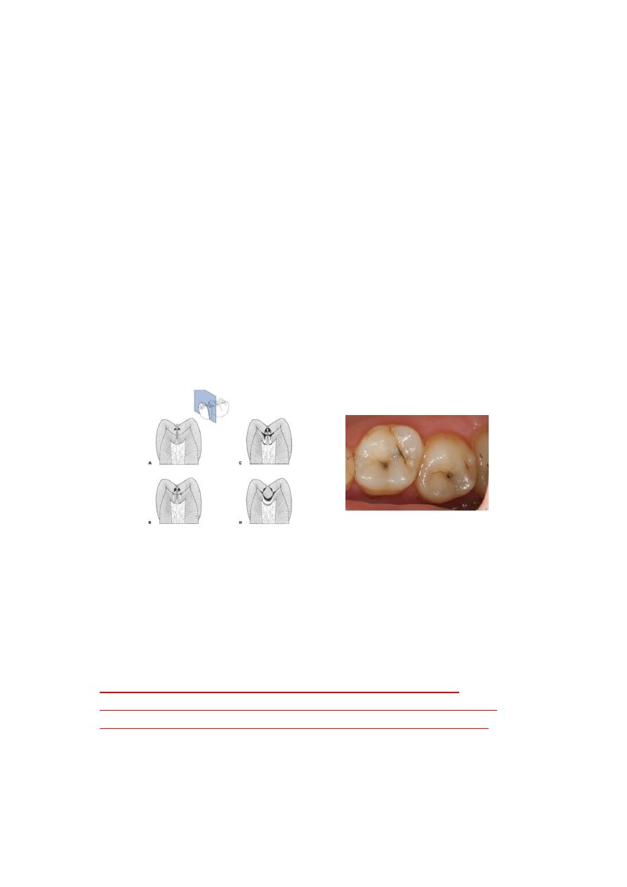
Fig 2-1a Occlusal caries.. The shadowing around the stained pits in second
molar indicates the presence of carious dentin at the base of fissure.
progression of caries to in pits and fissures
A. Initial lesions develop on the lateral wall of the fissure, demineralization
following the direction of the enamel rods.
B. Forceful probing of the lesion at this stage can result in damage to the
weakened porous enamel and accelerate the progression of the lesion.
clinical detection at this stage should be based on observation of
discoloration and opacification of enamel adjacent to the fissure. These
changes can be observed by careful cleaning and drying of the fissure.
C. Initial cavitation of the opposing walls of the fissure can not be seen on the
occlusal surface .
D. Extensive cavitation of the dentin and undermining of the covering enamel
wall darken the occlusal surface.
4
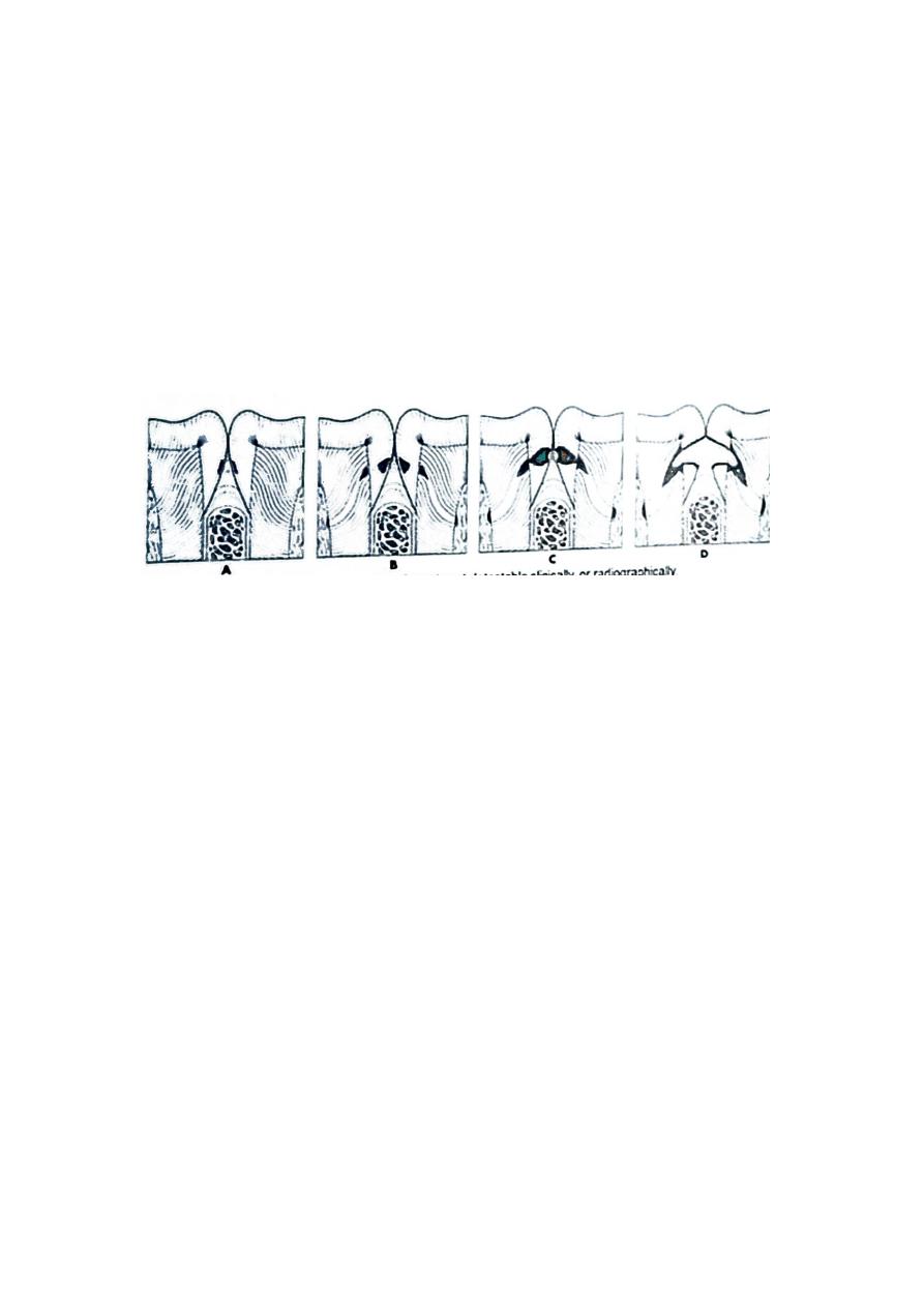
(2) Smooth enamel surfaces & interproximal region:
The smooth enamel surfaces of the teeth present a less favorable site
for plaque attachment. Lesions starting on smooth enamel surfaces have
a broad area of origin and a conical, or pointed, extension toward the
DEJ Smooth surface lesion shows a V shape with a wide area of origin
and the apex of the V directed toward the DEJ. The caries again spreads
at this junction in the same manner as in pit-and-fissure caries. Thus, the
apex of the cone of canes in the enamel contacts the base of the cone of
caries in the dentin.
A, Initial demineralization on the proximal surfaces is not detectable
clinically or radiographically .
B, When proximal caries first becomes detectable radiographically, the
enamel surface is likely to still be intact . An Intact surface is essential for
successful remineralization and arrest of the lesion
C, Cavitation of the enamel surface Is a critical event in the caries
process in proximal surfaces. Cavitation is an irreversible process and
requires restorative treatment / correction of the damaged tooth
surface. Cavitation can only be diagnosed by clinical observation.
The use of a sharp explorer to detect cavitation is problematic because
excessive force in application or the explorer tip during inspection of the
proximal surfaces can damage weakened and accelerate the caries
process by creating cavitation
What are the following terms mean ?
Backward Canes, Forward Caries, Residual Caries.

5
(3) Root surface.
Recently root caries get special attention due to the increase in the old
ago population. The cementum covering the root surface is extremely
thin and provides little resistance to caries attack. Root caries lesions
have less well defined margins, tend to be U-shaped in cross-section.
and progress more rapidly because of the lack of protection from an
enamel covering.
Characteristic of root caries
:
1. Rapidly progress
2. A symptomatic
3. Close to the pulp.
4. More difficult to restore.
Secondary (Recurrent) Caries. Secondary caries occurs at the junction of
a restoration and the tooth and may progress under the restoration.
Extent of Caries
Incipient Caries ( or remineralizable).
Incipient caries is the first evidence of caries activity in the enamel On
smooth surface enamel, the lesion appears opaque white when air-
dried, and will seem to disappear (not e distinguishable from contiguous
unaffected enamel) if wetted This lesion of demoralized enamel has not
extended to the DEJ and the enamel surface is fairly hard and still intact
(smooth to the touch) The lesion can be remineralized if immediate
corrective measures alter the oral environment,including plaque
removal and control .
Cavitated Caries ( or non remineralizable ).
In cavitated caries, the enamel surface is broken (not intact), and usually
the lesion has advanced into dentin Usually remineralization is not
possible (see Fig 9-5, E) and treatment by tooth preparation and
restoration is often Indicated.

6
Rate (Speed) of Caries
Acute (Rampant) Caries.
Acute caries, often termed rampant canes. is when the disease is rapid
in damaging the tooth II is usually in the form of many, soft light-colored
lesions in a mouth and is infectious (see Fig 9-5, E) Less time for extrinsic
pigmentation explains the lighter coloration .
Chronic (Slow or Arrested) Caries.
Chrome caws is slow. or it may be arrested following several active
phases. The slow rate results from periods when demineralized tooth
structure is almost remineralized (the disease is episodic over time
because of changes in the oral environment). The condition may be in
only a few locations in a mouth, and the lesion is discolored and fairly
hard. The slow rate of canes allows time for extrinsic pigmentation.
Progression of caries:
Decrease in caries progression have been reported recently possibly due
to improvement in the preventive measures like using of fluoride.
The progression of the carious lesion depending on the site of origin ( pit
and fissure or smooth surface and the conditions in the mouth (poor
oral hygiene and frequent sucrose-containing food, dry mouth).
Early detection of caries lesion is important to take necessary preventive
measures to arrest the progression and initiate the reminralization
process, this will imply us to apply what is call (Minimum intervention)