
GIT 3
Third year class
By Dr.Riyadh A. Ali
Department of pathology
TUCOM

Titles
CA stomach
Malabsorption syndrom

Ca Stomach
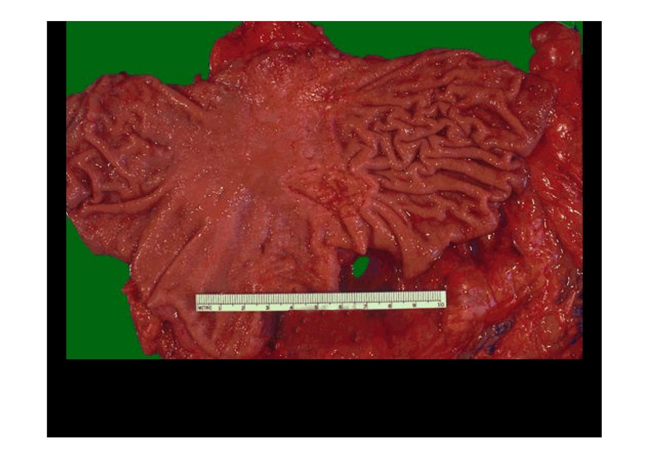
Here is a
gastric ulcer
in the center of the picture. It is shallow and is about 2
to 4 cm in size. This ulcer on biopsy proved to be
malignant
, so the stomach
was resected as shown here.
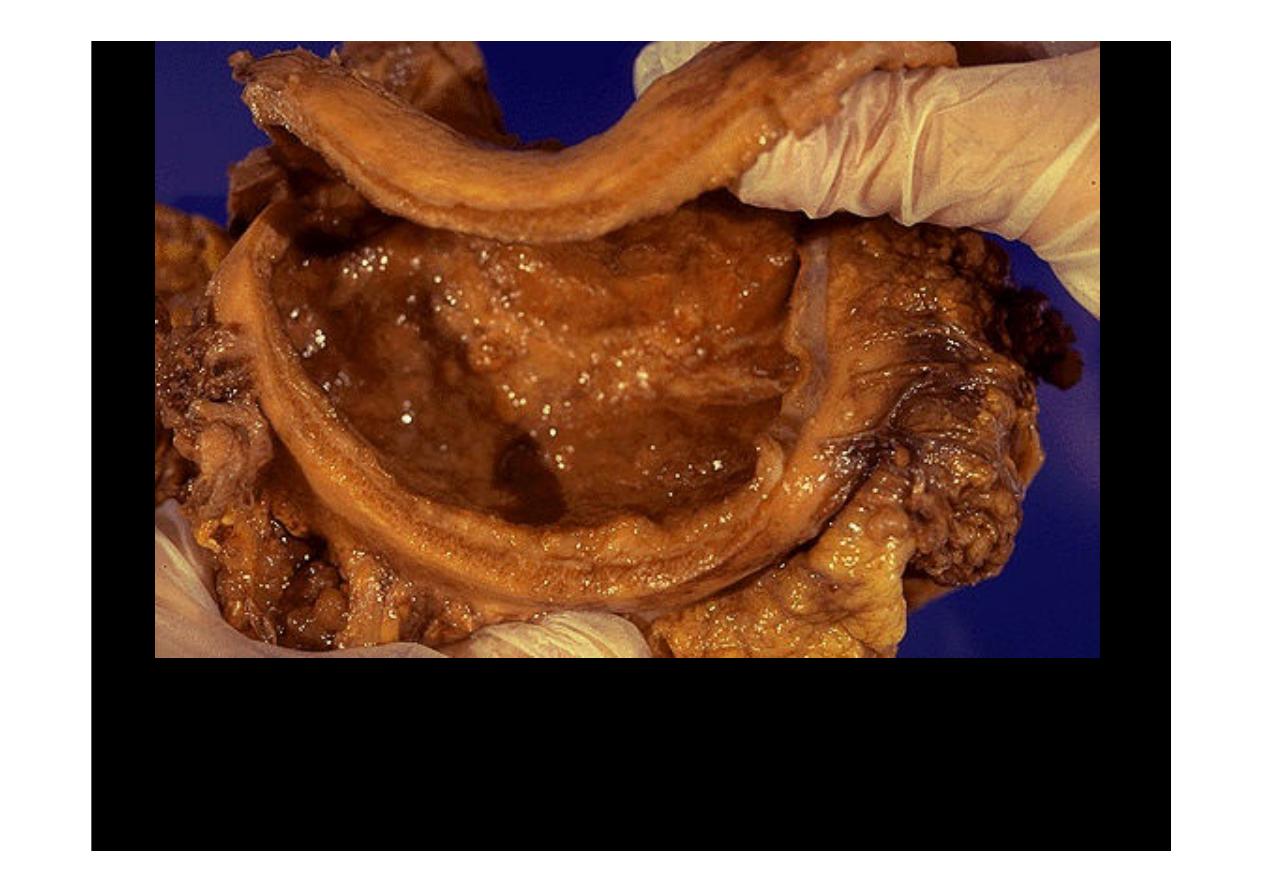
This is an example of
linitis plastica
, a diffuse infiltrative
gastric
adenocarcinoma
which turns the stomach into a shrunken "leather bottle"
appearance.
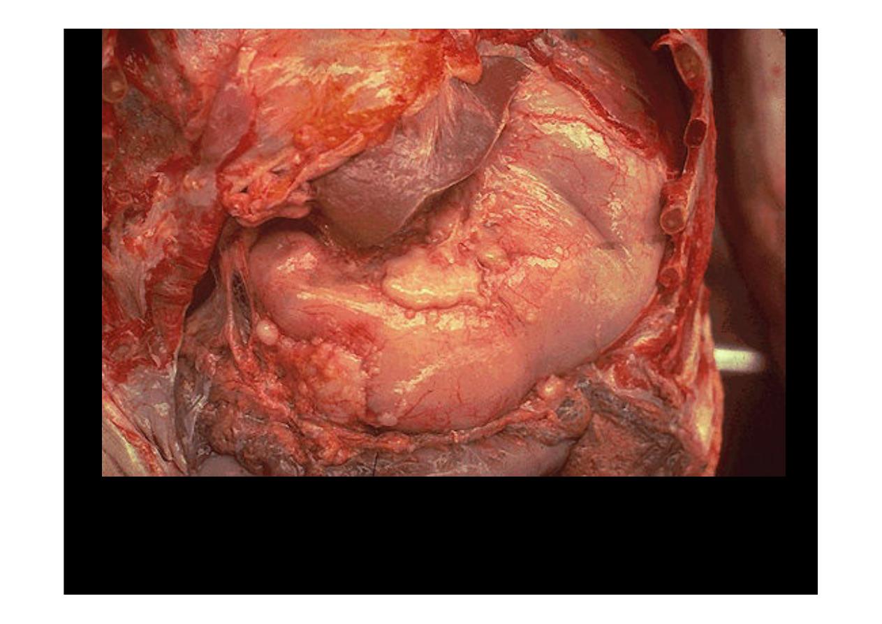
At autopsy, the thoracic cavity and abdominal cavity are both opened to reveal the
stomach just to the right and below the edge of liver in this photograph.
Gastric
adenocarcinoma
has infiltrated through the wall and appears on the surface as
irregular tan masses. The extensive tumor in this case caused gastric outlet
obstruction.
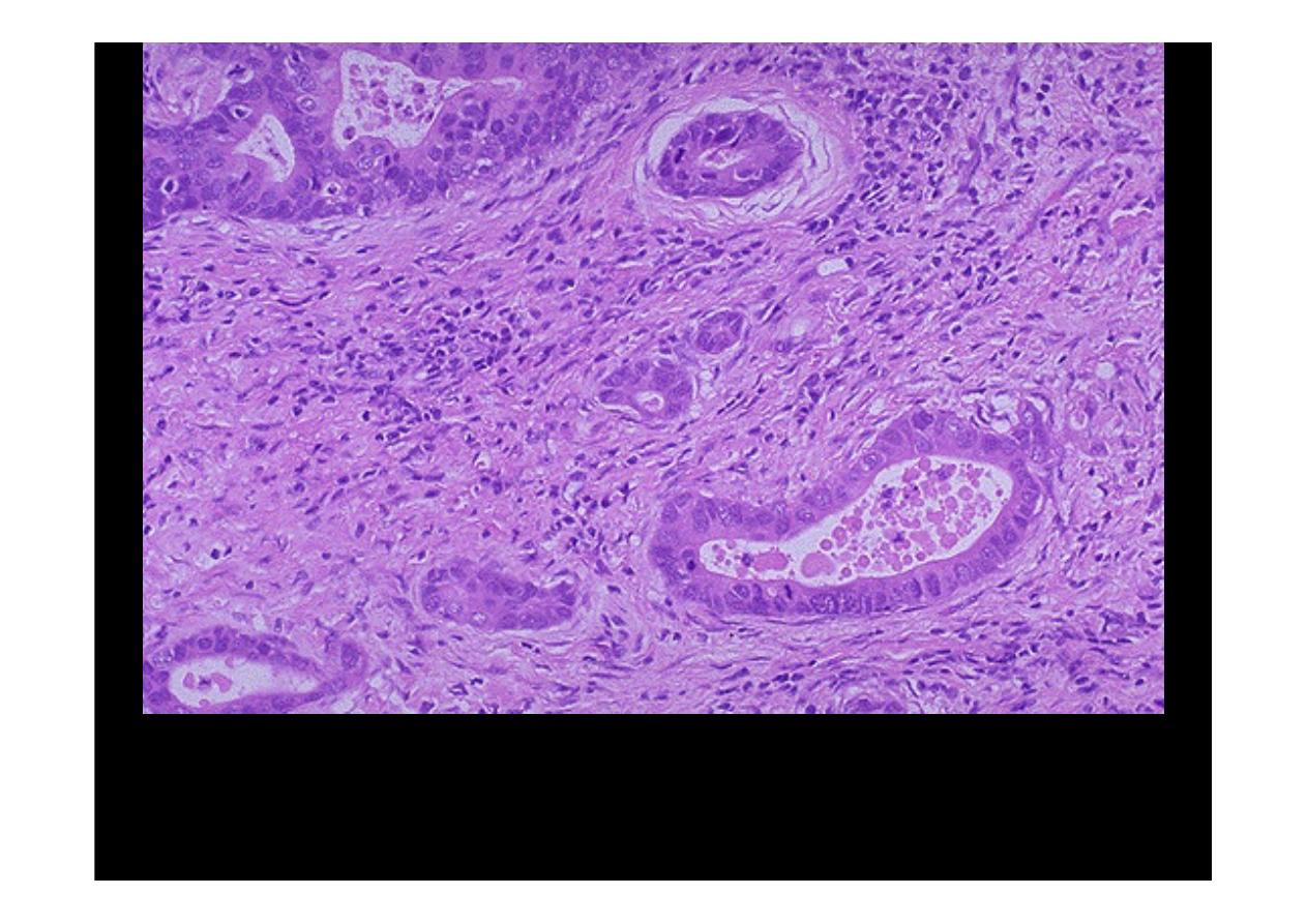
A
moderately differentiated gastric adenocarcinoma
is infiltrating up and into
the submucosa below the squamous mucosa of the esophagus. The neoplastic
glands are variably sized.
At higher magnification, the neoplastic glands of
gastric adenocarcinoma
demonstrate mitoses, increased nuclear/cytoplasmic ratios, and hyperchromatism.
There is a desmoplastic stromal reaction to the infiltrating glands.
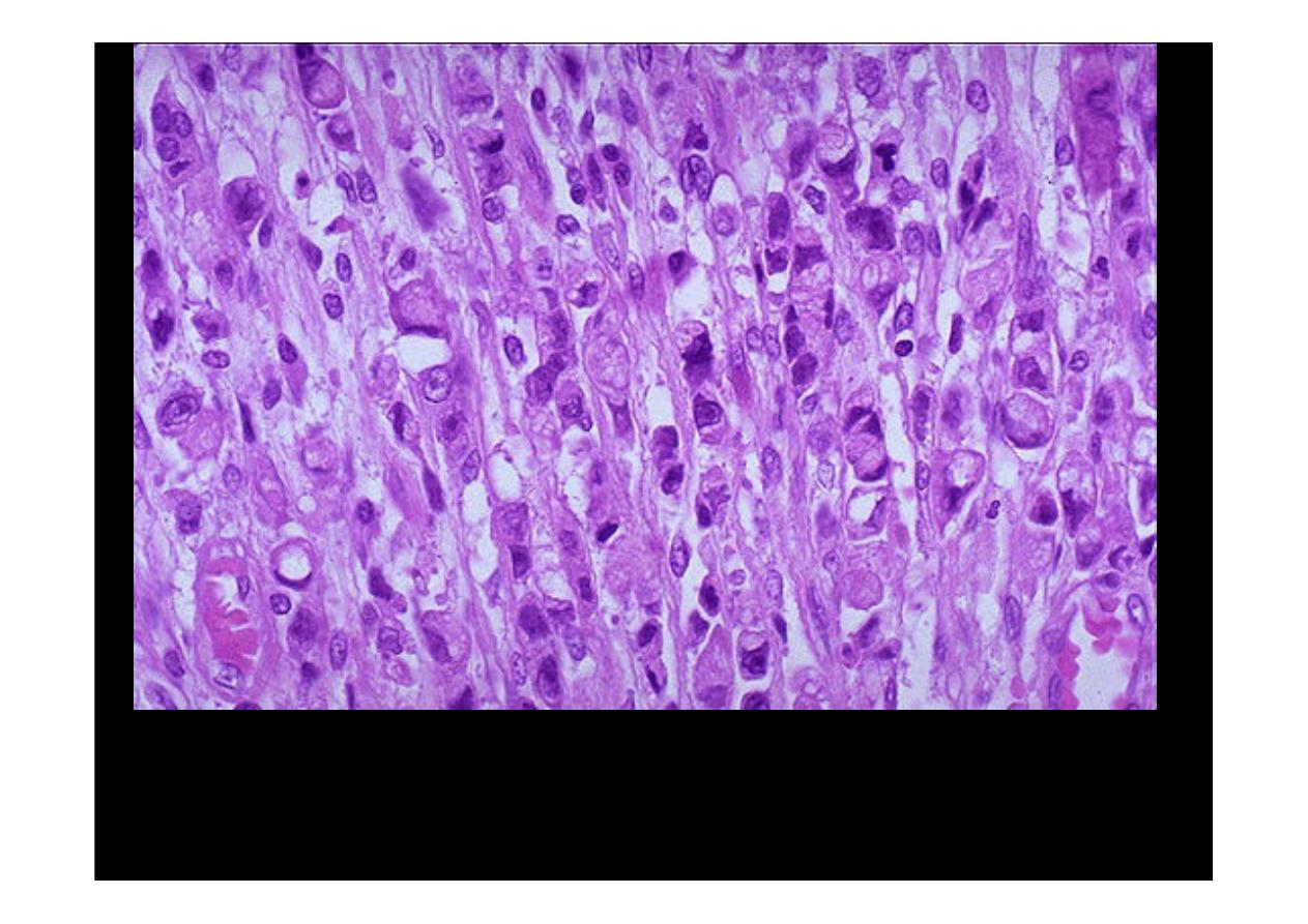
At high power, this
gastric adenocarcinoma
is so poorly differentiated that
glands are not visible. Instead, rows of infiltrating neoplastic cells with marked
pleomorphism are seen. Many of the neoplastic cells have clear vacuoles of
mucin.
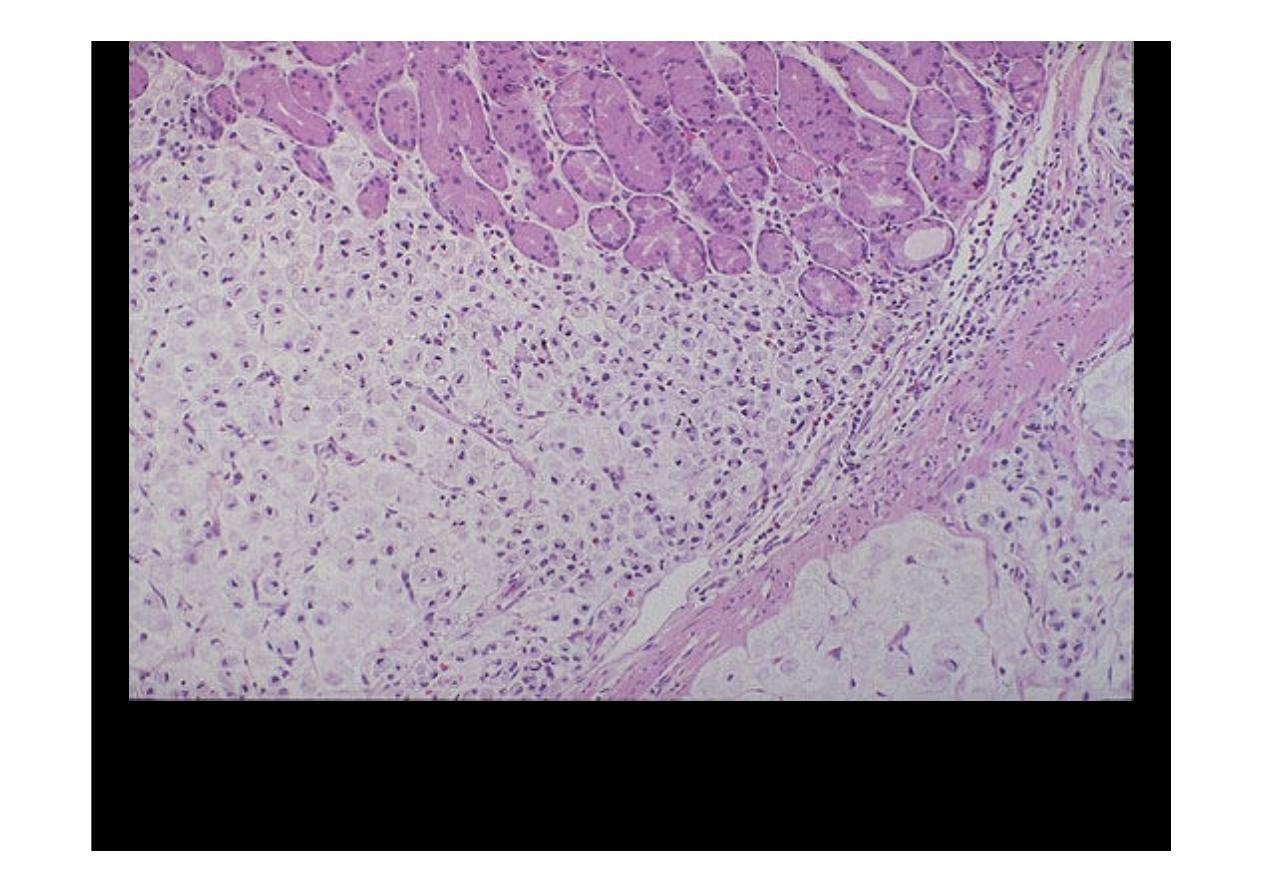
At medium power, normal gastric glands are seen at the top, and infiltrating
signet
ring cell adenocarcinoma
is seen below. This histologic pattern is typical for the
diffuse variant of gastric adenocarcinoma which is extensive and infiltrative.
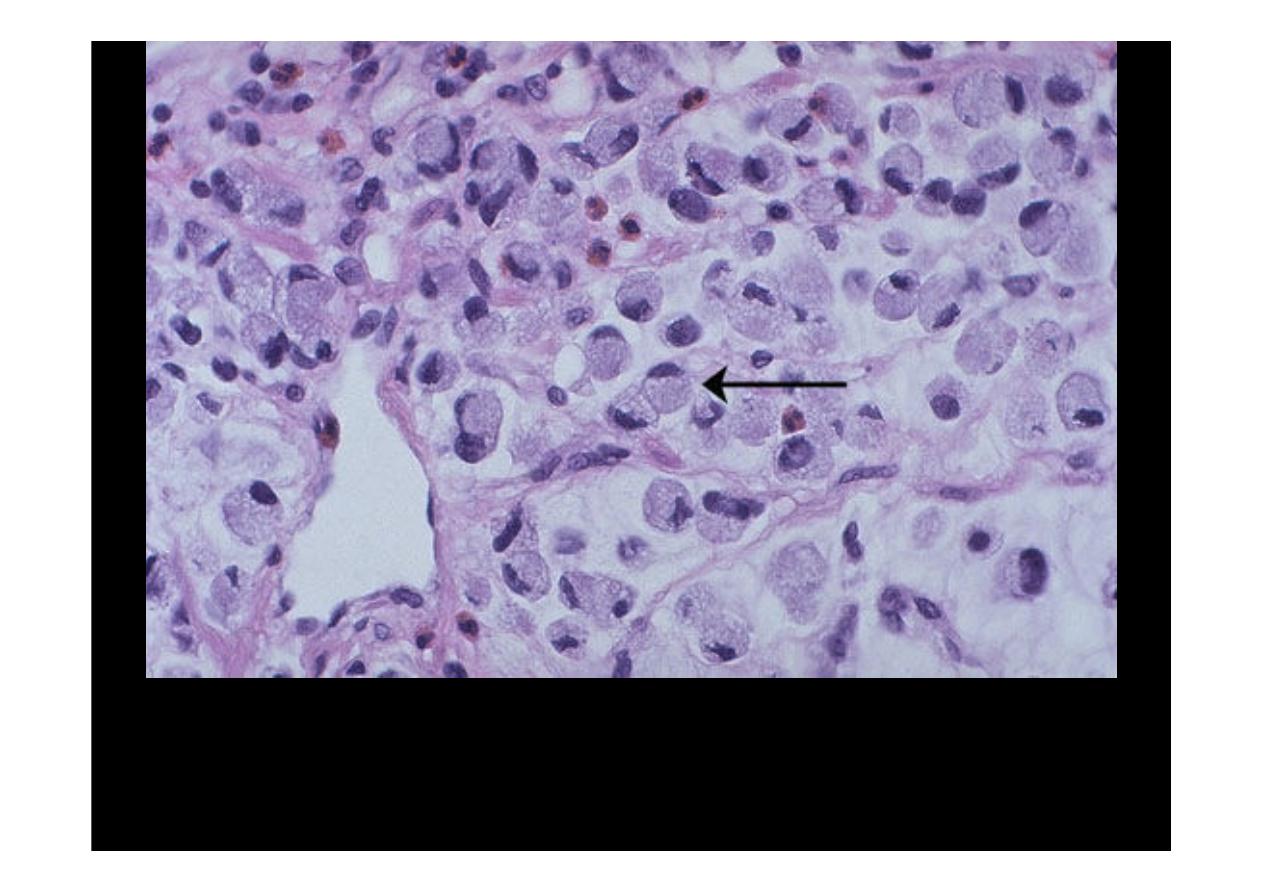
This is a signet ring cell pattern of
adenocarcinoma
in which the cells are filled
with mucin vacuoles that push the nucleus to one side, as shown at the arrow.

Malabsorption
syndrom
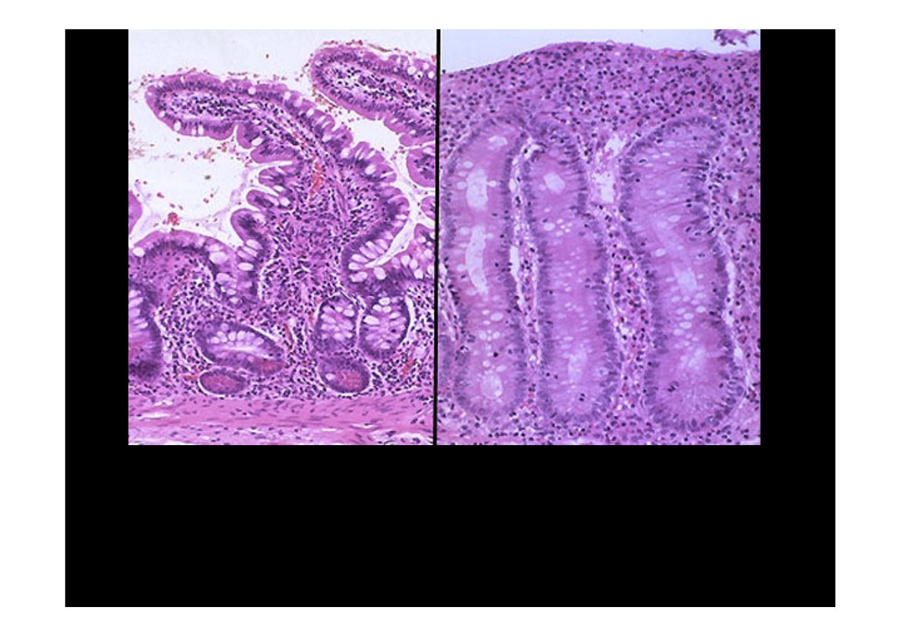
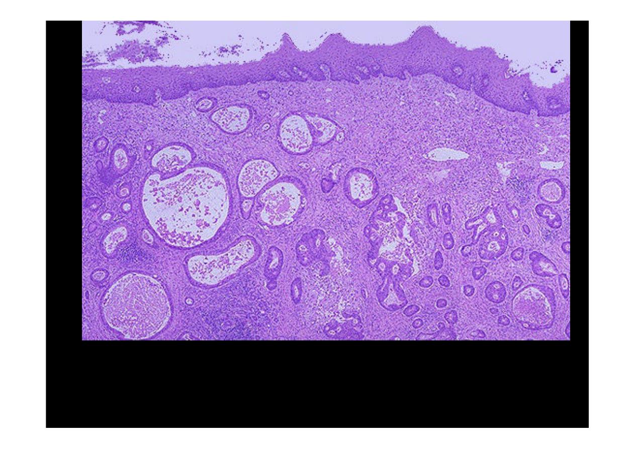
Normal small intestinal mucosa is seen at the left, and mucosa involved by
celiac
sprue at the right
. There is blunting and flattening of villi with celiac disease, and
in severe cases a loss of villi with flattening of the mucosa as seen here. Celiac
sprue has a prevalence of about 1:2000 Caucasians, but is rarely seen in other
races. Over 95% of affected patients will express the DQw2 histocompatibility
antigen, which suggests a genetic basis.
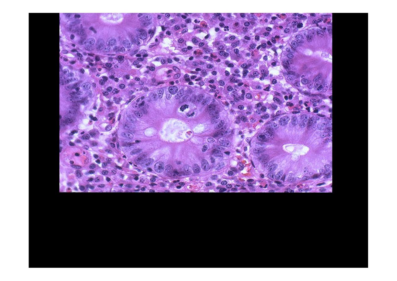
The small intestinal mucosa at high magnification shows marked chronic
inflammation in
celiac sprue
. There is sensitivity to gluten, which contains the
protein gliaden, found in cereal grains wheat, oats, barley, and rye. Removing foods
containing these grains from the diet will cause this gluten-sensitive enteropathy to
subside. The enteropathy shown here has loss of crypts, increased mitotic activity,
loss of brush border, and infiltration with lymphocytes and plasma cells (B-cells
sensitized to gliaden).