Endocrine System 2
Third Year Class
By Dr.Riyadh A. Ali
Department of Pathology
TUCOM
Pituitary Gland

Normal Gland
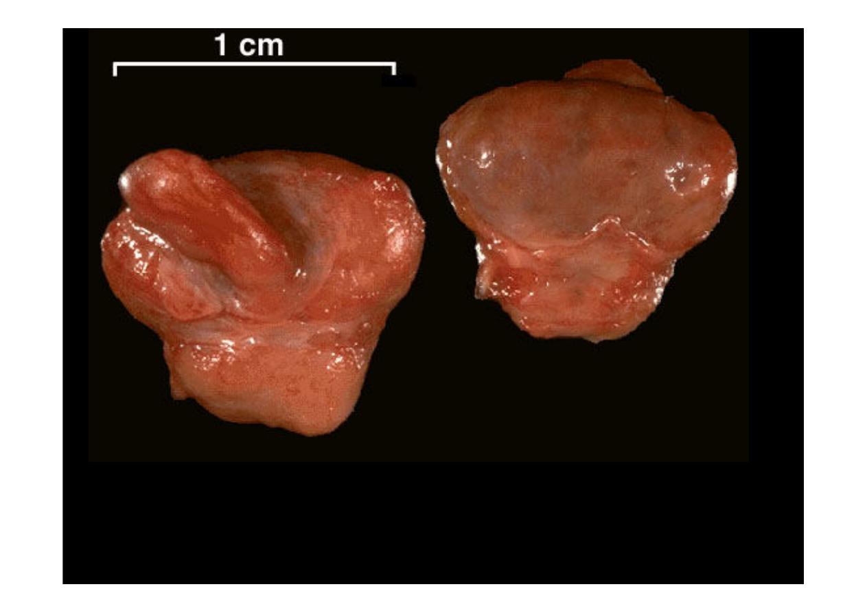
The normal gross appearance of the
pituitary gland
. The larger portion, the anterior
pituitary (adenohypophysis), is toward the top. The image at the left shows the
superior aspect of the pituitary with the stalk coming from the hypothalamus entering
it. The inferior aspect of the pituitary is shown at the right. The posterior pituitary
(neurohypophysis) is the smaller portion at the bottom

The normal microscopic appearance of the
pituitary gland
is
shown here. The adenohypophysis is at the right and the
neurohypophysis is at the left.
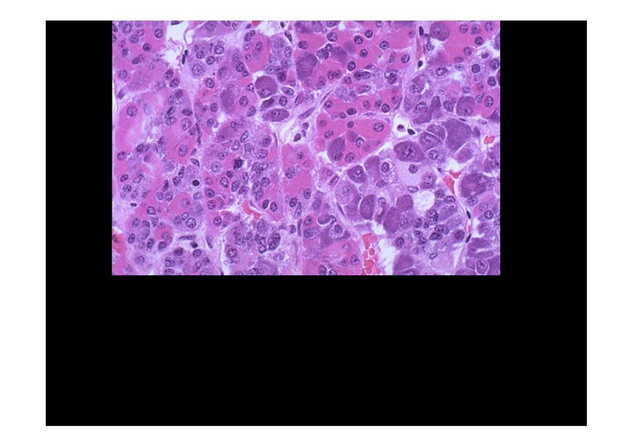
The
adenohypophysis
contains three major cell types: acidophils, basophils, and
chromophobes. A simplistic classification is as follows:
The pink acidophils secrete growth hormone (GH) and prolactin (PRL)
The dark purple basophils secrete corticotrophin (ACTH), thyroid stimulating
hormone (TSH), and gonadotrophins follicle stimulating hormone-luteinizing
hormone (FSH and LH)
The pale staining chromophobes have few cytoplasmic granules, but may have
secretory activity.
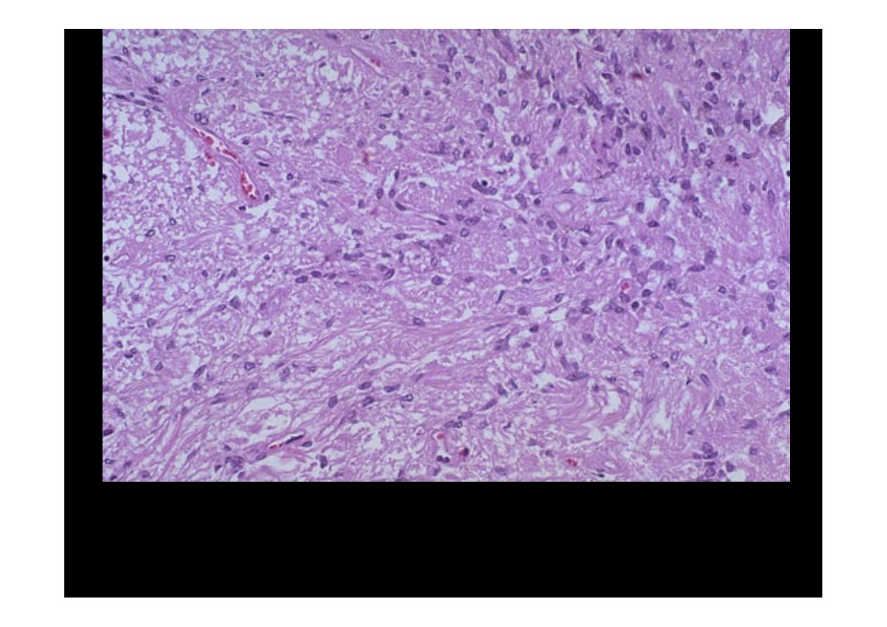
The neurohypophysis shown here resembles neural tissue, with glial cells, nerve
fibers, nerve endings, and intra-axonal neurosecretory granules. The hormones
vasopressin (antidiuretic hormone, or ADH) and oxytocin made in the
hypothalamus (supraoptic and paraventricular nuclei) are transported into the
intra-axonal neurosecretory granules where they are released.
Adenoma
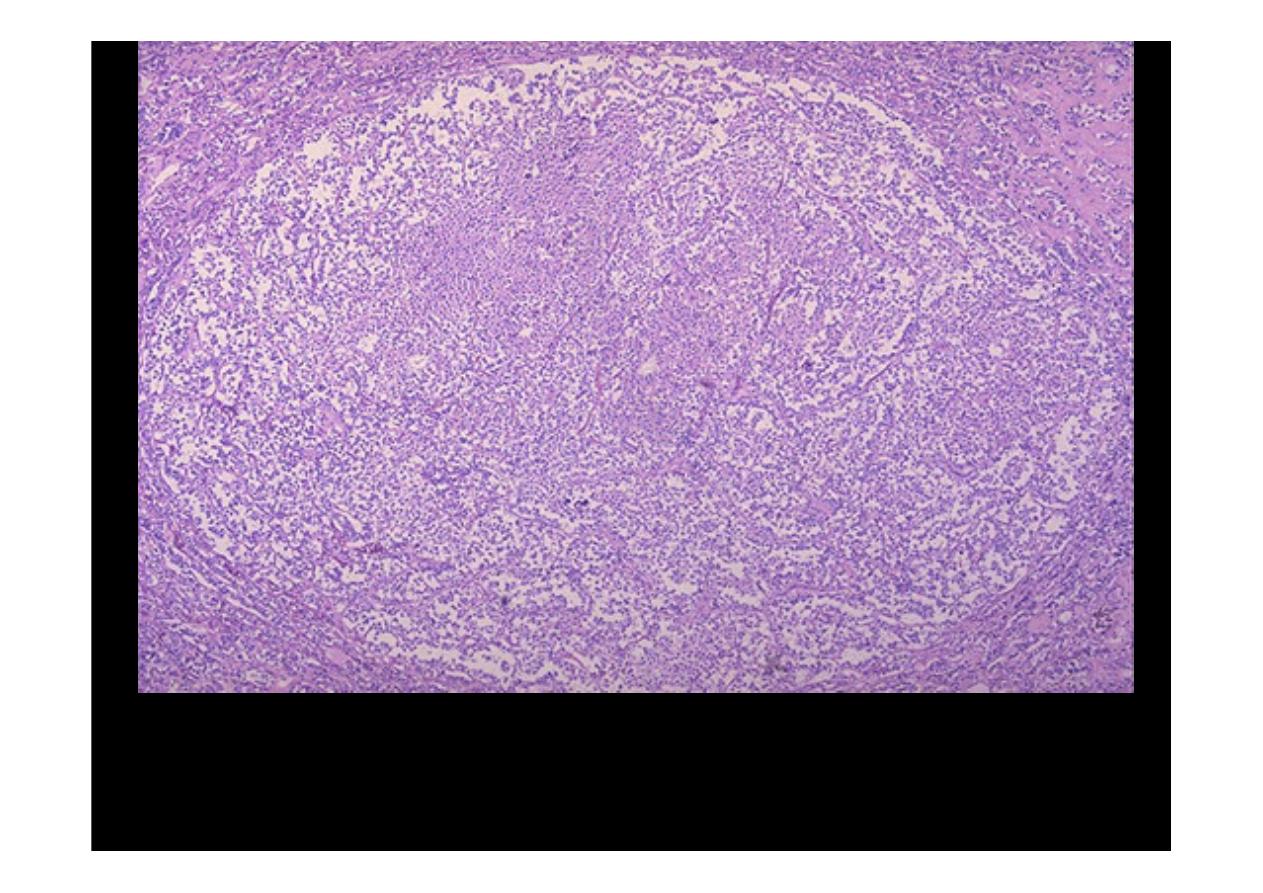
This is a
microadenoma of the anterior pituitary
. Such microadenomas
may appear in 1 to 5% of adults. These microadenomas rarely have a
significant hormonal output that leads to clinical disease
.
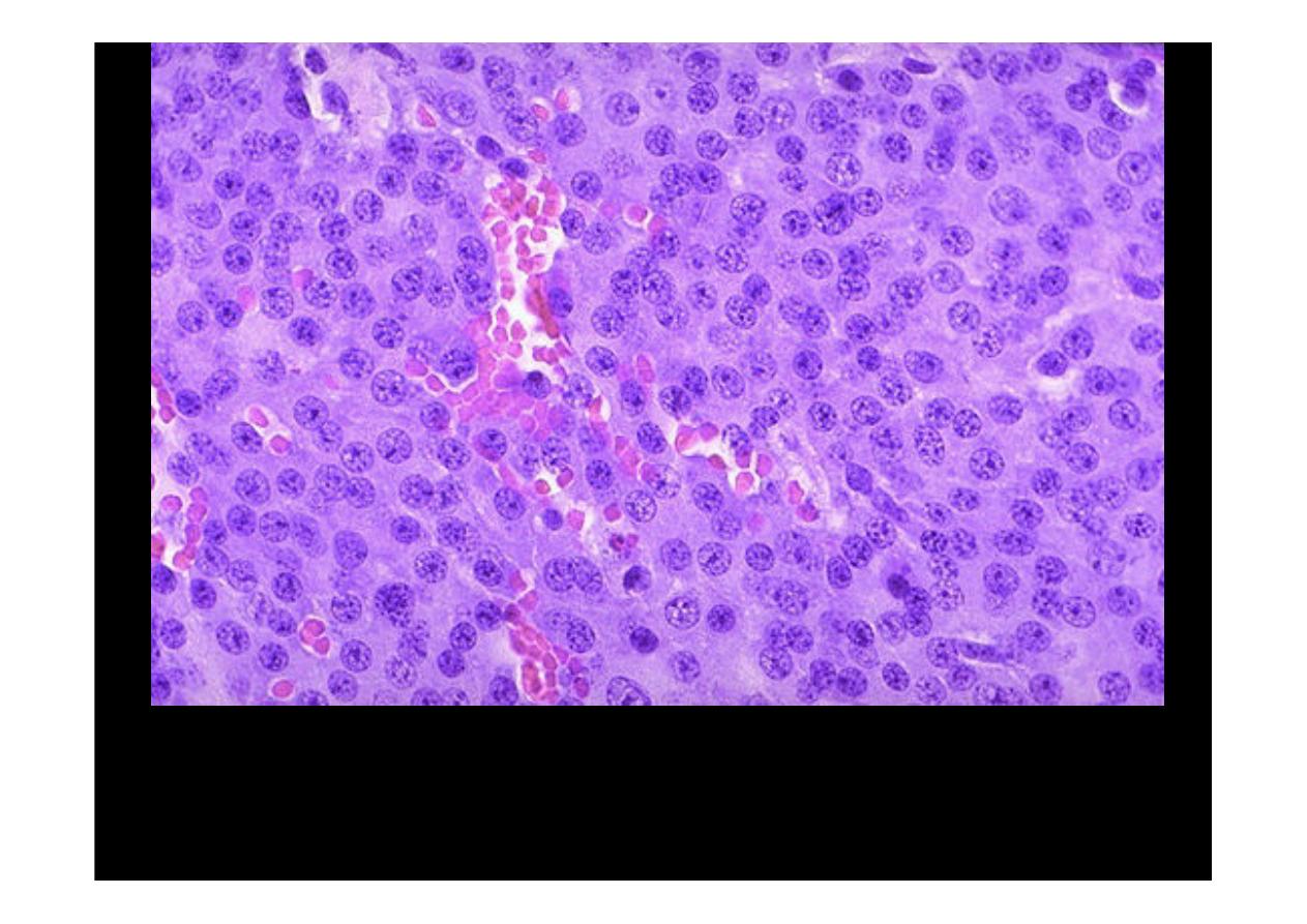
Here is a high power microscopic view of an
adenohypophyseal adenoma
.
Endocrine neoplasms are composed of small round cells with small round
nuclei and pink to blue cytoplasm. The cells may be arranged in nests or cords
and endocrine tumors also have prominent vascularity.