1
L5
Spinal Cord Disorder
D. Hazim
Introduction
Spinal cord is continuation of CNS contained within the bony spinal canal, from the
foramen magnum at base of skull caudally to conus medullaris at level of L1 .
The three meningeal layers that surround the spinal cord continues below level of Ll
as a fibrous tissue (filum teminale) that terminate at the coccyx.
The spinal cord is 45 cm length ,while the vertebral column length is about 70 cm
this is discrepancy is clinically important
WM located peripherally, while nerve cell cluster in an inner region shaped like a
four-leaf clover surround the central canal
Neuroanatomy of the spinal cord
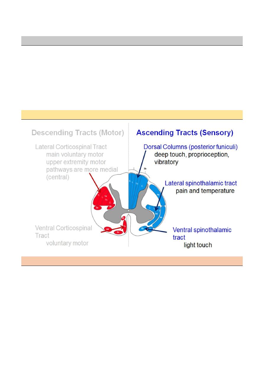
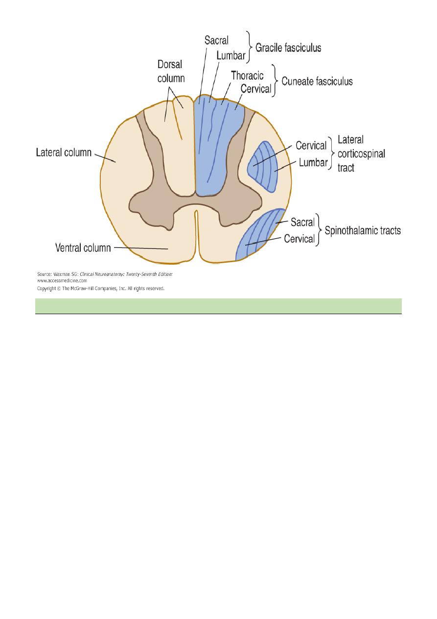
Distribution of fibers within the spinal cord
Both spinothalamic tract and corticospinal tract are arranged in a certain pattern, which is
laminated and looks like the layers of the onion and as follows:
C=cervical (the innermost)
T=thoracic
L=lumber
S=sacral(outermost)
This is important in localization of the lesion, for example, if the pathology is an extrinsic it will
affect the sacral fibers first, and if the pathology is intrinsic it will firstly affect the cervical fibers.
2
Spinal cord level relative to the vertebral bodies
This is clinically important, as when we find certain abnormality or suspect a lesion in certain
level, we should remember that discrepancy exits.
Site of suspected lesion the requested level of imaging
Upper cervical same as cord level
Lower cervical 1 levels higher
Upper thoracic 2 levels higher
Lower thoracic 2-3 levels higher
Lumber T10 _T12
Sacral T12_L1
Coccygeal Ll
3
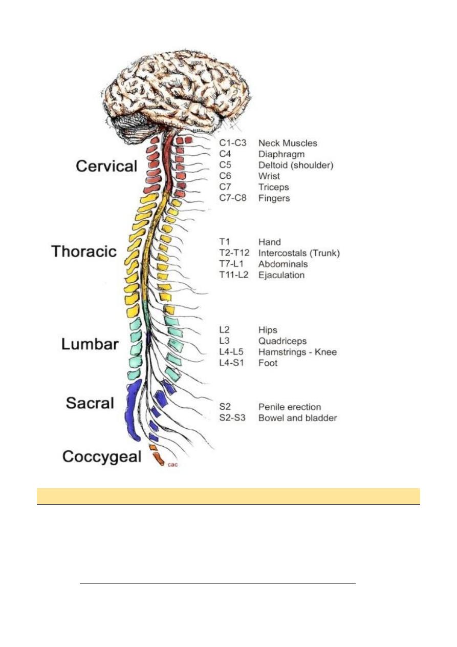
Injuries to spinal cord cervical cord
Junction of cervico-medullary usually fatal
C4-5= quadriplegia with diaphragm paralysis.
C5-6=loss of power and reflex in Biceps
C7=weakness in wrist ,finger extension &triceps
C8=finger &- wrist flexion
Horner syndrome may accompany a cervical cord lesion at any level .

4
Thoracic & lumber cord
o Lesion localized by sensory level
o T4=nipples, TlO=umbilicus
o Leg weakness and disturbance of bladder and bowel.
o L2-4=paralysis of flexion and adduction of the thigh, weakness of leg extension,
diminished patellar reflex
o L5-S I =paralysis of foot and ankle flexion and abolish ankle jerk(S 1) .
Basic Features of Spinal Cord disease
• UMN findings below the lesion
• Hyperreflexia and Babinski's
• Sensory and motor involvement that localizes to a spinal cord level
• Bowel and Bladder dysfunction common
Conus Medullaris Vs. Cauda Equina Lesion
Finding Conus CE
Motor Symmetric Asymmetric
Sensory loss Saddle Saddle
Pain Uncommon Common
Reflexes Increased Decreased
Bowel/bladder Common Uncommon
Investigation of Spinal Cord Disease
• Radiographic exam
o Plain films
o Myelography
o CT scan with Myelography
o MRI
• Spinal tap
If you suspect: inflammation, MS, rupture of a vascular malformation
Metabolic disease of the spinal cord
Subacute combined degeneration of the cord (SACD)
SACD, caused by vitamin B12 deficiency presents as a
Myelopathy with prominent dorsal column features.
Those typical are distal paraesthesiae and gait unsteadiness, mild upper motor neuron
lower limb weakness, depressed knee and ankle jerks, with extensor plantars – and
importantly, impaired joint position sense.
optic atrophy may develop with the macrocytic anaemia
The typical picture of SACD leads many experienced physicians to give therapy
immediately, even before serum levels are known.
5
Treatment is with hydroxocobalamin, a minimum of
1000 μg weekly by injection for the first 3 weeks followed by1000 μg monthly
injections for 6 months, and thereafter 1000 μg every 3 months, for life.
Spinal tuberculosis
TB typically first affects the intervertebral discs.
infection typically presents with local pain, fever, night sweats and general ill health
including weight loss.
If the disease spreads from the disc into the vertebral body osteomyelitis will occur
with epidural abscess formation and/or vertebral body collapse.
Pathological fractures will cause pain, deformity (kyphosis) and in some cases spinal
cord compression.
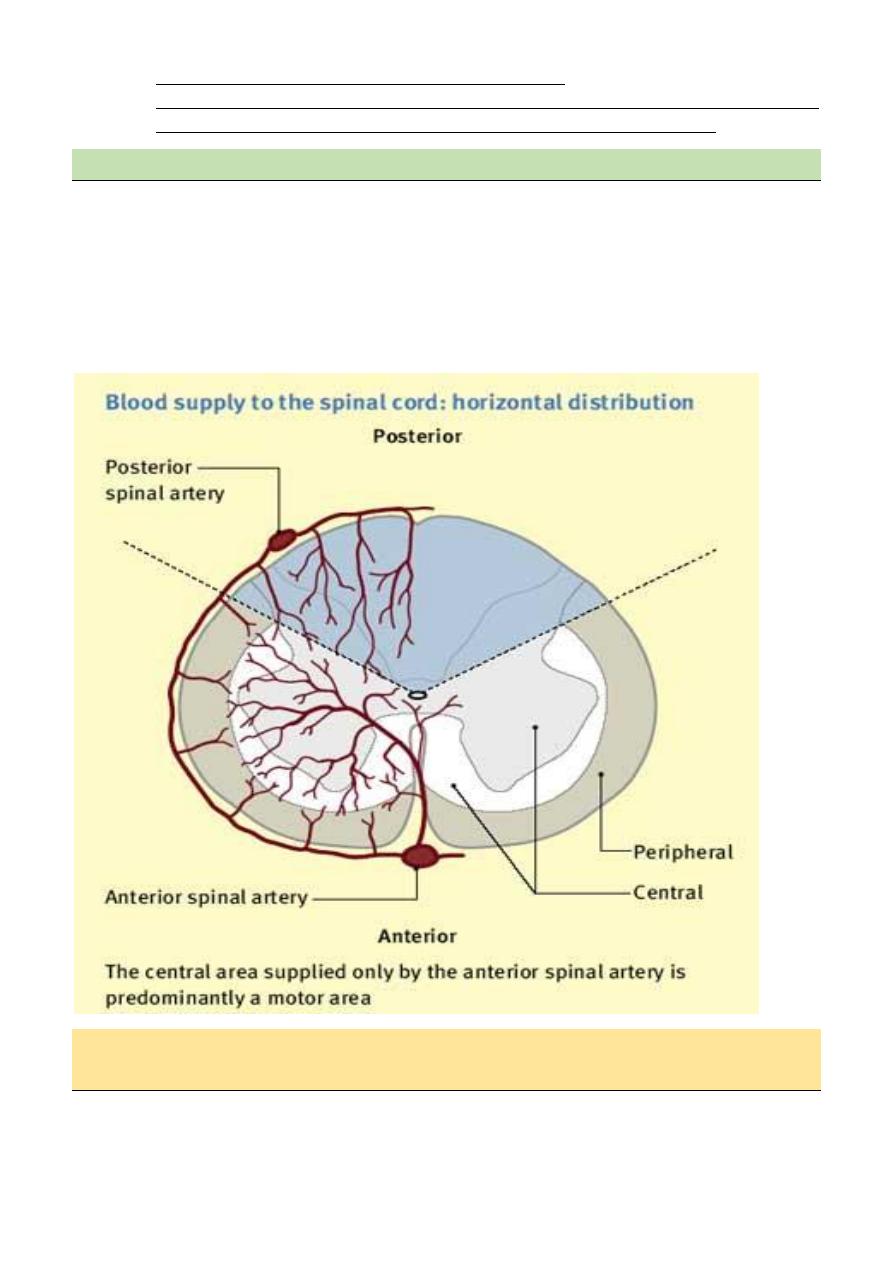
Spinal cord infarction
Anterior spinal artery occlusion
Spinal cord infarction usually presents acutely, often with pain followed by paralysis and
sensory loss.
In anterior spinal artery occlusion the anterior two-thirds of the spinal cord is affected.

6
The spinal level is determined by where in
Its course the supply from the anterior spinal artery is interrupted.
Clinical feature
The patient presents with an acute flaccid paraparesis with loss of sphincter control and
anesthesia to temperature and pain but classically with preservation of posterior column
functions of joint position and vibration sense. The most typical level is the upper thoracic
cord.
Diagnosis of spinal vascular disease
MRI is the primary diagnostic investigation.
MRI will detect over 90% of acute spinal cord ischaemic lesions.
MRI usually excludes compressive lesions
evidence of demyelination especially if cranial MRI is also performed (MS) lesion.
Management of spinalcord infarction
There is no curative treatment for acute spinal cord infarction.
The prognosis for functional recovery in established spinal infarction is poor.
Mubark A. Wilkins