Oral cavities &Gastrointestinal tract pathologyDr. Zahraa Marwanlecturer in Mosul medical college
ORAL CAVITY:Pathologic conditions of the oral cavity can be broadly divided into diseases affecting teeth their support structures, oral mucosa, salivary glands, and jaws.
Teeth And Supportive Structures
Dental Caries:
.focal degradation of tooth structure (enamel and dentin) due to mineral dissolution by acids generated during the fermentation of sugars by bacteria..the most common cause of tooth loss before the age of 35
.prevention by:
-improved oral hygiene
- Use of fluoride (Fluoride is incorporated into the crystalline structure of enamel, forming fluoroapatite, which is resistant to degradation by bacterial acids.
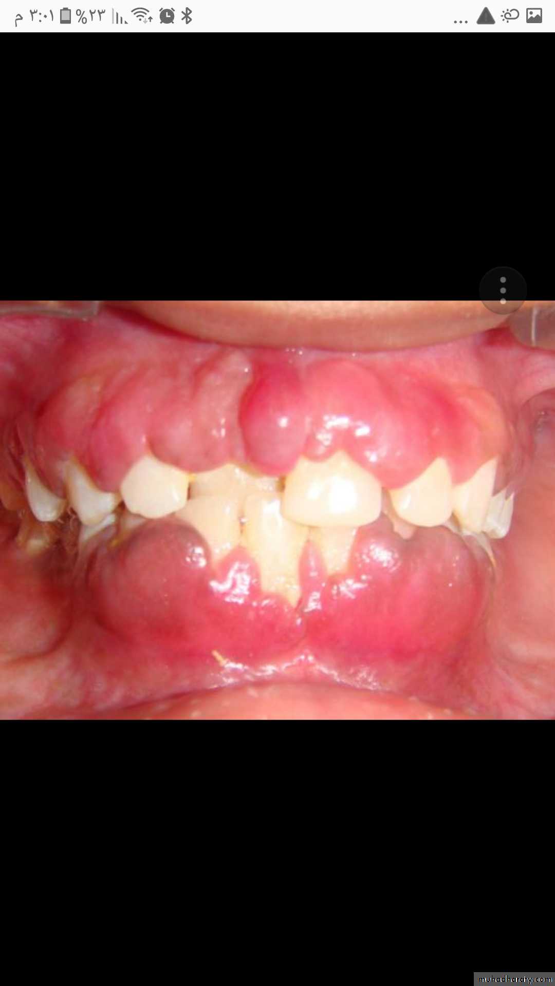
Gingival hyperplasia:
- Gingival hyperplasia is an overgrowth of gum tissue around the teeth.- There are a number of causes for this condition, but it’s often a symptom of poor oral hygiene or a side effect of using certain medications.
- Gingival hyperplasia is also referred to as:-
. gingival overgrowth
. Gum enlaregement
. hypertrophy
. hypertrophic gingivitis
One of the more common characteristics of this
condition is having red, tender and easily bleeding gums.Gingival hyperplasia bright red gingival overgrowth around the necks of the teeth
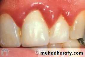
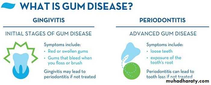
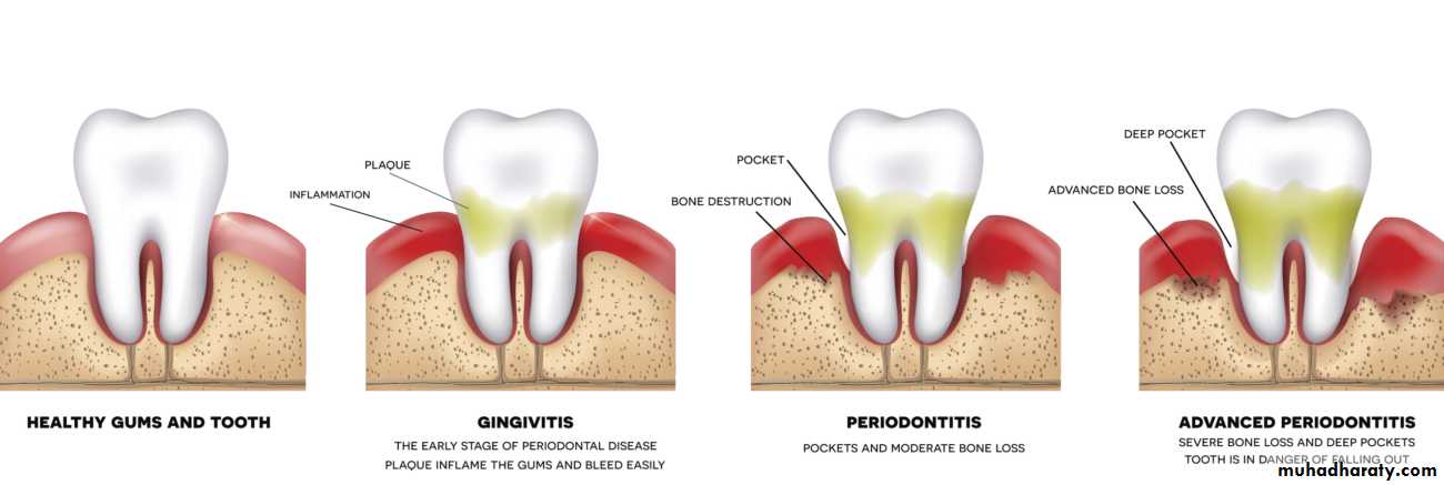
Gingivitis: .inflammation involving the squamous mucosa, or gingiva, and associated soft tissues that surround teeth, resulting from accumulation of dental plaques and calculi- Poor oral hygiene is the most frequent cause of gingivitis. .most prevalent in adolescence. gingivitis can be reversed by: - regular brushing and flossing of teeth which reduces accumulation of plaque and calculus.
- The symptoms of gingivitis are somewhat non-specific and manifest in the gum tissue as classic signs of inflammation i.e:
* Swollen gums which bleed easily.
* Bright red or purple gums.
* Gums are tender and in some cases, painful to the touch.
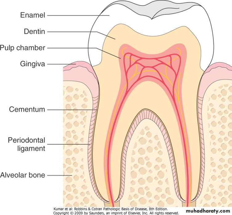
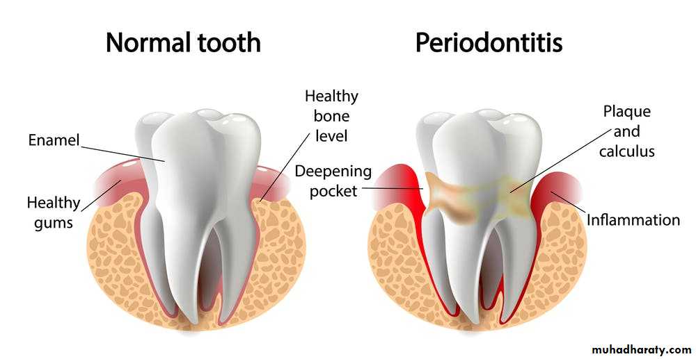
Periodontitis: .is an inflammatory process that affects: -periodontal ligaments -alveolar bone -cementum. Periodontitis is associated with poor oral hygiene that affects the composition of gingival bacteria and leads to shift in type and proportions of bacteria along the gums.
-Periodontitis associated with: -immune deficiency states -leukemia -Crohn’s disease -diabetes mellitus -defects in neutrophils.can be an etiologic factor in: -infective endocarditis -lung and brain abscesses -adverse pregnancy outcomes
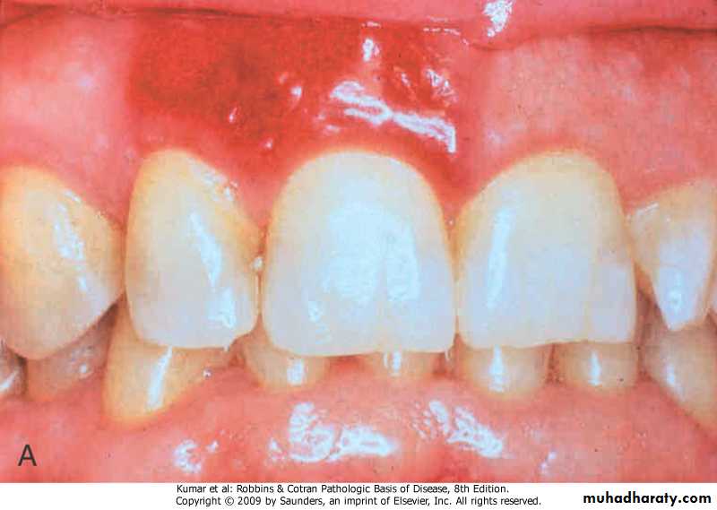
Periodontitis, the generalized inflammation, abnormal gingival anatomy owing to tissue destruction, gingival recession, swelling and inflammation, spontaneous bleeding and abundant plaque deposits.
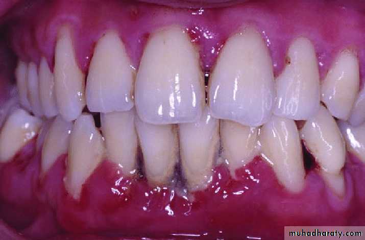
With progression, periodontitis may result in destruction of periodontal ligament and alveolar bone and eventual tooth loss.
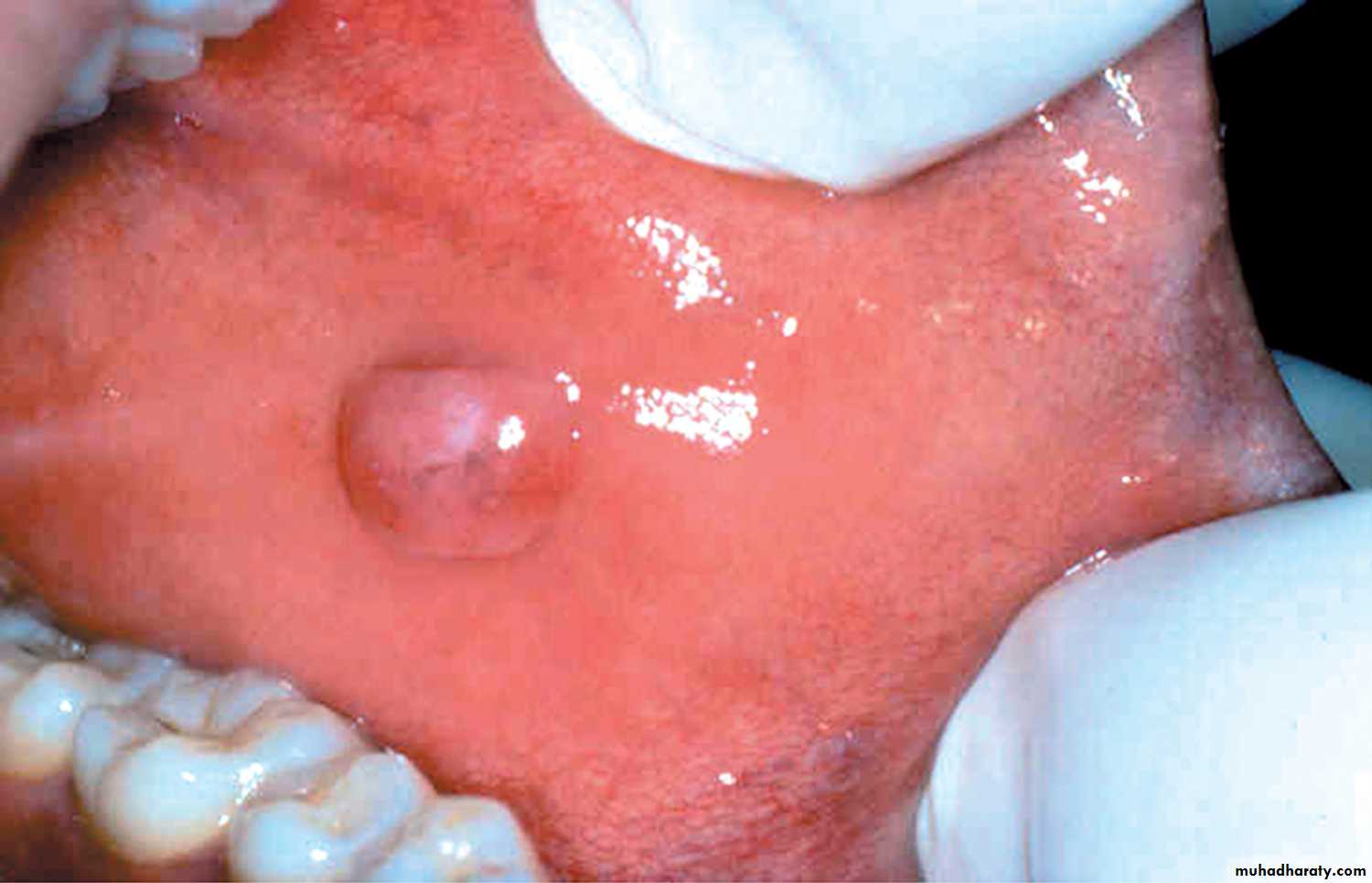
Fibrous proliferative lesions of the oral cavity:Irritation Fibroma:.location, buccal mucosa along bite line.treatment, surgical excision.Smooth pink nodule on the buccal mucosa.
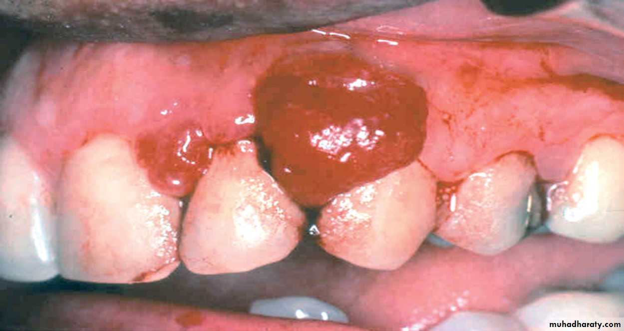
Pyogenic Granuloma:.location, gingiva of: -children and young adults -pregnant women. Erythematous hemorrhagic exophytic mass arising from the gingival mucosa.
Inflammatory lesions Of The Oral Cavity
Aphthus Ulcers (Canker Sores) .recurrent single or multiple, very painful ulcers most common in 1st two decades of life.. Although the cause of aphthus ulcers is unknown, they tend to be familial and may be associated with:
- celiac disease
- inflammatory bowel diseases
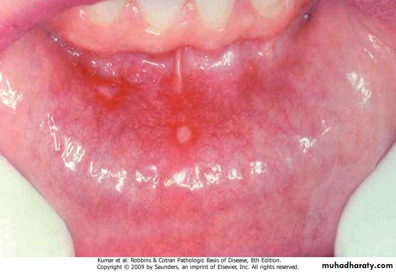
Aphthus ulcer. Single ulceration with an erythematous halo surrounding a yellowish fibrinopurulent membrane
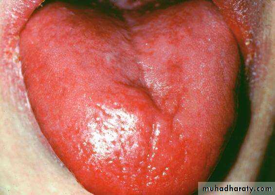
Glossitis: .characterized by beefy-red tongue and ulceration, seen in -certain deficiency states, (vitamin B12, riboflavin, niacin, pyridoxin, iron) -chronic irritation
• Infections Of The Oral Cavity:
• Herpes Simplex Infection:• .caused by HSV-1 and 2
• .primary infection occurs in children 2-4
• years old
• -asymptomatic
• -acute herpetic gingivostomatitis
• .Tzanck test, microscopic examination of
• vesicle fluid (nuclear inclusion, multi-
• nucleated cells (polykaryons))
• .adults harbor latent HSV-1, reactivation produce: - Mild cold sore - Recurrent herpetic stomatitis, (lips, nasal orifices, oral mucosa)

Oral Manifestations Of Systemic Diseases
• .infectious diseases (scarlet fever,• measles, diphtheria)
.dermatologic conditions (lichen planus, bullous diseases)
.hematologic disorders (leukemia, pancytopenia)
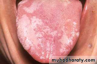
Hairy Leukoplakia: .seen in immunocompromised patients, (80% are infected with HIV) .presenting as white, confluent patch of fluffy hyperkeratotic thickening, almost always at lateral border of the tongue .EBV is thought to be the cause
Precancerous And Cancerous Oral Lesions:
Leukoplakia:.is a white patch or plaque that cannot be scraped off and cannot be categorized clinically or pathologically as any other disease
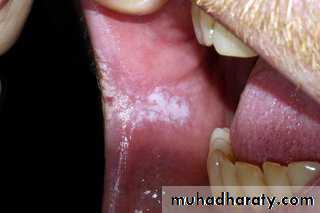
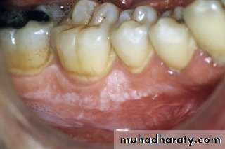
Leukoplakia patches often appear on the tongue, inside the cheeks, or on the gums.
The patches aren't painful ,are an irregular shape ,are slightly raised, may be slightly red within the patch.Erythroplakia:.a red, velvety area that is level with or slightly depressed in relation to the surrounding mucosa
- Both leukoplakia and erythroplakia affect adults 40-70 years, with 2:1 male preponderance, and caused by tobacco- Erythroplakia is associated with a much greater risk for malignant transformation than leukoplakia
Squamous Cell Carcinoma:
.Approximately 95% of cancers of the oral cavity are squamous cell carcinomas, with the remainder largely consisting of adenocarcinomas of salivary glands..an aggressive cancer (6th in the world)
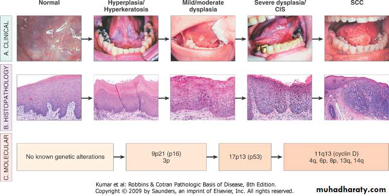
.Pathogenesis:
-abuse of tobacco and alcohol
-HPV infection
-genetic influence:
.mutation of p16>squamous hyperplasia
.mutation of p53>dysplasia
-actinic radiation (lower lip)
Oral cancer, clinical, histologic, and molecular progression
.Morphology:
-gross, plaque>ulcer at:
.ventral surface of the tongue
.floor of the mouth
.lower lip
.soft palate
.gingiva
-histology, (well differentiated, moderately differentiated, anaplastic)
.prognosis is poor, (diagnosis at late stage, multiple primary tumors)
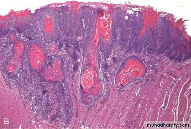
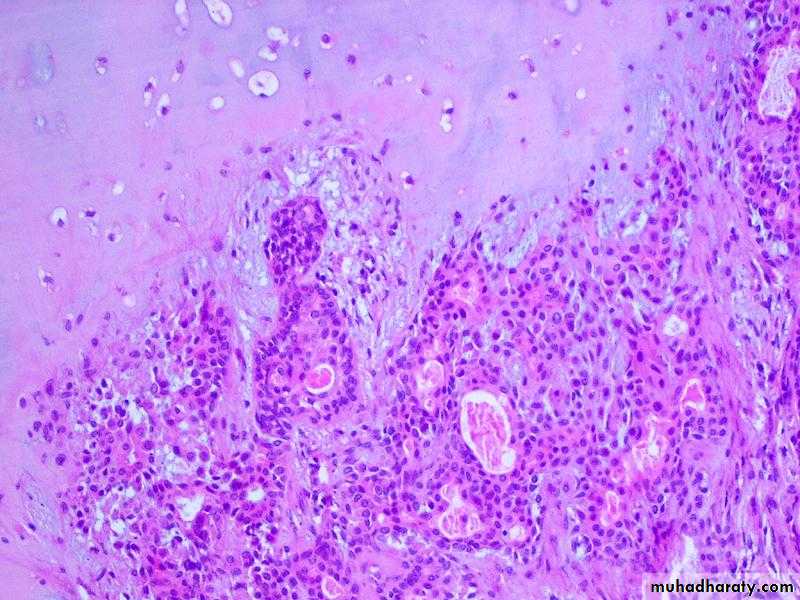
Oral squamous cell carcinoma. (A) Gross appearance demonstrating ulceration and induration of the oral mucosa. (B) Histologic appearance demonstrating numerous nests and islands of malignant keratinocytes invading the underlying connective tissue stroma.
Salivary Gland
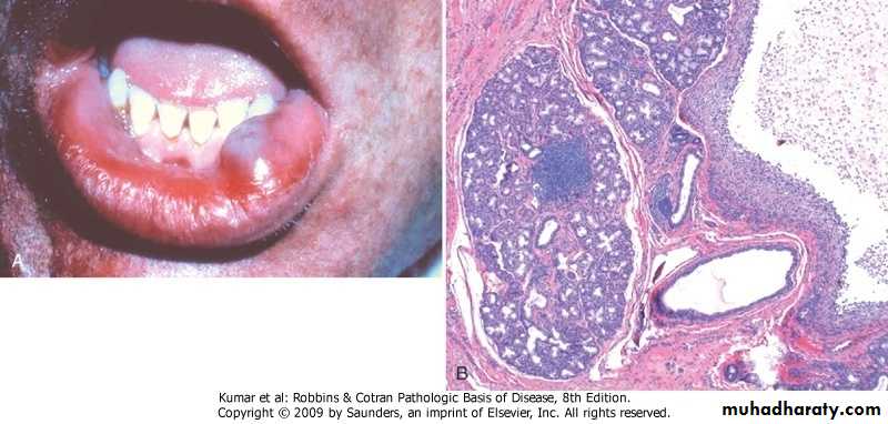
Xerostomia:.means dry mouth due to: -Sjögren syndrome -Radiation therapy -Drugs, (anticholinergics, diuretics, anti- depressants, antihistaminics, anti- hypertensives, sedatives, analgesics).Complications: -dental caries -candidiasis- difficulty in swallowing and speaking.Mucocele: - is the most common inflammatory lesion of the salivary glands and results from (either blockage or rupture) of a salivary gland duct, with consequent leakage of saliva into the surrounding connective tissue stroma.- Mucocele occurs most often in toddlers, young adults, and older adults, and typically manifests as a fluctuant swelling of the lower lip that may change in size, particularly in association with meal Ranula:.mucocele due to rupture of sublingual gland duct.
lower lip cyst lined by mucocele granulation tissue
Sialolithiasis:
.due to obstruction of salivary gland orifice by (impacted food, mucosal edema around the orifice)Sialadenitis:
. Inflammation of the salivary glands.
. May be induced by:
- stones, trauma, viral or bacterial infection, autoimmune disease.
- The most common form of viral sialadenitis is mumps, which may produce enlargement of all salivary glands but predominantly involves the parotids.
- Mumps produces interstitial inflammation marked by a mononuclear inflammatory infiltrate.
- While mumps in children is most often a self-limited benign condition, in adults it can cause pancreatitis or orchitis; the latter sometimes causes sterility.
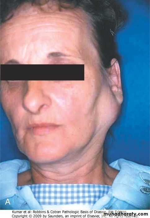
Mumps, there is a big swelling in the left cheek which located
anterior to the ear and extend to cover the angle of the jaw
Neoplasms:
.form 2% of all human tumors, usually affect adults with slight female predominance.age incidence:
-benign tumors (5th-7th decades)
-malignant tumors (somewhat later)
.the likelihood of a salivary gland tumor being malignant is inversely proportional to the size of the gland
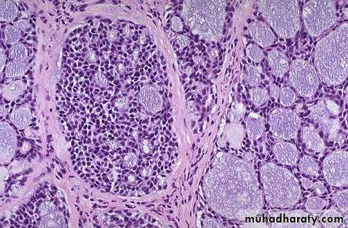
Pleomorphic Adenoma (Mixed Tumor): .the commonest salivary gland tumor, most commonly in the parotid..Pleomorphic adenomas are benign tumors that consist of a mixture of ductal (epithelial) and myoepithelial cells, so they exhibit both epithelial and mesenchymal differentiation. .risk factor is radiation exposure .presents as painless, slow-growing, mobile mass.
.gross, up to 6cm, well-defined, with grayish/whitish cut surface .histology, epithelial and mesenchymal components.treatment, excision with wide margin.frequencies of carcinoma is 2% and 10% for tumors present for less than 5 years and 15 years respectively
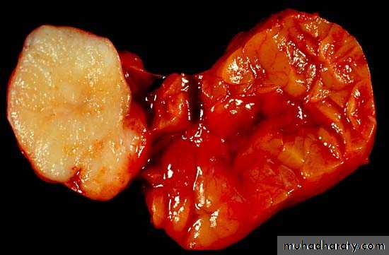
Parotid gland, pleomorphic adenoma
Pleomorphic adenoma –the tumor at the left side is white gray firm lobulated mass without hemorrhage or necrosis. note the normal lobulated gland at the right
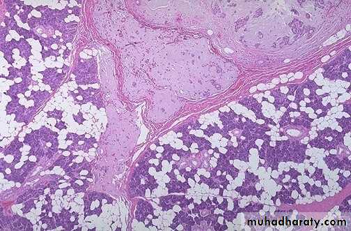
Pleomorphic adenoma –note the myxoid stroma with proliferated ducts & glands & scattered myoepithelial cells
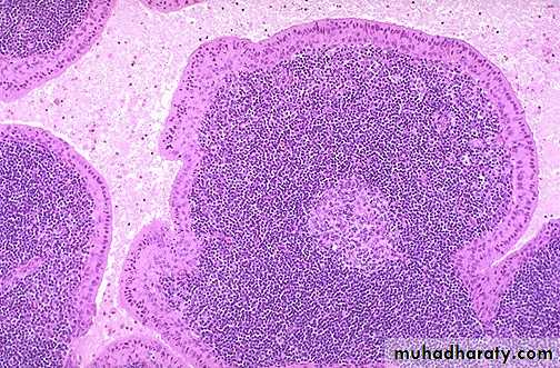
Benign papillary cystadenoma lymphomatosum (Warthin’s Tumor): .second most common salivary gland tumor, restricted to parotid.more common in males.may be bilateral or multifocal.more common in smokers .gross, up to 5cm, encapsulated.microscopy, clefts lined by double layer, resting on dense lymphoid stroma
Benign papillary cystadenoma lymphomatosum, (Warthin's tumor) cystic spaces filled with serous fluid with papillary projections which are covered by double layers of oncocytic epithelium overlying lymphoid stroma
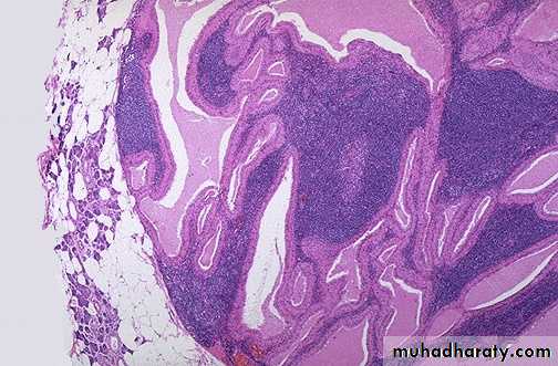
Warthin's tumor, there are cystic to cleft-like spaces filled with pale pink mucinous to serous secretions. The spaces are lined by a double layer of pink (oncocytic) cuboidal to columnar epithelial cells over papillary fronds. The fronds beneath the epithelium are filled with lymphocytes, sometimes with germinal centers.
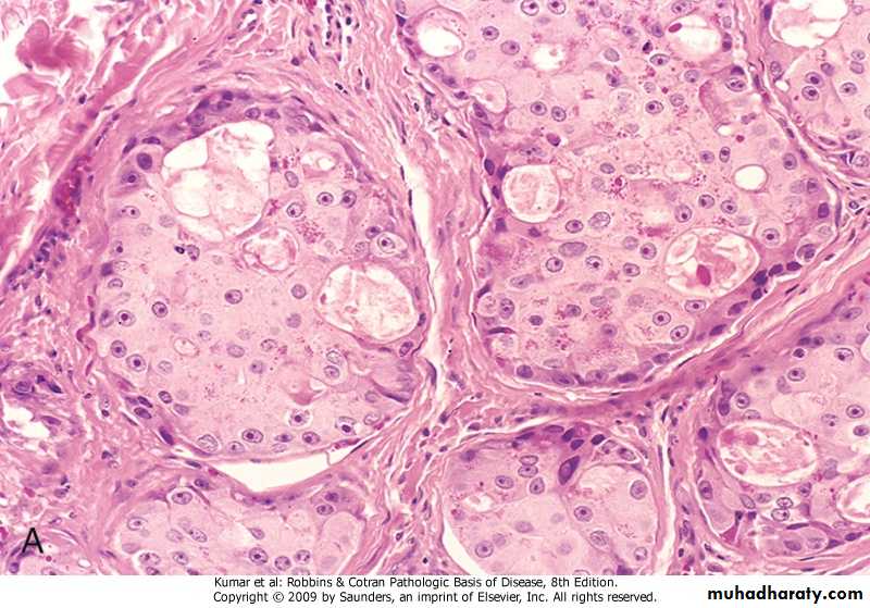
Mucoepidermoid Carcinoma:.Is the most common malignant neoplasm of salivary glands, especially minor ones. .gross, up to 8cm, infiltrative margins.histology, mixture of squamous, mucus-secreting, and intermediate cells.prognosis depends on grade of tumor
Salivary gland, mucoepidermoid carcinoma
Adenoid Cystic carcinoma:.relatively uncommon, slow growing, unpredictable, and recurrent neoplasm.those involving minor salivary glands . have worse prognosis .has infiltrative margins, consists of small dark cells with cribriform pattern, spaces are filled with hyaline material
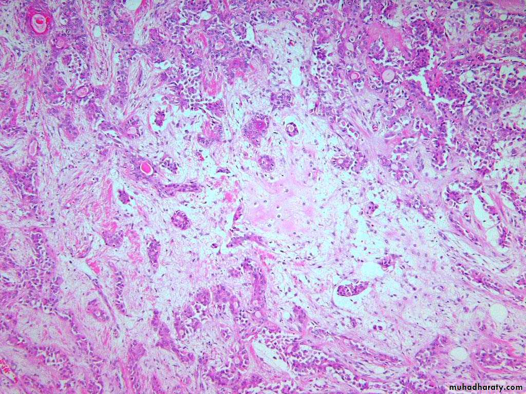
Adenoid cystic carcinoma of salivary gland, solid growth of small hyperchromatic neoplastic cells surrounding small (microcystic) spaces filled with mucinous secretions giving rise to cribriform appearance
Thank you