
Mucormycosis
(Zygomycosis)
Mucormycosis refers to infection caused by diverse fungal species, including
Rhizopus, Rhizomucor, and Mucor. Symptoms most frequently result from invasive
necrotic lesions in the nose and palate, causing pain, fever, orbital cellulitis, proptosis,
and purulent nasal discharge. CNS symptoms may follow. Pulmonary symptoms are
severe and include productive cough, high fever, and dyspnea. Disseminated infection
may occur in severely immunocompromised patients. Diagnosis is primarily clinical,
requires a high index of suspicion, and is confirmed by histopathology and culture.
Treatment is with IV amphotericin B and surgery to remove necrotic tissue. Even with
aggressive treatment, mortality rates are high.
Infection is most common among immunocompromised people, in patients with
poorly controlled diabetes (particularly those with ketoacidosis), and in patients
receiving the iron-chelating drug deferoxamine.
The most common form of mucormycosis is
Rhinocerebral
However, primary cutaneous, pulmonary, or GI lesions sometimes develop, and
hematogenous dissemination to other sites can occur. Cutaneous Rhizopus infections
have developed under occlusive dressings but more often result from trauma when the
injured areas are contaminated with soil.
Symptoms and Signs
Rhinocerebral infections are usually severe and frequently fatal unless diagnosed
early and treated aggressively.
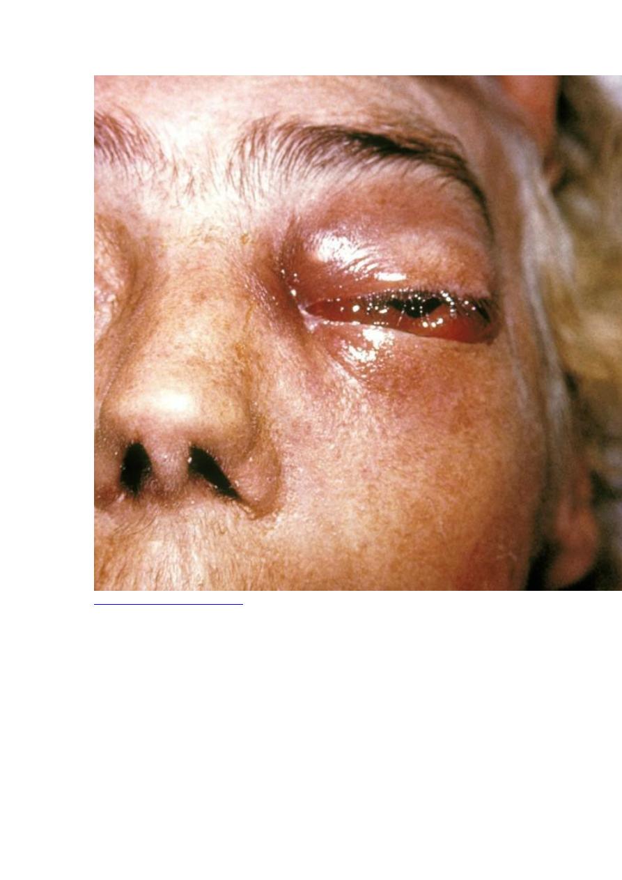
Necrotic lesions appear on the nasal mucosa or sometimes the palate. Vascular
invasion by hyphae leads to progressive tissue necrosis that may involve the nasal
septum, palate, and bones surrounding the orbit or sinuses. Manifestations may
include pain, fever, orbital cellulitis, proptosis, purulent nasal discharge, and mucosal
necrosis.
Progressive extension of necrosis to the brain can cause signs of cavernous sinus
thrombosis, seizures, aphasia, or hemiplegia.
Pulmonary infections resemble invasive aspergillosis. Pulmonary symptoms (eg,
productive cough, high fever, dyspnea) are severe.
Diagnosis
Examination of tissue samples for broad, ribbon-like, nonseptate hyphae
Culture
Diagnosis requires a high index of suspicion and painstaking examination of tissue
samples for large nonseptate hyphae with irregular diameters and right-angle
branching patterns; the examination must be thorough because much of the necrotic
debris contains no organisms. For unclear reasons, cultures may be negative, even
when hyphae are clearly visible in tissues.
CT and x-rays often underestimate or miss significant bone destruction.

Treatment
Control of underlying condition
Lipid amphotericin B formulations
Surgical debridement
Effective therapy requires that diabetes be controlled or, if at all possible,
immunosuppression be reversed or deferoxamine be stopped.
A high-dose lipid amphotericin B formulation (7.5 to 10 mg/kg IV once/day) is
recommended as initial therapy, although recent evidence suggests that posaconazole
may be effective, especially as consolidation therapy. Posaconazole has not been
studied as primary therapy.
Complete surgical debridement of necrotic tissue is critical.