Pedodontics
Lec.14 Dr. Sara Medhat Al-DabbaghManagement of root fractures
It is defined as fractures involving dentin, cementum and pulp, they are relatively uncommon. The mechanism of root fractures is usually a frontal impact, which creates compression zones labially and lingually. The resulting shearing stress zone then dictates the plane of fracture.
Notes:
1) Root fracture of primary teeth is relatively uncommon because the more pliable alveolar bone allows for displacement of the tooth. When root fracture does occur, it should be treated in the same manner as recommended for permanent teeth; however, the prognosis is less favorable.
2) The pulp in a permanent tooth with a fractured root has a better chance to recover, since the fracture allows immediate decompression and circulation is more likely to be maintained.
3) The prognosis is poor for any tooth with a fracture that extends below the gingival margin and involves the pulp in an immature tooth.
4) Root fractures occur in the apical half of the tooth are more likely to undergo repair. Fractures in the apical third are often repaired without treatment. In fact, many apparently are undetected until evidence of a calcified repair is seen radiographically sometime after the injury.
Clinical features
Root fractures involving the permanent dentition predominately affect the maxillary central incisor region.
Coronal fragments are displaced lingually or slightly extruded
Temporary loss of sensitivity
Radiographical features
Radiographic demonstration of root fractures is facilitated by the fact that the fracture line is most often oblique and at an optimal angle for radiographic disclosure. In this context it should be remembered that a root fracture would normally be visible only if the central beam is dictated within a maximum range of 15 to 20 of fracture line.Classification of root fractures:
Based on direction of fracture line with long axis of tooth
Horizontal transverse root fracture, intralveolar root fracture: fracture prepindicular to long axis of tooth.
Oblique: fracture is at an angle to long axis
Vertical: fracture parallel to long axis
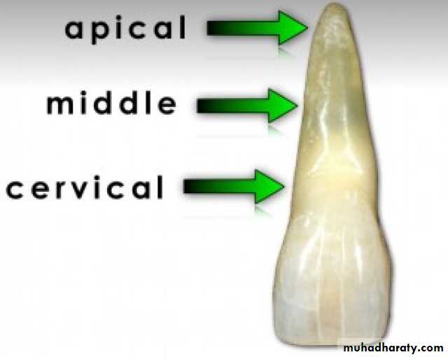
Based on location:
Cervical third
Middle third
Apical third
The position of the fracture line is an important factor in determining the treatment outcome.
Fractures, which occur in the apical third of the root, have an excellent prognosis if the coronal and apical segments can be maintained in close proximity. A tooth with a middle third fracture has fair prognosis for repair and requires endodontic therapy. If the root fracture is in the coronal third, approximation and stabilization of fractured segments is almost impossible. The tooth has to be extracted.
Tissue reactions after root fracture:
Four types of tissue reactions had been described after root fracture:Healing with calcified tissue, which is characterized by a uniting callus of hard tiisue that may consist of dentin, osteodentin, or cementum
Healing with interposition of connective tissue, in which the fractured root surfaces are covered by cementum with connective tissue fibers joining the two fragments.
Healing with interposition of bone and connective tissue, in which a bony bridge and connective tissue are positioned between the fragments.
Interposition of granulation tissue. It is the least favorable form of attempted repair, and the fracture will not heal spontaneously. The teeth usually present unfavorable symptoms that may be accompanied by fistulas resulting from necrosis of the coronal portion and also sometimes the apical portion of the pulp. These teeth require follow-up endodontic treatment or extraction. Gross separation of the root fragments invariably causes inflammation in the area and subsequent resorption of the approximating fractured surfaces. For repair to take place, the fragments must be maintained in apposition. Therefore splinting is usually necessary, particularly if the coronal fragment is mobile.
Treatment
The principle of treatment of permanent teeth is reduction of displaced coronal fragments and firm immobilization.Immobilization of teeth with root fractures is achieved with rigid fixation with an acid etch splint.
Following treatment modalities are recommended based on fracture line.
Coronal 3rd fractures of the root (cervical fracture):
If the remaining root is long enough, coronal portion can be removed, endodontic treatment completed on apical fragment and restored with post and core. Then cement it and take impression overall to make acrylic crown.If the remaining root is short do extraction.
Fracture of the middle third
If there is slight mobility, is treated by performing root canal treatment involving both the coronal and apical fragments.
If there is high mobility then the tooth should be extracted.
Fracture of apical third
High apical fractures require no treatment.
If there is vertical root fracture
It is also called as cracked tooth syndrome.It runs lengthwise from crown towards the apex.
It is mostly found in posterior teeth and its etiology is mostly iatrogenic like insertion of screws, after pulp therapy or due to traumatic occlusion.
Clinical features
Persistent dull pain
Pain is elicited by applying pressure
Radiographical feature
If the central line beam is the line of fracture it is visible as radiolucent line
Thickening of the PDL
Treatment
Single rooted tooth do extraction
Multi rooted tooth hemi section and the remaining tooth is endodontically treated and crown
Other displacement injuries of teeth requiring stabilization
Teeth subjected to less severe luxation injuries may also benefit from stabilization with a bonded resin and wire splint during the recovery period. The severity of the injury will help determine the length of time the splint should remain in place. Splinting times may vary from:
1 to 2 weeks, for teeth that have been discernibly loosened (subluxation),
4 to 6 weeks, for teeth that have been laterally displaced, fracturing the alveolar process.
As with all tooth injuries, frequent periodic evaluation is required for at least the first 6 months to afford the dentist the opportunity for early intervention if adverse sequelae develop; after this, evaluation at regular recall appointments should continue. Displaced teeth with closed apices and many with open apices will require follow up endodontic therapy. As with many of the other injuries, calcium hydroxide paste is the currently recommended material for initial canal filling, and the canal should be recleaned and refilled with calcium hydroxide periodically if signs or symptoms warrant retreatment. Placement of a permanent gutta-percha filling should be delayed for at least 1 year (arbitrarily determined), and the calcium hydroxide should be replaced at least once (again, arbitrarily determined) during this time. If the injured tooth had an open apex when endodontic therapy was initiated, the calcium hydroxide filling material should be used until the apexification process is complete or at least 1 year has elapsed, whichever is longer.
Types of fracture:
By Ellis and Davey (1960):Class I: Enamel fracture;
Class II: Enamel and dentin fracture;
Class III: Enamel and dentin fracture exposing dental pulp;
Class IV: The traumatized tooth that becomes non vital;
Class V: Avulsion;
Class VI: Fracture of the root;
Class VII: Displacement of tooth;
Class VIII: Fracture of crown en masse;
Class IX: Traumatic injuries of primary teeth.