1
THE IMMUNE SYSTEMLec. 2
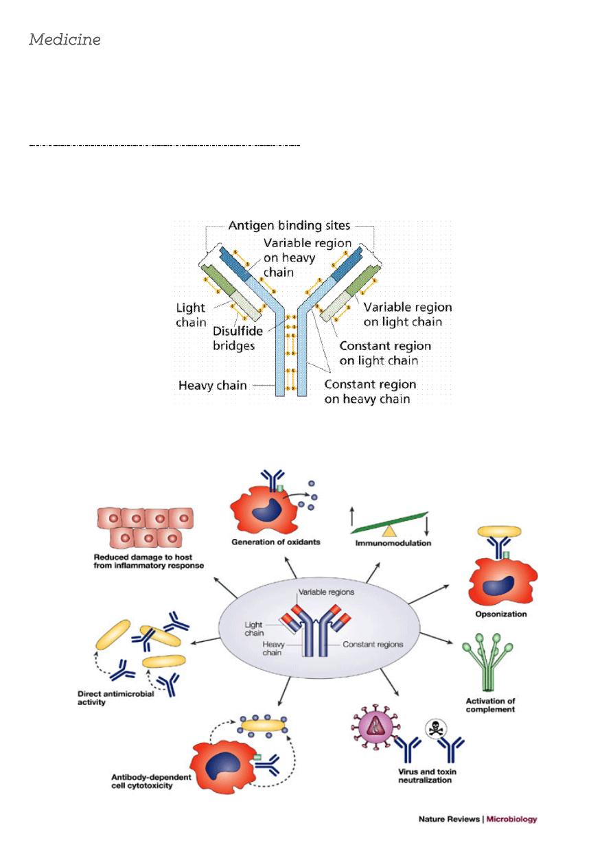
Antibody molecules (immunoglobulins)
Antibodies are glycoproteins. They consist of two heavy chains and two light chains (either κ
or λ polypeptides) . The heavy chain determines the antibody isotype or class, i.e. IgG, A, M, D
or E.
The major functions of antibody are:
2
Immune regulation:
Ab acting as the antigen receptor on B cells and presenting the antigen to helper T cells,
plays
a part in antigen presentation.
Antigen:
is any substance capable of generating an immune respons, Ag react with T.cells and B.cells to
induce the formation of Ab, then Ag react with these Ab and cells.
In primary immune respons when Ag first introduce in to body there is lag phase of several
days during which no Ab .detected ,then several day later(5-10 days)IgM Ab appear.
IN secondary immune respons A group of B.cell called memory cell ,enhance immune
response to previously encountered Ag (the lag phase IS decrease) , the first antibody to be
produced is IgM, which appears in the serum after 5-10 days., other antibody classes (IgG, IgA
and IgE) are produced 3-7 days later. If, some time later, a memory B cell is re-exposed to
antigen, the lag time between antigen exposure and the production of antibody is decreased (to
2-3 days
HUMAN LEUCOCYTE ANTIGENS (HLA)
Antigen presentation
. The immune system has the ability to recognize between 'self' and 'non-self' antigens. This
process is facilitated by a
recognition system
called the major histocompatibility complex
(MHC) which dictates the way that antigen is recognized as foreign. In man, the products of
MHC are termed human leucocyte antigens (HLA).
The HLA system :
is cluster of genes is located on the short arm of chromosome 6. The system comprises six
genetic loci - HLA-A, -B, -C, -D, -DR and –DQ.The gene encodes
3
The HLA molecules
(cell surface antigen presenting proteins)which are distributed throughout the body tissues
and it is through differences in HLA that cells are classified as self or non-self. The possibility of
two different individuals having the same combination of HLA molecules is very remote.. The
HLA genes code for cell-surface glycoproteins that extend from the plasma membrane to the
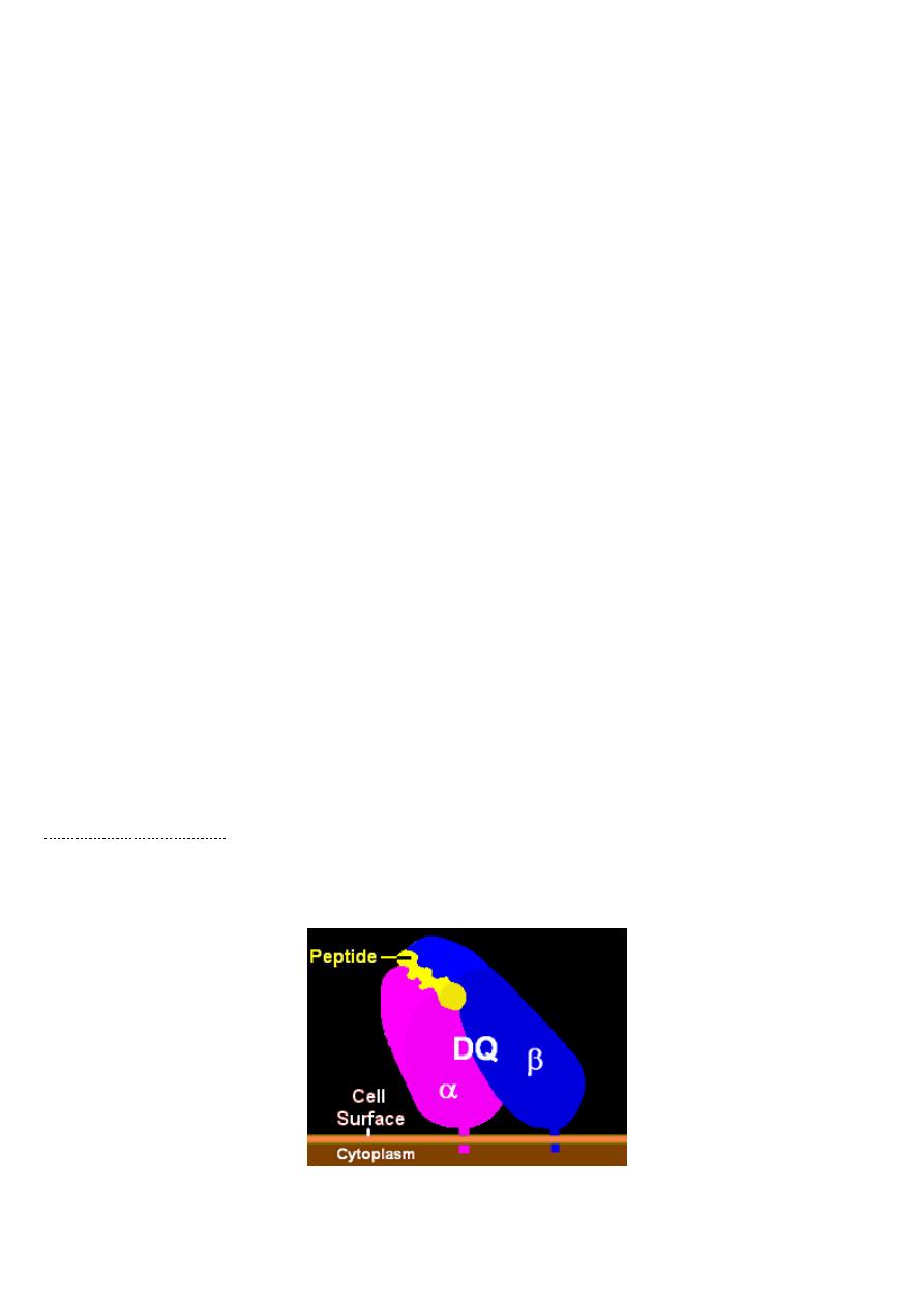
cytoplasm and are known as class I and class II molecules. These glycoproteins consist of two
chains of unequal size (α and β chains). The chains form a groove in which an antigenic
peptide sits ready for presentation to T cells.
Class I (HLA-A, -B and -C) are expressed on all cell types except erythrocytes and
trophoblasts.. Class I molecules interact with CD8 T cells during antigen presentation and
therefore are involved in driving mainly cytotoxic reactions.
Class II (HLA-D and -DR, D-related) are expressed only on professional antigen-presenting
cells (B cells, monocytes/macrophages, Langerhans' cells, dendritic cells) and activated T cells.
Class II antigens link with CD4 molecules during antigen presentation and the reaction induced
by cells bearing this molecule is therefore of the helper type.
T cells respond to protein antigens, but they cannot recognise these in their native form.
Instead, intact protein must be processed into component peptides which can bind to the cell
surface HLA . This process is known as antigen processing and presentation, and it is
the
peptide/HLA complex
which is recognised by individual T cells.
T lymphocytes are classified into
1. Helper/inducer cells
Bear CD4 cell surface molecule ( cytokine-secreting cells), making up about 75% of peripheral
blood T cells) and the ability to recognize antigen only when the Ag expressed with HLA class II
on antigen-presenting cells.
2. Cytotoxic/suppressor cells
Bear CD8cell surface molecule (mainly cytotoxic suppressor cells), which account for the
remainder.able to recognize antigen only when presented with HLA class I molecules.These
cell types are indistinguishable morphologically, but can be separated by the presence of cell-
surface moleculesCD(specific target molecule on a cell that is recognized by one or more
antibodies).

4
Components of the immune response.
Antigen is presented to T-helper cells (Th cells) by an antigen-presenting cell. Th cells secrete
lymphokines(cytokine), which
1. activate cytotoxic T cells (Tc cells) that are involved in antiviral and anti-tumour activity.
2. activate NK cells and macrophage, which are involved inantitumor activity.
3. induction of antibody responses by B cells
Helper T cells are unable to destroy pathogens or cells directly, but through cytokine
production are able to activate macrophages to kill organisms within them and further
activate cytotoxic T cells and NK cells.
Some diseases show a close association with HLA type.
HLA - associations with disease
Sara Abdulbasit
A1, B8, DR3
Polymyositis and dermatomyositis
A3, B14
Hereditary haemochromatosis
A28
Schizophrenia
B5
Behçet's syndrome
Polycystic kidney disease
Ulcerative colitis
B8, DR3, DR7, DQ2
Coeliac disease
B18
Hodgkin's disease
B27
Acute anterior uveitis
Ankylosing spondylitis
Psoriatic arthropathy
Reiter's syndrome
Juvenile arthritis