Lab -11-Intestinal Nematodes
Enterobius vermicularis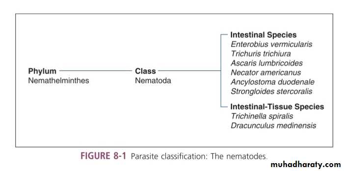
Introduction
Enterobius vermicularis, commonly known as pinworm or seatworm.Disease: enterobiasis,pinworm infection.
Humans are the only host.
Adults worms reside in the colon.
World-wide distribution,particulary in temperate area, it is commonly found in kindergarten and primary school students.
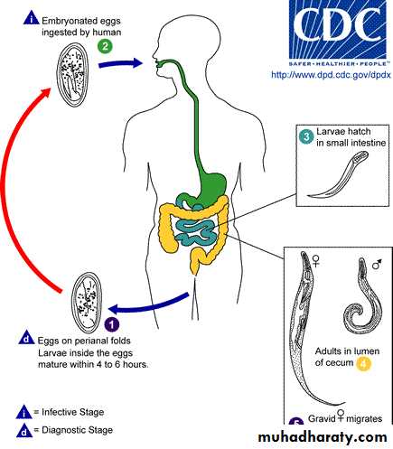
The infective stage is the egg.
Morphology
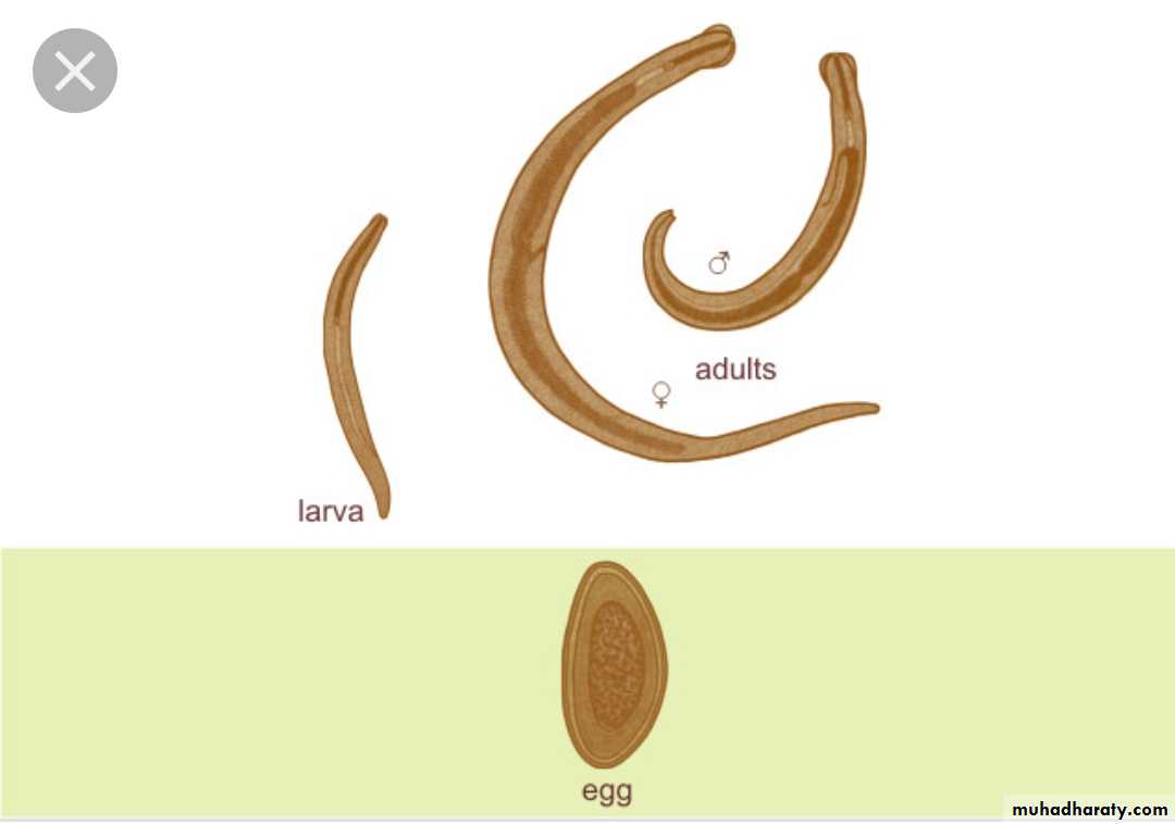
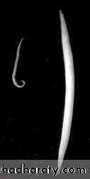
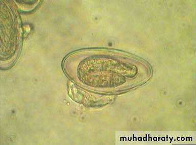
Morphology -- Adult
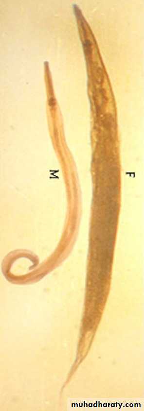
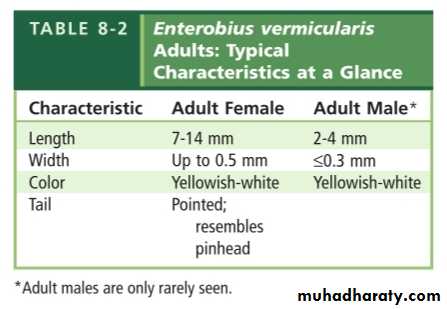
Female -- fusiform body with a long, thin, tapering tail,7-14mmMale -- “6” shape, curved tail, 2-4. Males die right after mating, thus are rarely seen
White in color
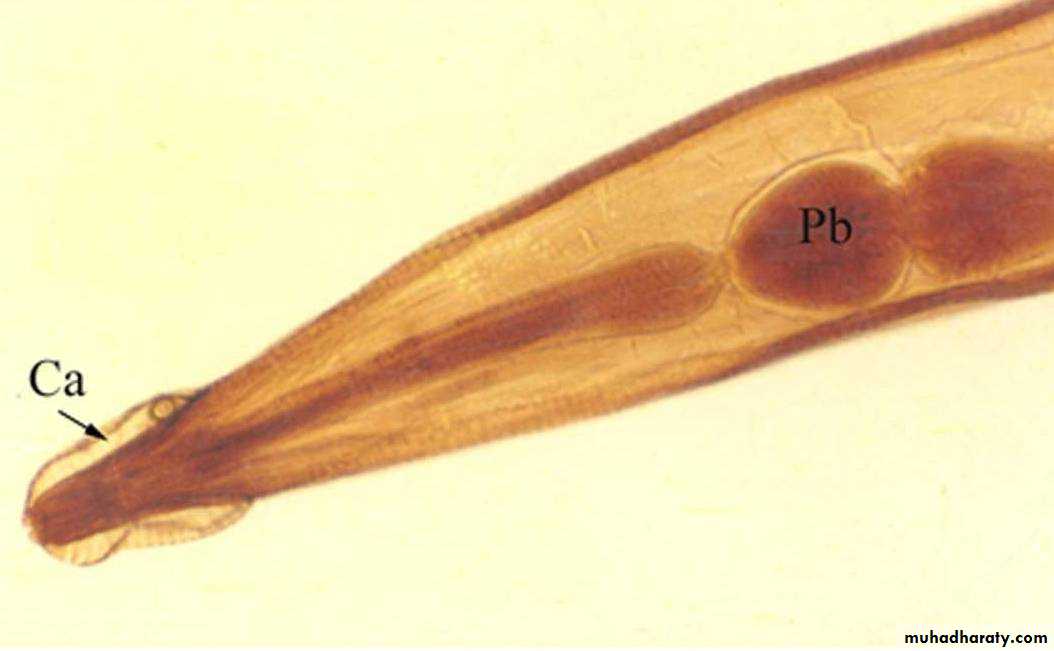
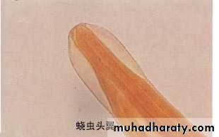
• Cephalic alae
The anterior end tapers and is flanked on each side by cuticular extensions of headPharyngeal bulb
The esophagus is slender, terminating in a prominent posterior bulb
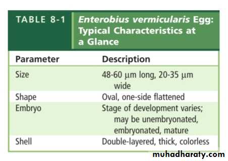
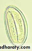
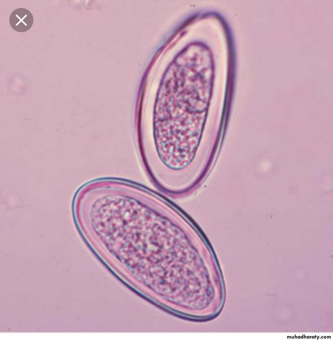
Morphology -- Egg
Life cycle

Life cycle note
Ingestion of the infective eggs.The eggs migrate through the digestive tract into the small intestine,where they hatch and release young larvae.
The larvae grow and mature into adult worms.
The adult worms reside in the colon.
Following copulation,the resulting pregnant(gravid) femal worm migrates outside the body to the perianal region where she may deposit up to 15,000 eggs.
Characteristics of life cycle
Humans are the only host in natureNo intermediate host (direct life cycle)
No larval migration between organs
Transmission
Ingestion of infective eggs.Retroinfection.
Autoreinfection(hand to mouth contamination)
Clinical Symptoms
Asymptomatic .The most common symptoms include intense itching and inflammation of the anal and vaginal areas.
Laboratory Diagnosis
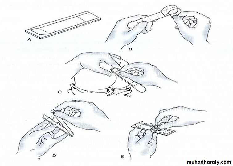
Microscopic identification of eggs collected in the perianal area by cellophane (Graham Scotch) tape method or anal swabs. This must be done in the morning, before defecation and washing