Lab 5
Cryptosporidium parvum
common associated disease and condition names: cryptosporidiosis.
World wide distribution.Infect human and mammles.
Infection occur by water and food contaminated with infected feces with oocyst.
Immunocompromized persons (AIDS) are at risk of contracting this parasite.
Immunocompetent children in tropical areas also at risk.
Morphology
The parasite shows six distinct morphological forms during its life cycle:• oocyst
• sporozoite
• trophozoite
• meront
• microgamont
• macrogamont
Life cycle
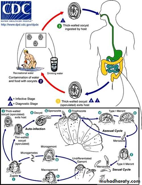
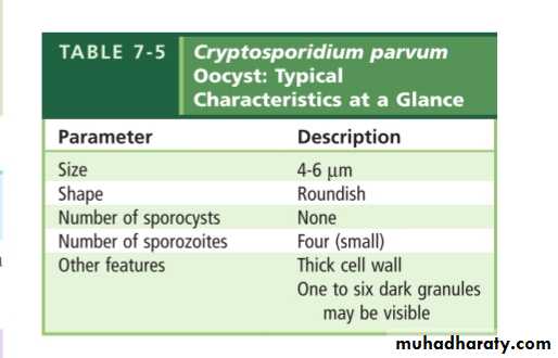
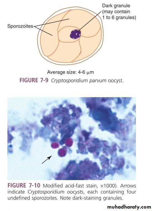
The infective stage is oocyst containing sporozoites.
The diagnostics stage is oocyst containing 4 sporozoites.Thin shelled oocyst is responsible for autoreinfection.
Thick shelled oocyst usually remains intact and is passed out of the body.
Habitat is the small intestine(brush border of epithelial cell).
Clinical symptoms
Cryptosporidiosis:healthy persons infected with cryptosporidium typically complain of diarrhea,which is self limiting and lasts approximately 2 weeks.Immunocompromized individuals(AIDS) usually suffer from sever diarrhea.
Malabsorption.
Infection may migrate to the other body areas,such as stomach and respiratory tract.
Lab diagnosis
• Stool examination (iodine or modified acid-fast stain).
• Enterotest.
• ELISA.
• Indirect immunofluorescence.
• Intestinal biopsy(merozoites and gametocytes).
• PCR.
Prevention and control
Proper treatment of water supplies.Proper hand washing.
Handling known infected material by using gloves.
Properly disinfecting equipment potentially with 5% to 10% amonia.