• 23-12-2018
Medical Parasitology3rd class
Intestinal NematodesASCARIASIS
• Dr. Abdul Rahman Dahham, PhD
• Department of Microbiology
• College of Medicine
• Nineveh University
1
ASCARIASIS
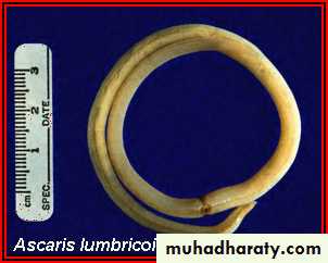
Ascaris Lumbricoides is the largest roundworm of human, belonging to the phylum Nematoda. It is responsible for the disease ascariasis in human. One sixth of the human population is estimated to be infected by this parasite.Ascariasis is prevalent worldwide and more in tropical and subtropical countries. Ascariasis can occur at all ages, but is more prevalent in 5-9 years old group. The incidence is higher in poor rural population.
2
The adult females of this species can measure up to 45 cm long (males are generally shorter. The adult worms live in the small intestine and eggs are passed in the feces. A single female can produce up to 200,000 eggs each day.

Round worms
Taxonomy
Kingdom : AnimaliaPhylum : Nematoda
Class : Rhabditea
Order : Ascaridida
Family : Ascarididae
Genus : Ascaris
Species : lumbricoides
Largest of intestinal nematodes .
Distribution:- world wide, Asia,Iraq.Habitat:- small intestine, mainly jejunum.
larva: in lung due to larval migration stage, sputum sample may be collected on persons suffering from infection with A. lumbricoid.
Common name:- Giant Intestinal round worms.
Disease name:-Ascariaisis
Host:- The human is considered an intermediate and final host.
Introduction
Infective stage: Embryonated eggs.
Three diagnostic stages in stool: adult, Fertilized eggs, & Unfertilized eggs.
Transmission occurs mainly by ingestion of contaminated food or water with eggs. Occasionally by inhalation of contaminated dust with eggs. Children playing in contaminated soil may acquire the parasite from their hands.
Accounts for 60,000 deaths per year, mainly in children and estimated that 1 billion people are infected worldwide. Children seem to be affected more severely than adults
6
Parasite morphology
AdultsAscaris lumbricoides are also called round worms because of their shape.
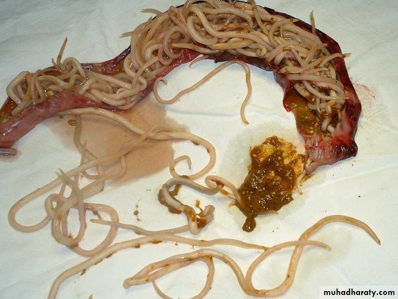
Adult worms live in the lumen of the small intestine.
Adult worms can live 1 to 2 years.
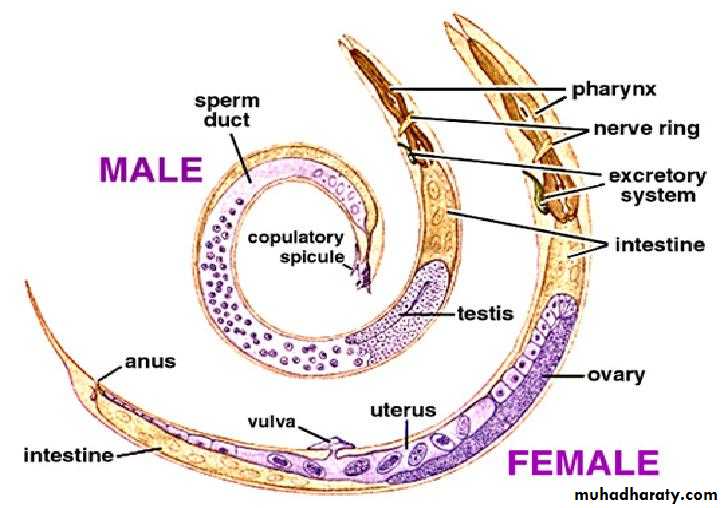
Male: has a coiled posterior end, 15-30 cm in length.
Female: oviparous have straight, pointed tail and, 20-40 cm in length.
7
The mouth at the anterior end of both male and female has three finely denticulate lips, one dorsal and two ventro-lateral.
They are pale pink or flesh colored when freshly passed in stool, but become white outside the body.
Female worms without males produce infertile eggs that are markedly sub-spherical, internally they contain a mass of disorganized granules that completely fill the shell
8
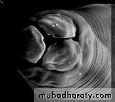
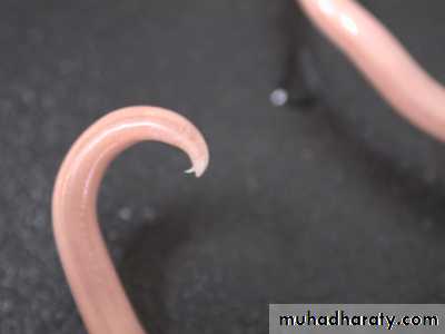
The three lips are seen at the anterior end. The margin of each lip is lined with minute teeth which are not visible at this magnification.
The Eggs
EggsFertilized
Unfertilized
Decorticated
Corticated
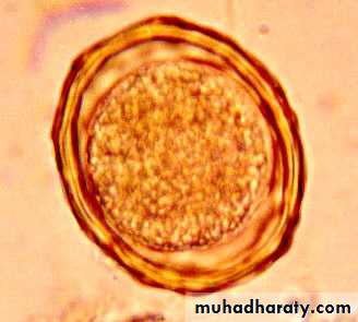
Fertilized egg( corticated egg)
Size:- 45-75 mm by 35-50 mm.
Shape:-Rounded or oval
Shell:-Thick, consisting of 3 layers : (Inner layer ) consist of thin yolk Membrane ,(Mid layer) thick, (Outer layer ) coarse consist of regular Albuminous.
Color:-Golden brown.
A. lumbricoides eggs are extremely resistant to strong chemical, drying, and low temperature. The eggs can remain viable in the soil for several months or even years.
( outer layer) coarse
( regular Albuminous )(Inner layer)
the thin yolk membrane( mid layer) thick
Embryo
Crescent space
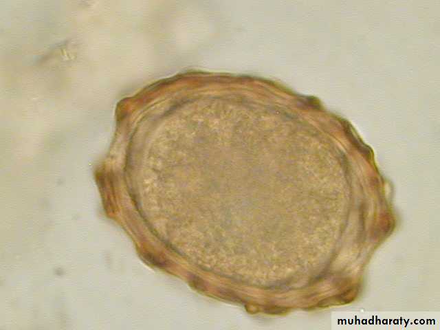
Decorticated Egg
• decorticated fertilized eggs sometimes may lack the outer albuminous layer and are colorless.•
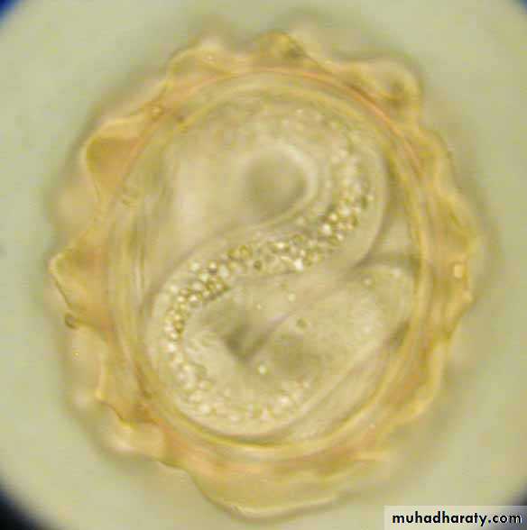
Unfertilized eggs: elongated and larger than fertile egg
Size:- 85-95 mm by 43-47 mm.Shape: Elongated oval
• Shell: Unfertilized eggs has a thin shell protects the inner a morphous mass of protoplasm, Irregular albuminous layer, not has crescent space. The shell layers of the egg provide a very resistant structure which can withstand many chemicals which make them ideal parasites of the intestine.
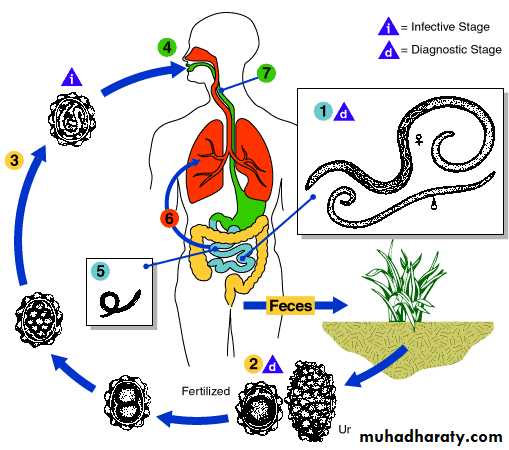
Life cycle
The worm lives in the lumen of small intestine, feeding on the intestinal contents, where the fertilized female lays eggs. An adult female can produce approximately 200,000 eggs per day, which are passed in feces. When passed, the eggs are un-segmented and require outside development of about three weeks until a motile embryo is formed within the egg.
After the ingestion of embryonated eggs in contaminated food or drink or from contaminated fingers, host digestive juices acts on the egg shell and liberate the larva into the small intestine. These larvae penetrate the intestinal mucosa and enter lymphatics and mesenteric vessels.
They are carried by circulation to the liver, right heart and finally to the lungs where they penetrate the capillaries into the alveoli, stay for 10-14days and then they are carried, or migrate, up the bronchioles, bronchi, and trachea to the epiglottis.
When swallowed, the larvae pass down into the small intestine where they develop into adults. The time from the ingestion of embryonated eggs to ovi-position by the females is about 60-75 days. The adult worms live for about one year.
18
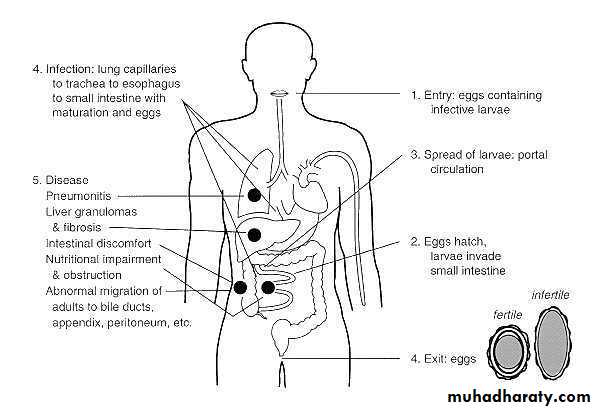
Disease Pathogenesis & symptoms
There are two phase in ascariasis:The blood-lung migration phase of the larvae: During the migration through the lungs, the larvae may cause a pneumonia. The symptoms of the pneumonia are low fever, cough, blood-tinged sputum, asthma. Large numbers of worms may give rise to allergic symptoms. Eosionophilia is generally present. These clinical manifestation is also called Loeffler’s syndrome.
The intestinal phase of the adults:
The presence of a few adult worms in the lumen of the small intestine usually produces no symptoms, but may give rise to unclear abdominal pains or intermittent colic, especially in children.A heavy worm problems can result in malnutrition, more serious manifestations have been observed. Moving adults may block the appendicle lumen or the common bile duct and even perforate the intestinal wall.
Thus complications of ascariasis, such as intestinal obstruction, appendicitis, biliary ascariasis, perforation of the intestine, cholecystitis, pancreatitis and peritonitis, may occur, in which biliary ascariasis is the most common complication.
22
23
Diagnosis
In the larval migration phase of the infection, diagnosis can be made by finding the larvae in sputum or in gastric washings.
During the intestinal phase, the diagnosis can be made by finding the eggs (unfertilized or fertilized) or adult worms in the stool. The eggs are most easily seen on a direct wet smear or a wet preparation of the concentration sediment.
Both fertilized and unfertilized eggs can easily be recovered using the sedimentation concentration. (Unfertilized eggs will not float using zinc sulfate flotation concentration) (eggs too heavy).
Eggs might be difficult to identify on a permanent stained smear due to stain retention and asymmetrical shape. Intestinal disease can often be diagnosed from radiographic studies of the gastrointestinal tract where the worm intestinal tract may be visualized.
24
Treatment
Infections with A. lumbricoides are easily treated with a number of anthelmintic drugs:The drugs of choice for treatment of ascariasis are albendazole, mebendazole, and pyrantel pamoate.
Mebendazole 100 mg PO 12 hourly for 3 days.
Albendazole 400 mg given as single oral dose (contraindicated during pregnancy and children under 2 years).
Pyrantel pamoate 11 mg/kg as a single dose.
Piperazine 75 mg/kg as a single oral dose.
25
PREVENTION
Keeping good sanitation conditions is the only way to prevent the infection of Ascaris.Pollution of soil with human faeces should be avoided.
Vegetable should be thoroughly washed in a mild solution of Pottasium permanganate and properly cooked before use.
Finger nails should be regularly cut to avoid the collection of dirt and eggs below them.
Hands should be properly washed with some antiseptic soap before touching edibles or eating.