Secret Lectures
(9)
/ Diagnostic Imaging / Dr.Riyadh A. Al-Kuzzay (M.B.Ch.B – FICMS-RD)
P a g e
1
Plain films of the Abdomin
What to Examine
•
Gas pattern
•
Extraluminal air
•
Soft tissue
masses
•
Calcifications
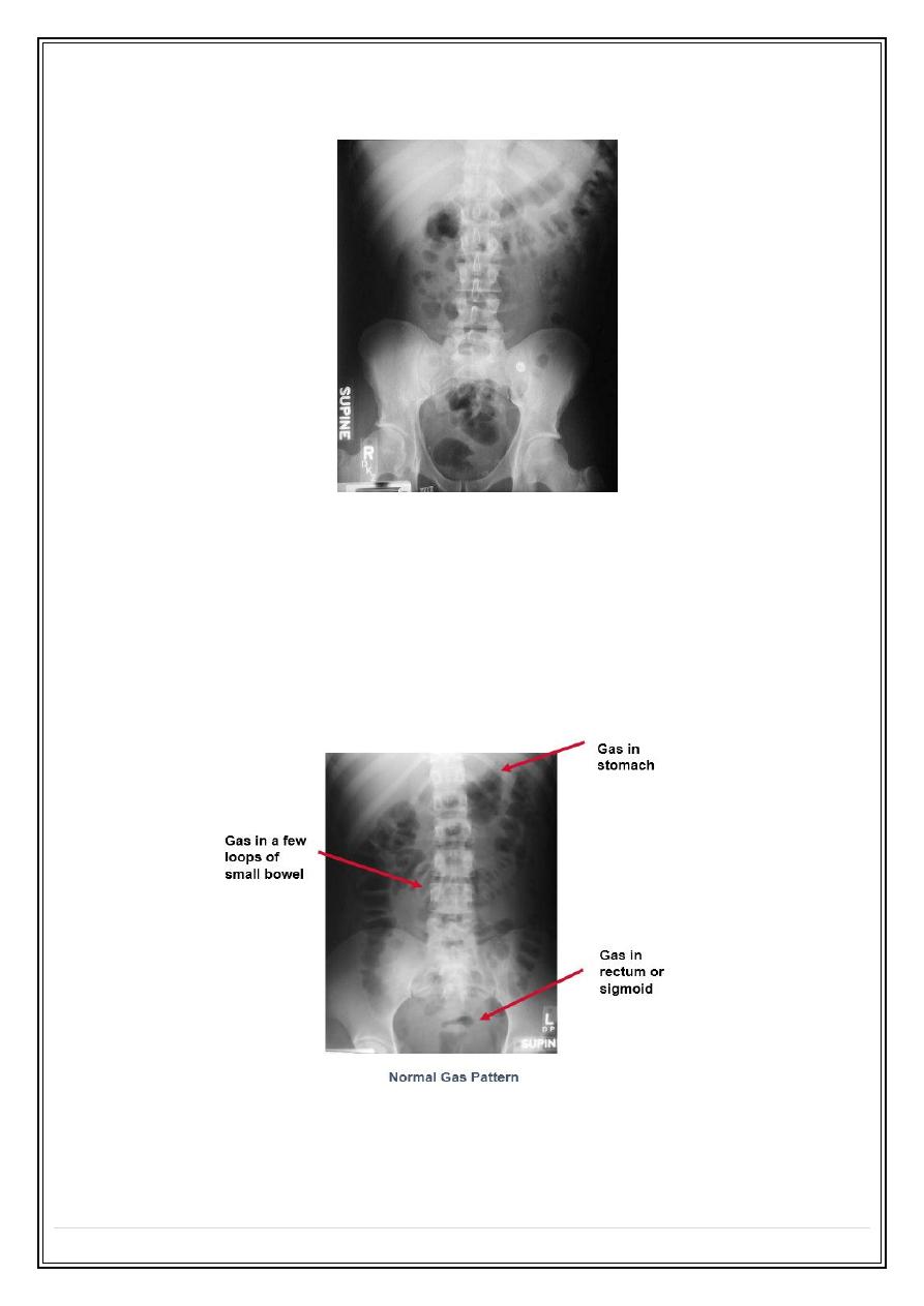
Normal Gas Pattern
•
Stomach
o
Always
•
Small Bowel
o
Two or three loops of non-distended bowel
o
Normal diameter = 2.5 cm
•
Large Bowel
o
In rectum or sigmoid – almost always
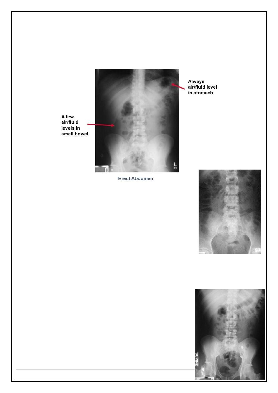
Normal Fluid Levels
•
Stomach
o
Always (except supine film)
Secret Lectures
(9)
/ Diagnostic Imaging / Dr.Riyadh A. Al-Kuzzay (M.B.Ch.B – FICMS-RD)
P a g e
2
•
Small Bowel
o
Two or three levels possible
•
Large Bowel
o
None normally
Large vs. Small Bowel
•
Large Bowel
o
Peripheral
o
Haustral markings don't extend from wall to wall
•
Small Bowel
o
Central
o
Valvulae extend across lumen
o
Maximum diameter of 2"
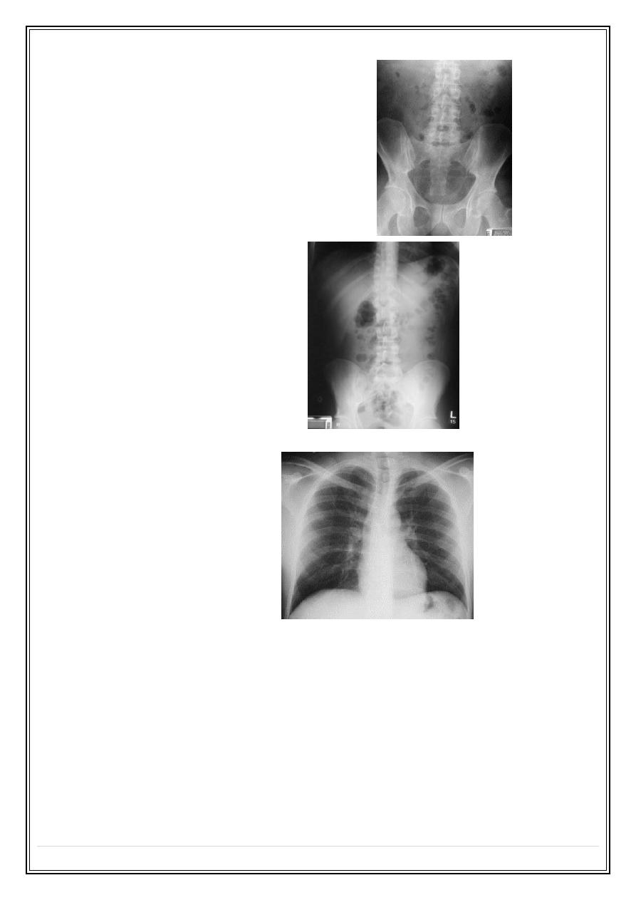
Complete Abdomen Obstruction Series
•
Supine
•
Prone or lateral rectum
•
Erect or left decubitus
•
Chest - erect or supine
Supine
•
Looking for
o
Scout film for gas pattern
o
Calcifications
o
Soft tissue masses
•
Substitute – none
Secret Lectures
(9)
/ Diagnostic Imaging / Dr.Riyadh A. Al-Kuzzay (M.B.Ch.B – FICMS-RD)
P a g e
3
Prone
•
Looking for
o
Gas in rectum/sigmoid
o
Gas in ascending and descending colon
•
Substitute – lateral rectum
Erect
•
Looking for
o
Free air
o
Air-fluid levels
•
Substitute – left lateral decubitus
Erect Chest
•
Looking for
o
Free air
o
Pneumonia at bases
o
Pleural effusions
•
Substitute – supine chest
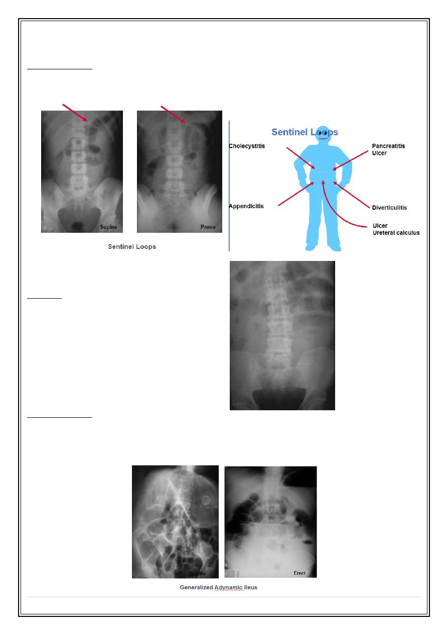
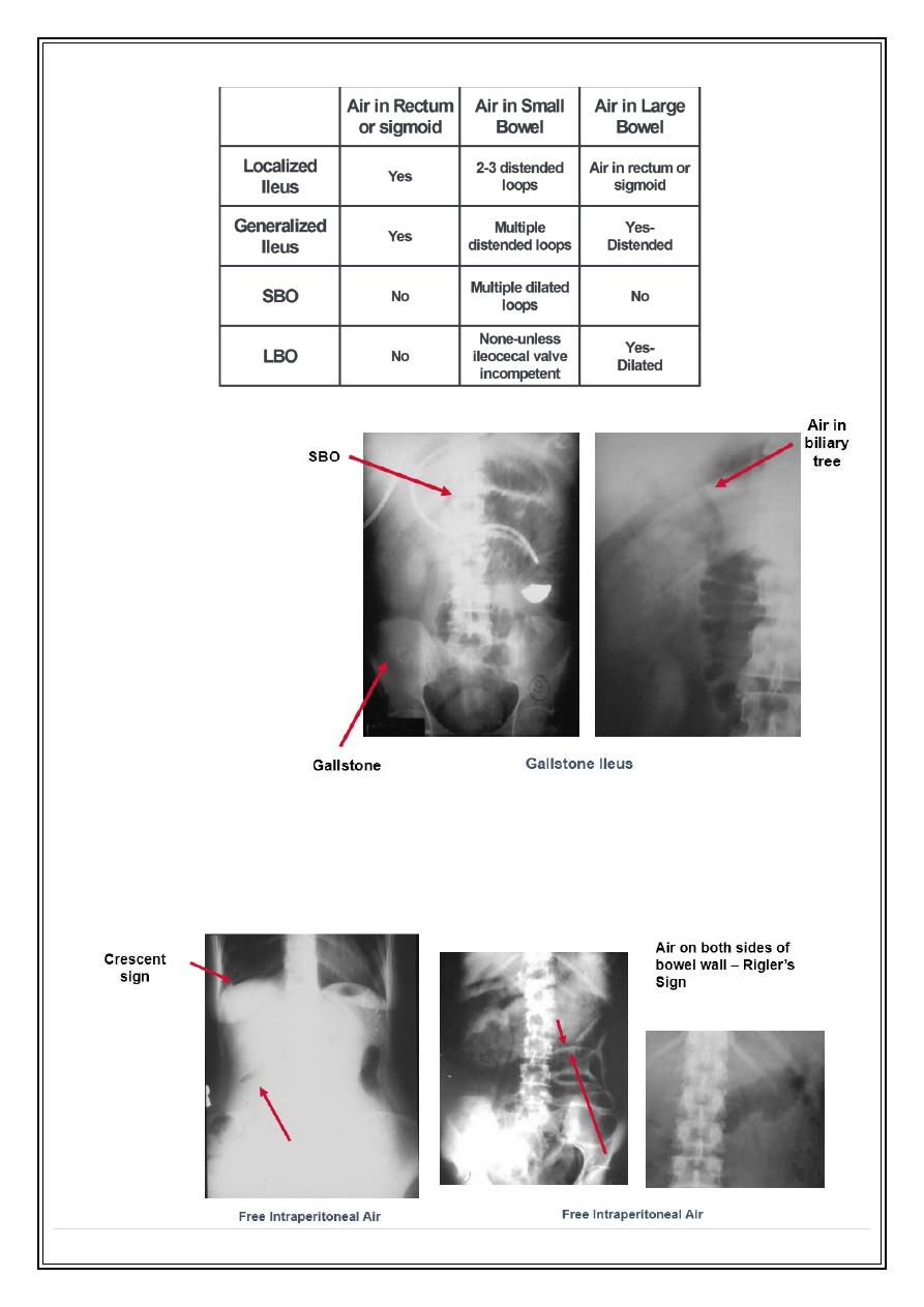
Abnormal Gas Patterns
•
Functional Ileus
o
Localized (Sentinel Loops)
o
Generalized adynamic ileus
•
Mechanical Obstruction
o
SBO
o
LBO
Secret Lectures
(9)
/ Diagnostic Imaging / Dr.Riyadh A. Al-Kuzzay (M.B.Ch.B – FICMS-RD)
P a g e
4
Localized Ileus
Key Features
•
One or two persistently dilated loops of large or small bowel
•
Gas in rectum or sigmoid
Pitfalls
•
May resemble early mechanical SBO
o
Clinical course
o
Get follow-up
Generalized Ileus
Key Features
•
Gas in dilated small bowel and large bowel to rectum
•
Long air-fluid levels
•
Only post-op patients have generalized ileus
Secret Lectures
(9)
/ Diagnostic Imaging / Dr.Riyadh A. Al-Kuzzay (M.B.Ch.B – FICMS-RD)
P a g e
5
Is It An Ileus?
•
Is the patient immediately post-op?
•
Are the bowel sounds absent or hypoactive?
o
If “no,” then it isn’t an ileus
•
Patients don’t present to the ER with a generalized adynamic ileus!
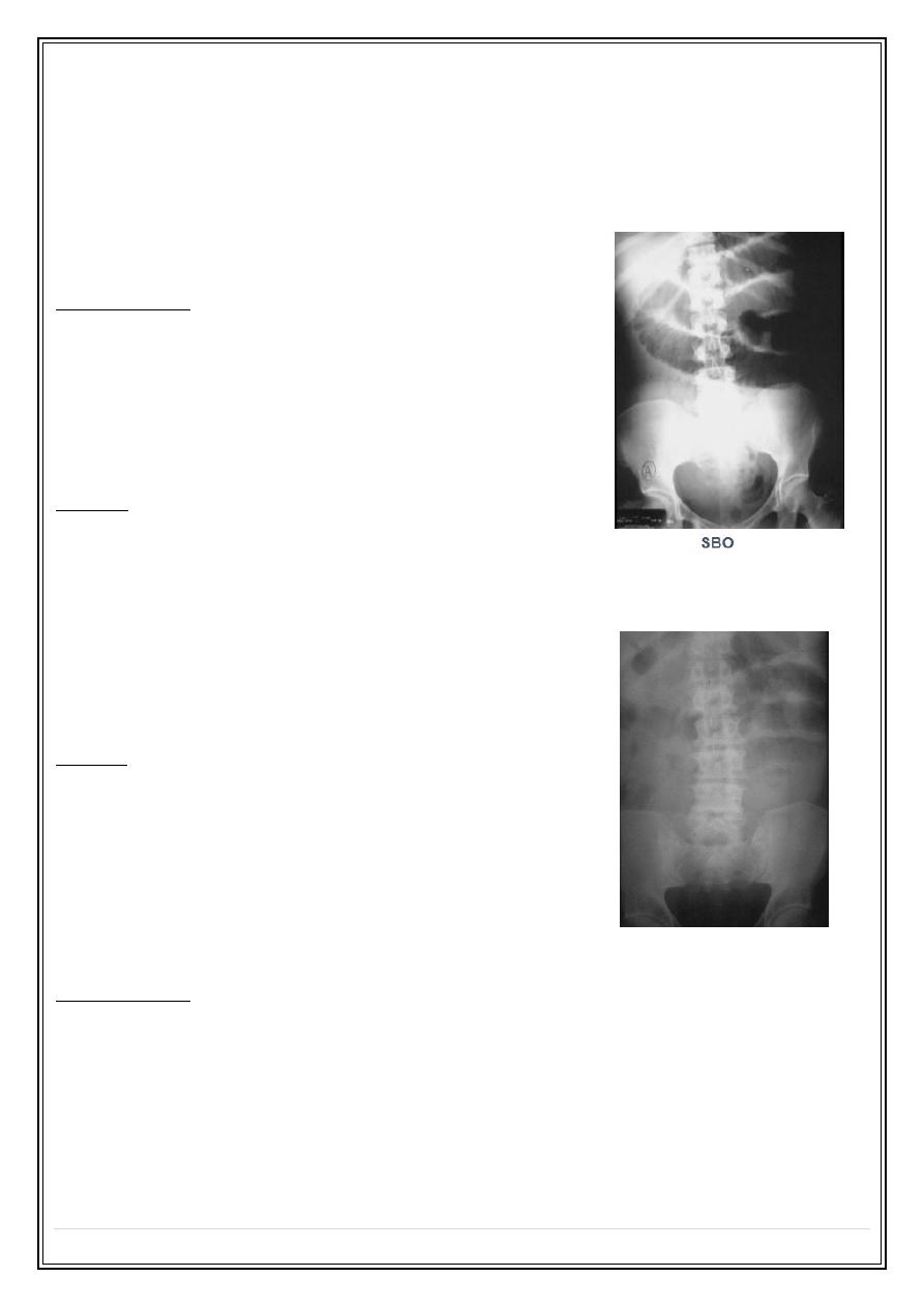
Mechanical SBO
Key Features
l Dilated small bowel
l Fighting loops
l Little gas in colon, especially rectum
l Key: disproportionate dilatation of SB
Causes
•
Adhesions
•
Hernia*
•
Volvulus
•
Gallstone ileus*
•
Intussusception
*Cause may be visible on plain film
Pitfalls
•
Early SBO may resemble localized ileus -get F/O
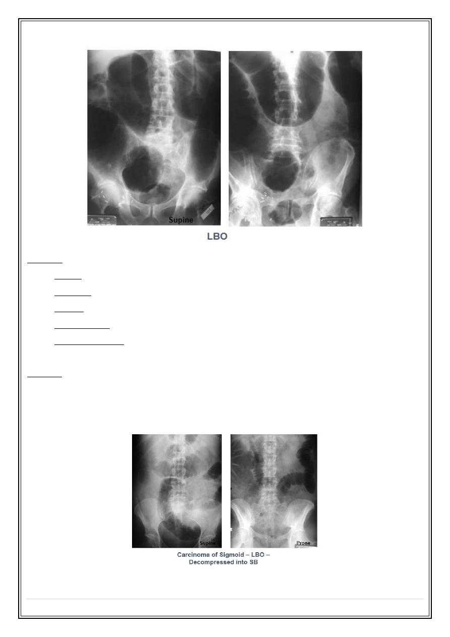
Mechanical LBO
Key Features
•
Dilated colon to point of obstruction
•
Little or no air in rectum/sigmoid
•
Little or no gas in small bowel, if…
o
Ileocecal valve remains competent
Secret Lectures
(9)
/ Diagnostic Imaging / Dr.Riyadh A. Al-Kuzzay (M.B.Ch.B – FICMS-RD)
P a g e
6
Causes
l Tumor
l Volvulus
l Hernia
l Diverticulitis
l Intussusception
Pitfalls
•
Incompetent ileocecal valve
o
Large bowel decompresses into small bowel
o
May look like SBO
o
Get BE or follow-up
Secret Lectures
(9)
/ Diagnostic Imaging / Dr.Riyadh A. Al-Kuzzay (M.B.Ch.B – FICMS-RD)
P a g e
7
What is the Diagnosis ?
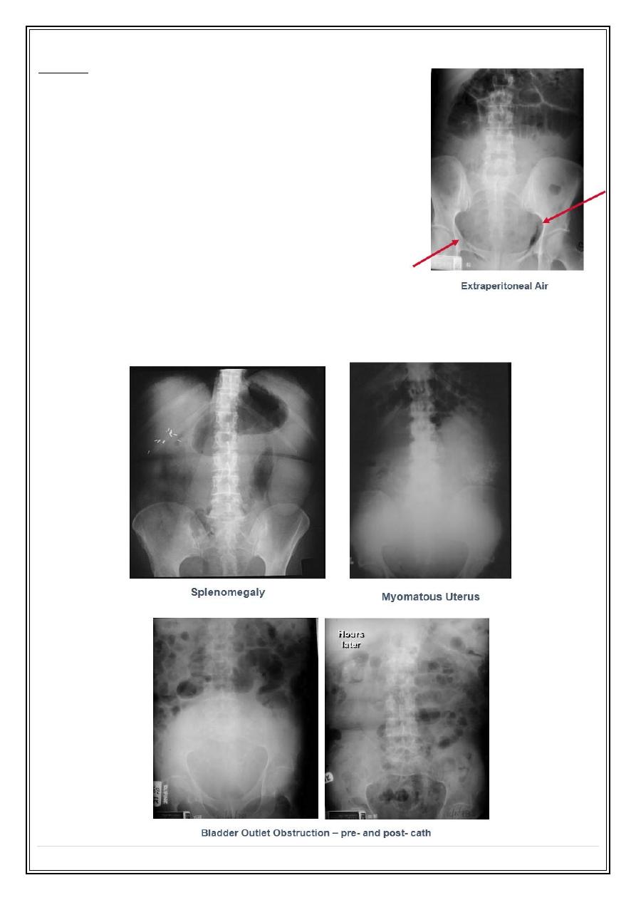
Extraluminal Air (Free Intraperitoneal Air)
Signs of Free Air
•
Air beneath diaphragm
•
Both sides of bowel wall
Secret Lectures
(9)
/ Diagnostic Imaging / Dr.Riyadh A. Al-Kuzzay (M.B.Ch.B – FICMS-RD)
P a g e
8
Causes
•
Rupture of a hollow viscus
o
Perforated ulcer
o
Perforated diverticulitis
o
Perforated carcinoma
o
Trauma or instrumentation
•
Post-op 5–7 days
•
NOT perforated appendix
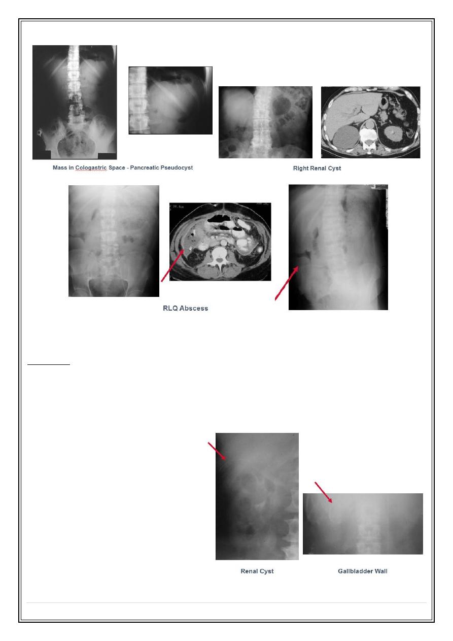
Soft Tissue Masses
•
Hepatosplenomegaly
o
Plain films poor for judging liver size
•
Tumor or cyst
o
Bowel displacement
Secret Lectures
(9)
/ Diagnostic Imaging / Dr.Riyadh A. Al-Kuzzay (M.B.Ch.B – FICMS-RD)
P a g e
9
Abdominal Calcifications
Patterns
•
Rimlike
•
Linear or track-like
•
Lamellar
•
Cloudlike
Rimlike Calcification
•
Wall of a hollow viscus
o
Cysts
▪
Renal cyst
o
Aneurysms
▪
Aortic aneurysm
o
Saccular organs e.g. GB
▪
Porcelain
Gallbladder
Secret Lectures
(9)
/ Diagnostic Imaging / Dr.Riyadh A. Al-Kuzzay (M.B.Ch.B – FICMS-RD)
P a g e
10
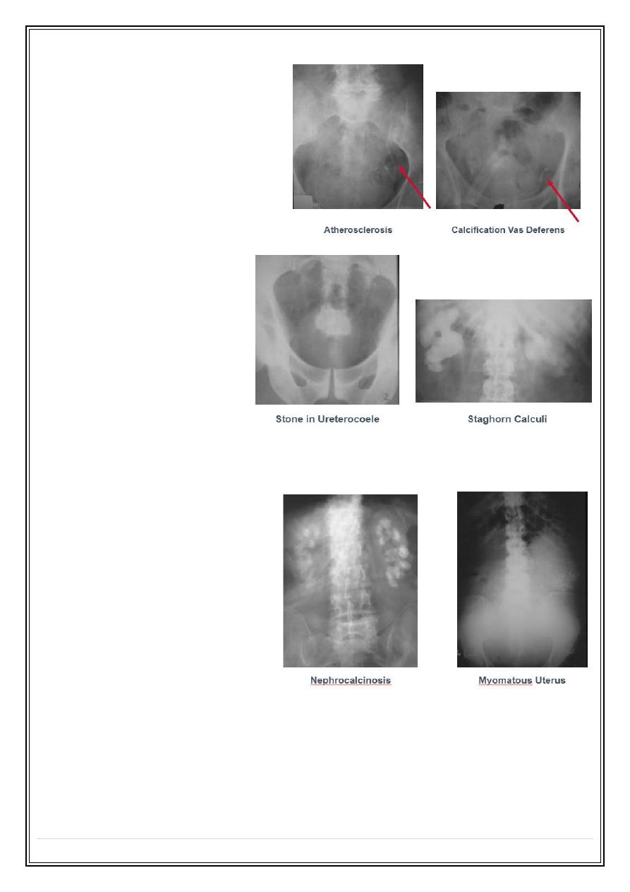
Linear or Track-like
•
Walls of a tube
o
Ureters
o
Arterial walls
Lamellar or Laminar
•
Formed in lumen of a
hollow viscus
o
Renal stones
o
Gallstones
o
Bladder stones
Cloudlike, Amorphous, Popcorn
•
Formed in a solid organ or
tumor
o
Leiomyomas of uterus
o
Ovarian cystadenomas
Thank you,,,