1
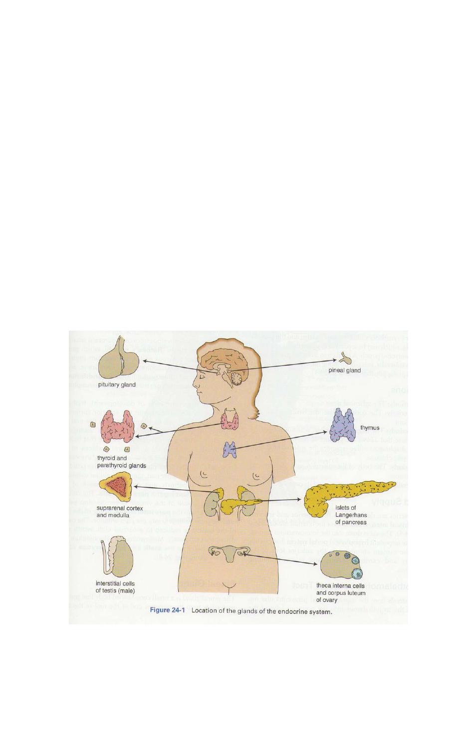
The Endocrine System
The endocrine system is made up of several glands:-
the pituitary gland (Cerebral hypophysis), pineal, thyroid, thymus, parathyroids,
suprarenals, islets of Langerhans of the pancreas, testes, ovaries, and, when
present, the placenta. In addition, there are groups of cells that form a minor part
of the system and are not considered in this chapter: the gastroenteroendocrine
cells, kidney cells, and certain cells the lung that store and secrete amines.
The endocrine glands have no ducts and consist of masses of cells richly supplied
by blood vessels, which pour their secretions (hormones) directly into the blood
stream.
The medical professional needs a sound grounding in the structure and function
of the endocrine system to be able to apply physiology lid hormone therapy in
daily clinical practice, moreover, it must be remembered that disease y affect
more than one endocrine gland at same time in an individual patient, a condition
own as multiple endocrine neoplasia. It should ) be remembered that patients
with advanced
malignant disease sometimes produce hormones that are not indigenous to the
tissue from which the tumor arose (paraneoplastic syndromes).

2
Pituitary Gland (Hypophysis Cerebri)
Location and Description
The pituitary gland is a small, oval structure attached to the undersurface
of the brain by the infundibulum. During pregnancy, it doubles in size. The
gland is well protected by virtue of its location in the sella turcica of the
sphenoid bone. Because the hormones produced by the gland influence the
activities of many other endocrine glands, the hypophysis cerebri is often
referred to as the master endocrine gland. For this reason, it is vital to life.
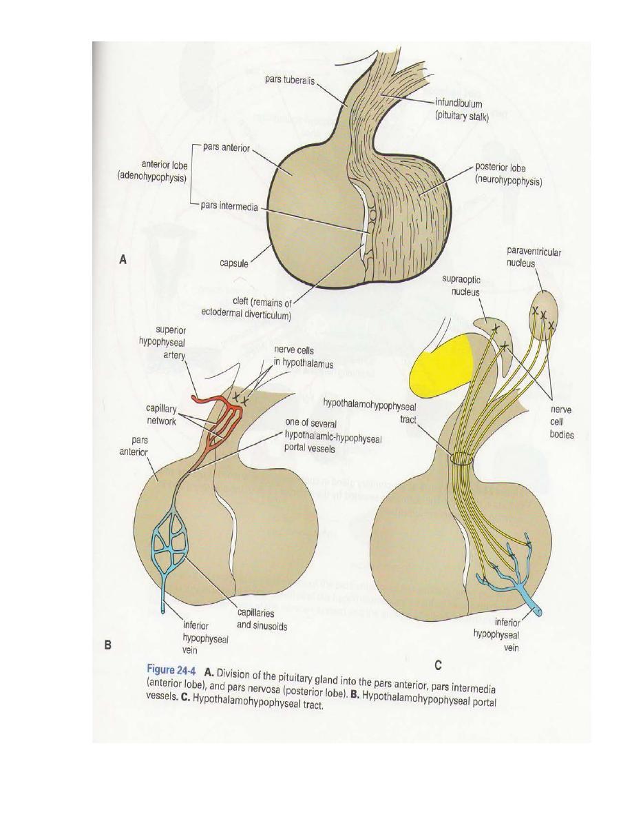
The pituitary gland is divided into an anterior lobe, or adenohypophysis,
and a posterior lobe, or neurohypophysis. The anterior lobe is subdivided
into the pars anterior (sometimes called the pars distalis) and the pars
intermedia, which may be separated by a cleft that is a remnant of an
embryonic pouch. A projection from the pars anterior, the pars tuberalis,
extends up along the anterior and lateral surfaces of the pituitary stalk.
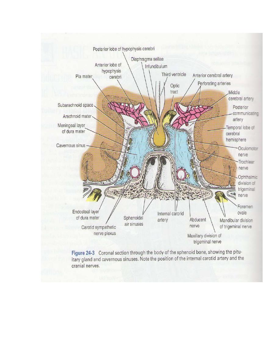
Relations
Anteriorly: The sphenoid sinus.
Posteriorly: The dorsum sellae, the basilar artery, and the pons.
Superiorly: The diaphragma sellae, which has a central aperture that allows
the passage of the infundibulum. The diaphragma sellae separates the
anterior lobe from the optic chiasma.
Inferiorly: The body of the sphenoid, with its sphenoid air sinuses.
Laterally:
The cavernous sinus and its contents.
Blood Supply
The arteries are derived from the superior and inferior hypophyseal
arteries, branches of the internal carotid artery. The veins drain into the
intercavernous sinuses. Note the important hypophyseal portal system that
extends from the median eminence to the anterior lobe of the pituitary and
carries releasing and release-inhibiting hormones.
Hypothalamohypophyseal Tract
This extends from the supraoptic and paraventricular nuclei of the
hypothalamus into the posterior lobe of the pituitary. The hormones
vasopressin and oxytocin are released at the axon terminals in the posterior
lobe
of the pituitary.
3
4

5
Pineal Gland
The pineal gland is a small cone-shaped body that projects posteriorly from
the posterior end of the roof of third ventricle of the brain. The pineal
consists essentially of groups of cells, the pinealocytes,
supported by glial
cells. The gland has a rich blood supply and is innervated by postganglionic
sympathetic nerve fibers.
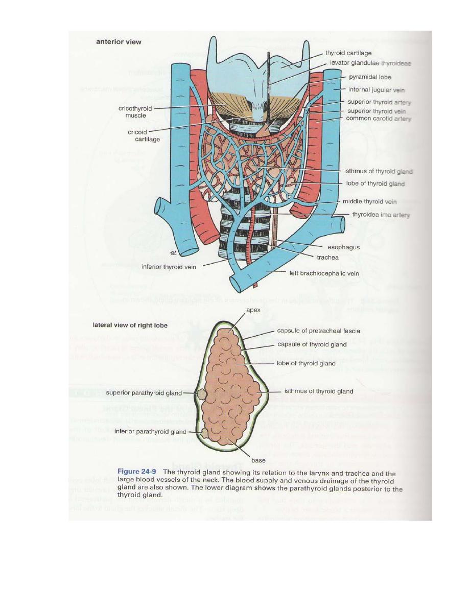
Thyroid Gland
The thyroid gland consists of right and left lobes connected by a narrow
isthmus. It is a vascular organ surrounded by a sheath derived from the
pretracheal layer of deep fascia. The sheath attaches the gland to the larynx
and the trachea.
Each lobe is pear shaped, with its apex being directed upward as far as the
oblique line on the lamina of the thyroid cartilage; its base lies below at the
level of the fourth or fifth tracheal ring.
The isthmus extends across the midline in front of the second, third and
fourth tracheal rings. A pyramidal lobe is often present, and it projects
upward from the isthmus, usually to the left of the midline. A fibrous or
muscular band frequently connects the pyramidal lobe to the hyoid bone; if
it is muscular, it is referred to as the levator glandulae thyroideae.
Relations of the Lobes
Anterolaterally: The sternothyroid, the superior belly of the omohyoid,
the sternohyoid, and the anterior border of the sternocleidomastoid.
Posterolaterally: The carotid sheath with the common carotid artery, the
internal jugular vein, and the vagus nerve.
Medially: The larynx, the trachea, the pharynx, and the esophagus.
Associated with these structures are the cricothyroid muscle and its nerve
supply, the external laryngeal nerve. In, the groove between the esophagus
and the trachea is the recurrent laryngeal nerve.
The rounded posterior border of each lobe is related posteriorly to the
superior and inferior parathyroid glands and the anastomosis between the
superior and inferior thyroid arteries.

6
Relations of the Isthmus
Anteriorly: The sternothyroids, sternohyoids, anterior jugular veins, fascia,
and skin.
Posteriorly: The second, third, and fourth rings of the trachea.
The terminal branches of the superior thyroid arteries anastomose along
its upper border.
Blood Supply:
The arteries to the thyroid gland are the superior thyroid artery, the inferior
thyroid artery, and sometimes the thyroidea ima. The arteries anastomose
profusely with one another over the surface of the gland.
1- The superior thyroid artery, a branch of the external carotid artery,
descends to the upper pole of each lobe, accompanied by the external
laryngeal nerve.
2- The inferior thyroid artery, a branch of the thyrocervical trunk, ascends
behind the gland to the level of the cricoid cartilage. It then turns medially
and downward to reach the posterior border of the gland. The recurrent
laryngeal nerve crosses either in front of or behind the artery, or it may pass
between its branches.
3- The thyroidea ima,
if present, may arise from the brachiocephalic artery
or the arch of the aorta. It ascends in front of the trachea to the isthmus.
The veins from the thyroid gland are:
1- The superior thyroid vein, which drains into the internal jugular vein.
2- The middle thyroid vein which drains into the internal jugular vein.
3- The inferior thyroid vein, which receives its tributaries from the isthmus
and the lower poles of the gland. The inferior thyroid veins of the two sides
anastomose with one another as they descend in
front of the trachea. They
drain into the left brachiocephalic vein in the thorax.
Lymph Drainage:
The lymph from the thyroid gland drains mainly laterally into the deep
cervical lymph nodes. A few lymph vessels descend to the paratracheal
nodes.
Nerve Supply
Superior, middle, and inferior cervical sympathetic ganglia.
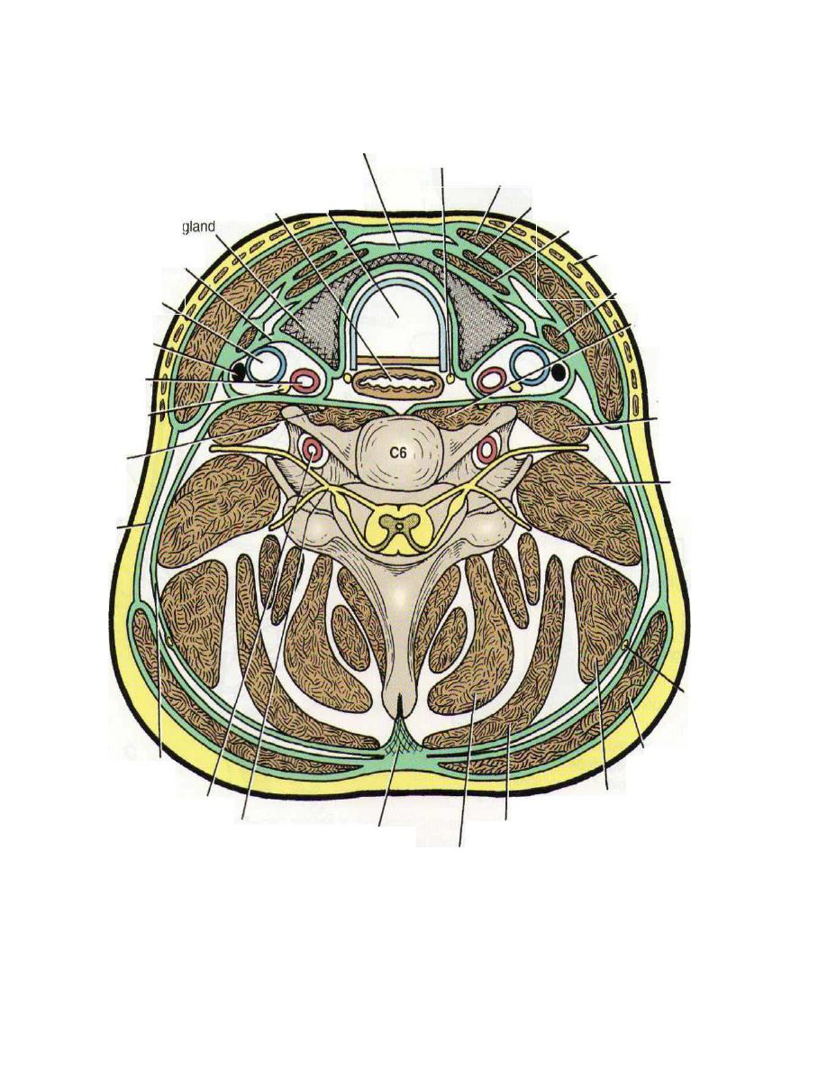
7
pretracheal fascia
trachea
esophagus
recurrent laryngeal nerve
sternocleidomastoid muscle
thyroid
carotid sheath
internal jugular
vein.
sternohyoid muscle
sternothyroid muscle platysma
omohyoid muscle
spinal nerve
ligamentum
nuchae
semispinalis capitis
Figure24-10 Cross section of the neck at the level of the sixth cervical vertebra.
8
9
Parathyroid Glands
The parathyroid glands are ovoid bodies measuring about 6 mm long in
their greatest diameter. They are four in number and are closely related to
the posterior border of the thyroid gland, lying within its fascial capsule.
The two superior parathyroid glands are the more constant in position
and lie at the level of the middle of the posterior border of the thyroid gland.
The two inferior parathyroid glands usually lie close to the inferior
poles of the thyroid gland. They may lie within the fascial sheath, embedded
in the thyroid substance, or outside the fascial sheath. Sometimes they are
found some distance caudal to the thyroid gland, in association with the
inferior thyroid veins; or they may even reside in the superior
mediastinum
in the thorax.
Blood Supply
The arterial supply to the parathyroid glands is from the superior and
inferior thyroid arteries. The venous drainage is into the superior, middle,
and inferior thyroid veins.
10
Thymus
The thymus is a flattened, bilobed structure lying between the sternum
and the pericardium in the anterior mediastinum. In the newborn infant, the
thymus reaches its largest size relative to the size of the body, at which time
it may extend up through the superior mediastinum in front of the great
vessels into the root of the
neck. The thymus continues to grow until puberty
but thereafter undergoes involution. It has a pink, lobulated appearance and
is the site for the development of thymus processed lymphocytes, T (thymic)
lymphocytes, which are distributed to the whole body.
Blood Supply
The blood supply of the thymus is from the inferior thyroid and internal
thoracic arteries.
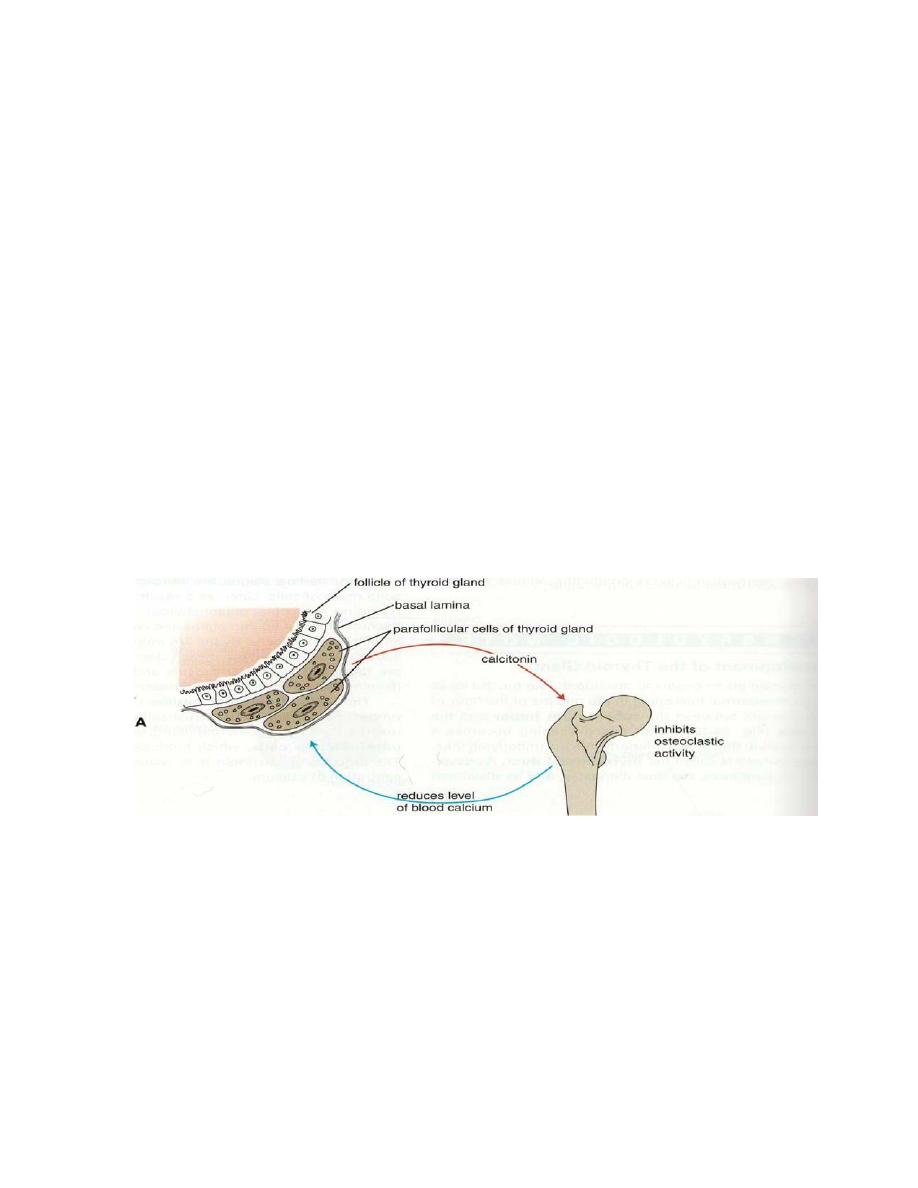
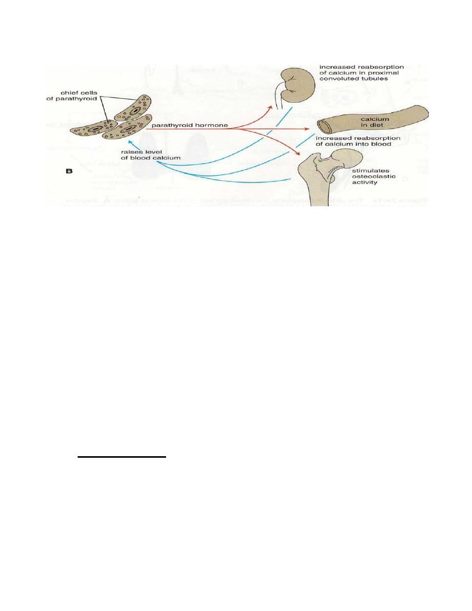
Figure 24-13
Actions of thyrocalcitonin
(A)
and the parathyroid hormone
(B)
on calcium metabolism.

11
Suprarenal Glands
Location and Description
The two suprarenal glands are yellowish retroperitoneal organs that lie
on the upper poles of the kidneys. They are surrounded by renal fascia (but
are separated from the kidneys by the perirenal fat). Each gland has a yellow
cortex; dark brown medulla.
The right suprarenal gland is pyramid shaped and caps the upper pole
of the right kidney. It lies behind the right lobe of the liver and extends
medially behind the inferior vena cava. It rests posteriorly on the diaphragm.
The left suprarenal gland is crescentic in shape and extends along the
medial border of the left kidney from the upper pole to the hilus. It lies
behind the pancreas, the lesser sac, and the stomach and rests posteriorly on
the diaphragm.
Blood Supply:
Arteries:
1-
The arteries supplying each gland are three in number:
2-
inferior phrenic artery,
3-
aorta,
renal artery.
Veins:
A single vein emerges from the hilum of each gland drains into the inferior
vena cava on the right and into renal vein on the left.
Lymph Drainage:
The lymph drains into the lateral aortic nodes.
Nerve Supply:
Preganglionic sympathetic fibers derived from the splanchnic nerves supply
the glands. Most of the nerves end in
medulla of the gland.