Histology
LAB.10
Endocrine System
Contains of this LAB.
Thyroid gland (
MH151
)
Para-thyroid (
MH154
)
Adrenal gland (
MH155a
)
Pituitary gland (
MH149
)
Edited by Fahad A.
Mosul Medical College
2018-2019
2th stage
Get it from
www.muhadharaty.com
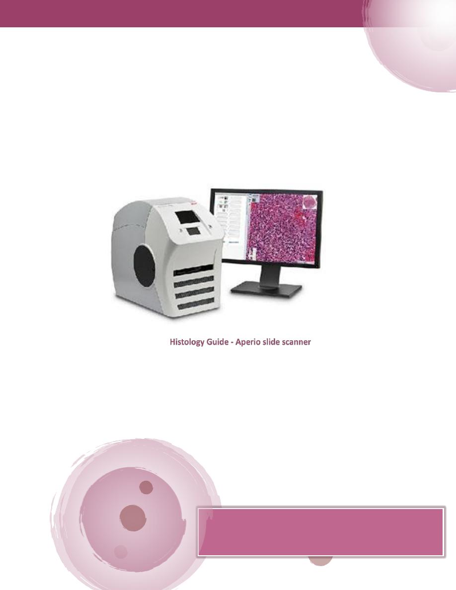
This copy is used to study tissues with Histology Guide website
to get a better result and a deeper understanding.
Do not hesitate to ask the doctors of the histology branch.
Histology
MMC 2019 – By Fahad. A.
1
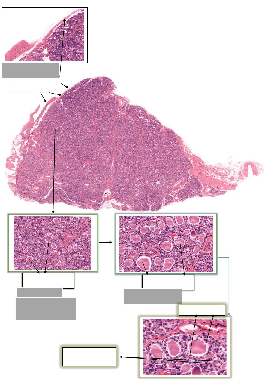
1 - Thyroid gland
Capsule
Enclosed by a thin layer
of connective tissue.
صورة مكبرة
Follicles
Spherical in shape
Colloid
gel-like mass fill lumen
of each follicle
هي البصيالت فقط
-
تحتوي
داخلها المادة الشبيهة بالجل
تسمى
Colloid
Follicular Cells
C cells
"parafollicular cells"

MMC 2019 – By Fahad. A.
2
Notes
Structure
Enclosed by a thin layer of connective tissue.
Capsule
connective tissue extends inwards
from the capsule to partially
outline irregular lobes and lobules.
Trabeculae
spherical follicles
of varying size (50 to 500 µm) in which
thyroid hormones are stored.
Follicles
lumen of each follicle is filled with the
gel-like mass
called
colloid. It is mostly the
protein thyroglobulin (pink)
and
bound thyroid hormones (triiodothyronine and
tetraiodothyronine (or thyroxin)). The clear space around
the colloid is a shrinkage artifact.
Colloid
follicles are lined by a
simple cuboidal to columnar
epithelium depending on functional activity
. Secrete
thyroid hormones when active.
Follicular Cell
small numbers of larger cells located at the periphery of
follicles
that secrete calcitonin. They stain poorly with H&E
making identification difficult.
C - cells
"
Parafollicular
cells
"
Thyroid gland on histologyguide.com
151
MH
VS Code
–
.com
guide
istology
H
MMC 2019 – By Fahad. A.
3
2 - Parathyroid gland
Capsule
Trabeculae
Chief Cells
Oxyphil cells
Chief Cells
ذات لون احمر كثيرة السايتوبالزم
تحتوي نواة
الحظ الفرق بين النوعين من
الخاليا المتواجدة في الغدة

MMC 2019 – By Fahad. A.
4
Notes
Structure
Enclosed by a thin layer of connective tissue.
Capsule
Connective tissue extends inwards
from the capsule to partially
outline irregular lobes and lobules.
Trabeculae
The
majority
of cells in the parathyroid.
-
خاليا صغيرة (من
5
إلى
8
ميكرومتر) مع نواة داكنة وحافة رقيقة من
السيتوبالزم الملون بخفة
They secrete parathyroid hormone (
PTH
).
Chief Cells
خاليا أكبر (من
8
إلى
21
ميكرومتر) مع نواة داكنة وسيتوبالزم قوي
بصبغة
الحمراء
They appear after the first decade of life and are thought to
be non-secretory cells.
Oxyphil Cells
هي الفجوات ذات اللون االبيض المتواجدة
بالنسيج وهي كبيرة الحجم وتزداد
بالحجم مع تقدم العمر
Adipose Cells
Larger cells (8 to 12 µm diameter) with dark nuclei and a
watery, clear cytoplasm.
Clear Cells
Parathyroid gland on histologyguide.com
MH 154
VS Code
–
.com
guide
Histology
MMC 2019 – By Fahad. A.
5
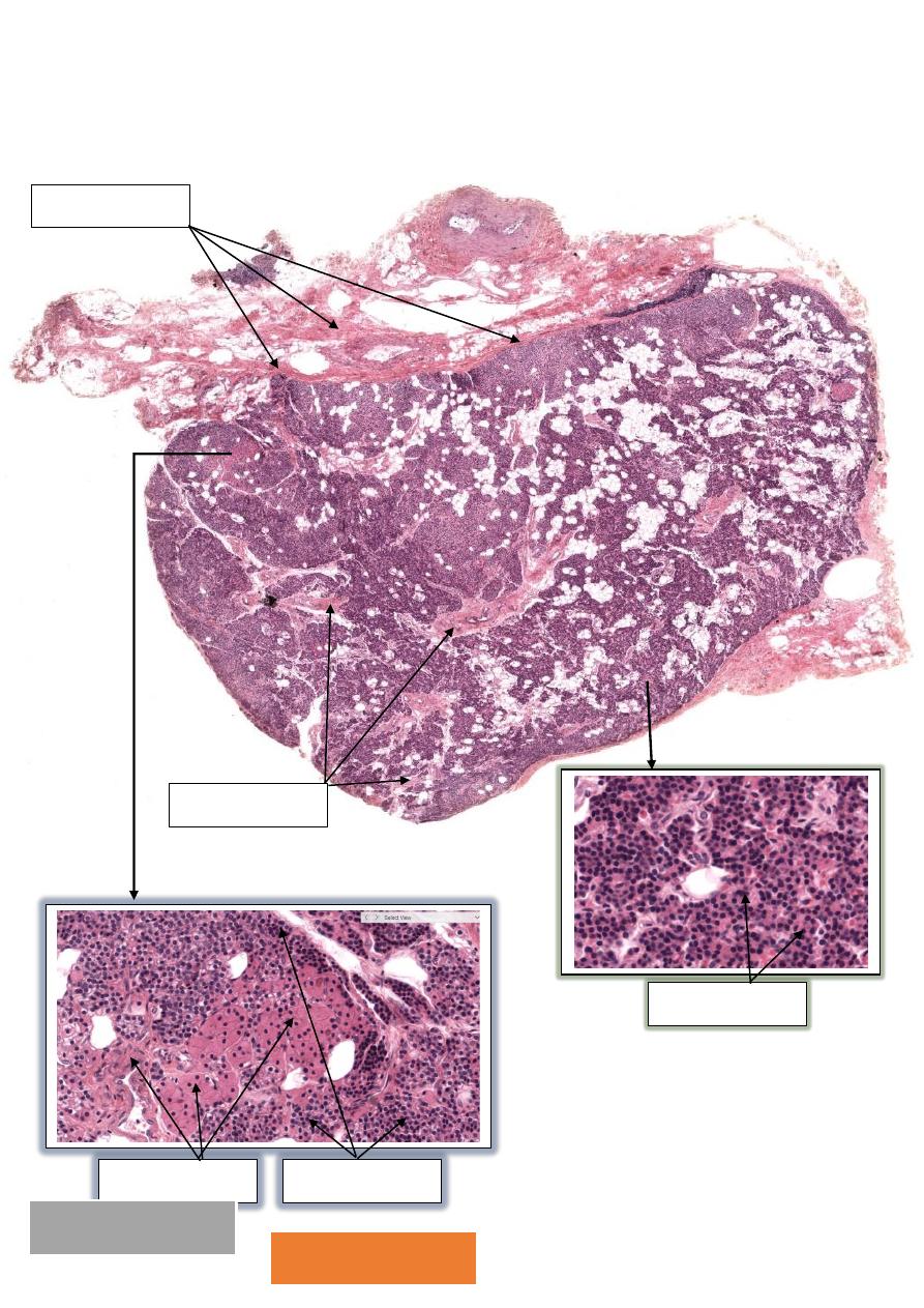
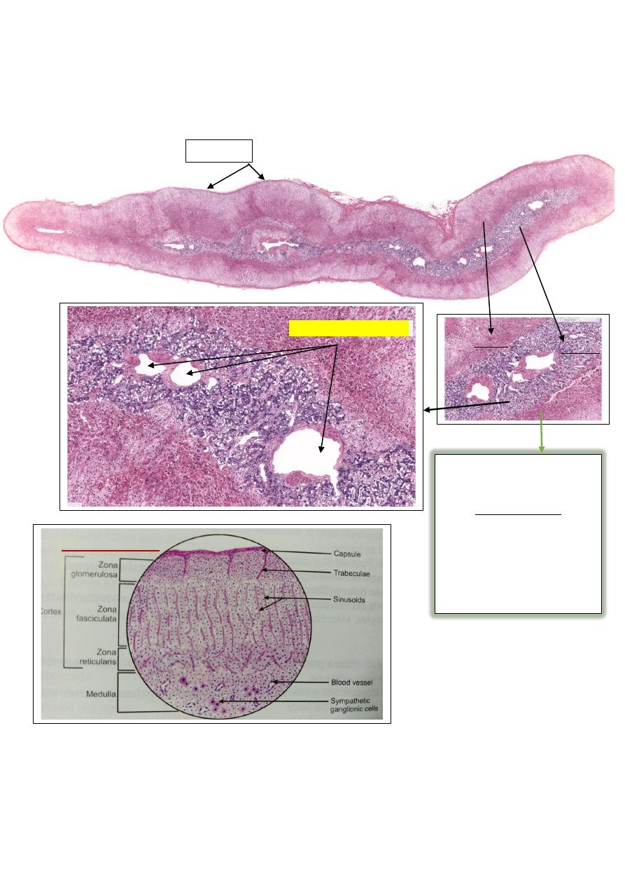
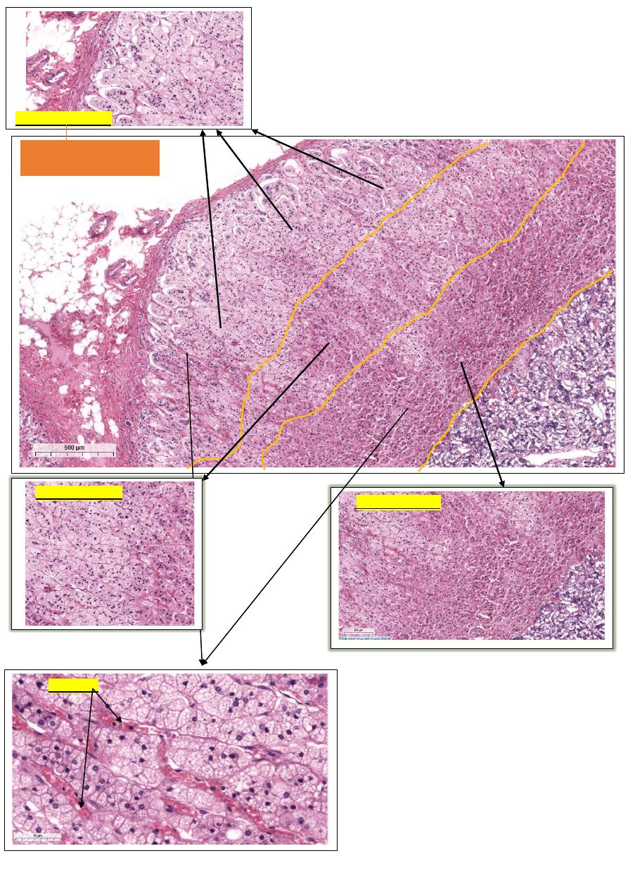
3 – Adrenal gland ( MH 155a)
Cortex
Medulla
تقسم منطقة ال
Cotext
القشرة الى ثالث
: مناطق
-
وهي من الخارج للداخل
1- Zone Glomerulosa
2- Zone Fasciculata
3- Zone Reticularis
G-F-R
Capsule
صورة ( مخطط ) توضيحة
MMC 2019 – By Fahad. A.
6
Adrenal gland
Cortex
Zone Glomerulosa
Zone Reticularis
Sinusoids
Zona Fasciculata
Arranged in round to oval
clusters

MMC 2019 – By Fahad. A.
7
Notes
Structure
Enclosed by a thin layer of connective tissue.
Capsule
Cross-sections are seen in the capsule.
Nerves
Penetrate the capsule and branch into sinusoids that
supply the cortex and medulla.
Blood Vessels
Cells that synthesize and secrete steroid hormones.
- Zona Glomerulosa
- outer zone (15%) of glomerular-like
clusters of cells that secrete aldosterone. The cells have a
central nucleus and lipid filled ("foamy") cytoplasm
- Zona Fasciculata
- middle zone (80%) of two-cell wide
vertical cords that secrete cortisol. The cells have a central
nucleus and lipid filled ("foamy") cytoplasm
- Zona Reticularis
- inner zone (7%) of one-cell wide
anastomosing rows that secrete precursors of testosterone.
The cells have a central nucleus and eosinophilic cytoplasm
Cortex
Chromaffin Cells - modified postganglionic sympathetic
neurons that secrete catecholamines (epinephrine or
norepinephrine)
Ganglion Cells - infrequent sympathetic ganglion cells
Medulla
Sinusoidal capillaries (or sinusoids) are one type with a
larger diameter (15 to 30 µm) and more irregularly shaped.
sinusoids
Adrenal gland on histologyguide.com
155a + MHS 216
MH
VS Code
–
Histologyguide.com
8
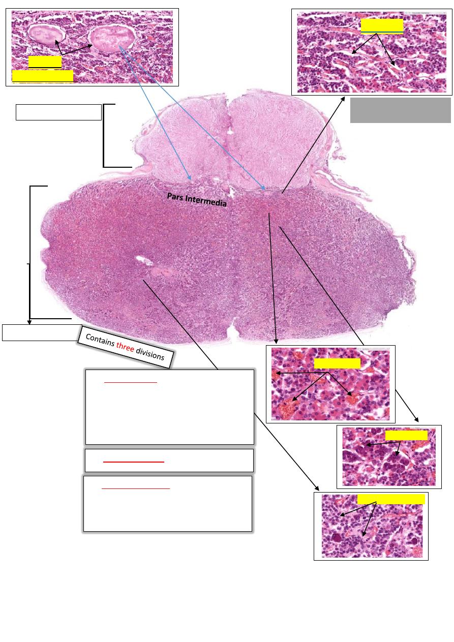
4 – Pituitary gland
Posterior Pituitary
Anterior Pituitary
تسمى ايضا
Adenohypophysis
تسمى ايضا
Neurohypophysis
تسمى ايضا
Pars Nervosa
comprises most of the
-
Pars Distalis :
(1)
anterior lobe (~75%) and contains five
types of endocrine cells.
1- Chromophobes %50
2- Acidophils %40
3- Basophils %10
not shown.
-
:
Tuberalis
Pars
(2)
thin remnant (<2%)
-
:
Pars Intermedia
)
3
(
at interface between the anterior and
posterior lobes that contains numerous
colloid (protein)-filled cysts.
Acidophils
Basophils
Chromophobes
تعمل على استقبال الهرمونات من
كال
Acidophils
+
Basophils
Colloid
"
cysts
Rathke's
"
MMC 2019 – By Fahad. A.
9
تكملة
-
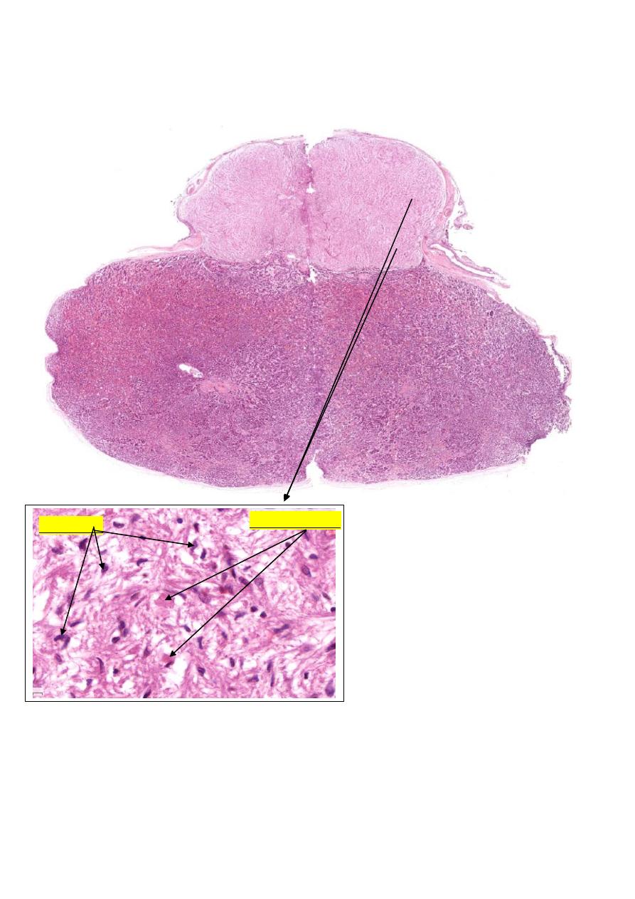
Posterior Pituitary
Pituicytes
Herring Bodies
MMC 2019 – By Fahad. A.
11
The pituitary gland is composed of an anterior and posterior lobes.
Notes
Structure
Contains three divisions:-
1)
Pars Distalis
- comprises most of the anterior lobe (~75%)
and contains five types of endocrine cells.
a)
Chromophils
- stain with H&E and secrete hormones
b)
Acidophils
- stain pinkish-red with H&E.
c)
Basophils
- stain bluish-purple with H&E.
d)
Chromophobes
stain poorly with H&E and do not secrete
hormones.
e)
Sinusoidal Capillaries
- extensive network that receives
hormones from acidophils and basophils.
2)
Pars Tuberalis
3)
Pars Intermedia
- thin remnant (<2%) at interface between the
anterior and posterior lobes that contains numerous
colloid
(protein)-filled cysts
Anterior
Pituitary
Axons from the hypothalamus that release hormones into the
capillaries of the pars nervosa.
1)
Pituicytes
- most nuclei belong to glial cells.
2)
Herring Bodies
- dilations of axons filled with neuro-secretion
vesicles.
Posterior
Pituitary
Pituitary gland on histologyguide.com
149
MH
VS Code
–
Histologyguide.com

MMC 2019 – By Fahad. A.
11
Examine the VS slide MHS 209 to recognize the relation between Thyroid, parathyroid
and thymus.
Task: With the help of a diagram summarize the effect of the hypothalamus / pituitary
complex on the rest of the body i.e. hormones secreted and their action on target
organs and tissues.

MMC 2019 – By Fahad. A.
12
List all the hormones secreted by the pituitary gland