بسم الله الرحمن الرحيم
In oral and maxillofacial surgery
BIOPSY
Principles and techniques
What is a Biopsy?
Biopsy is derived from a Greek word(By-op-see) = Bio – meaning LIFE and
Opsy – TO LOOK(Vision)
Biopsy is the removal of tissue from a living organism for the purpose of microscopic examination and diagnosis.
introduction
A systematic approach should be developed in evaluating a patient with an oral and maxillofacial lesions, which include the following steps:1. A detailed history
2. Clinical examination:
Extraoral
Intraoral
3.Special investigations: (as appropriate)
Radiography or other imaging techniques
Biopsy for histopathology( including immunoflourescence, immunohistochemistry, electron microscopy etc…)
Specimens for microbial cultures
Haematological or biochemical tests.
BIOPSY : When, why, where?

INDICATION
CONTRAINDICATIONSUSPICIOUS OF MALIGNANCY
OBJECTIVE
WHEN IS ORAL BIOPSY NOT NEEDED?
Characteristics of lesions that raise the suspicion of malignancy
Growth rate– lesion exhibits rapid growthBleeding— lesion bleeds on gentle manipulation
Induration– lesion and surrounding tissue is firm to the touch
Fixation– lesion feels attached to adjacent structures
Characteristics of lesions that raise the suspicion of malignancy
Erythroplakia—lesion is totally red or has speckled red appearance
Ulceration—lesion is ulcerated or presents as an ulcer
Duration— lesion has persisted for more than 2 weeks
Indication for Biopsy
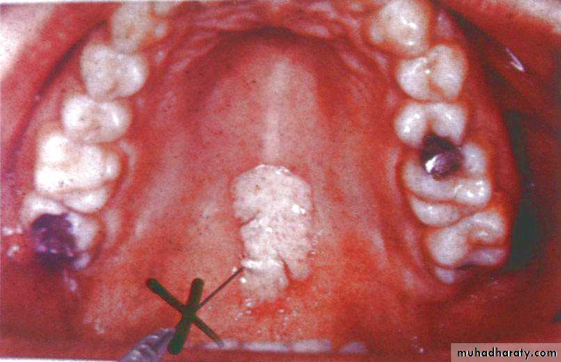
Persistent hyperkeratosis changes in surface tissue (ex: lips or oral mucosa)Lesion that interfere with local function (ex: fibroma)
Any inflammatory lesion that does not respond to local treatment after 10 to 14 days (that is after removing local irritant)Indication for Biopsy
Bone lesions not specifically identified by clinical and radiographic finding.Any lesion persists for more than 2 weeks with no apparent etiology basis.
Any lesion that has the characteristics of malignancy .WHEN IS ORAL BIOPSY NOT NEEDED?
There is no need to biopsy normal structures.There is no need to biopsy for inflammatory or infectious lesions that respond to specific local treatments, as pericoronitis, gingivitis or periodontal abscesses.
No incisional biopsies should be performed on suspected angiomatous lesions.
CONTRA-INDICATIONS
Anticoagulant therapy
Over-whelming sepsis
Severe impaired lung function
Uncontrolled bleeding.
Uncooperative patient
Local infection near the site
To confirm a diagnosis made on clinical findings.
To determine the treatment planAs a medical record
OBJECTIVES OF BIOPSYCLASSIFICATION OF BIOPSY
According to the procedures applied, oral biopsies can be classified by:a) Features of the lesion:
• Direct biopsy: when the lesion is located on the oral mucosa and can be easily accessed with a scalpel from the mucosal surface.• Indirect biopsy: when the lesion is covered by an apparently normal oral mucosa.
b) Area of surgical removal:
• Incisional biopsy: consists of the removal of a representative sample of the lesion and normal adjacent tissue in order to make a definitive diagnosis before treatment.• Excisional biopsy: is aimed at the complete surgical removal of the lesion for diagnostic and therapeutic purposes. This procedure is elective when the size and location of the lesion allows for a complete removal of the lesion and a wide margin of surrounding healthy tissue.
c) By the timing of the biopsy/ Clinical timing of sampling:
• Pre-operative• Intra-operative
• Post-operative
Types of Biopsy
Surgical biopsy- Incisional Biopsy ,Excisional Biopsy and Punch Biopsy.Fine Needle Aspiration Cytology(FNAC) and CT guided FNAC.
Exfoliative Cytology.
Brush Biopsy.
Frozen Section Biopsy.
Core Needle Biopsy. trucut biopsy
STEPs OF BIOPSY
1.SELECTION OF AREA OF BIOPSY
2.PREPARATION OF SURGICAL FIELD
3.LOCAL ANASTHESIA
4.INCISION
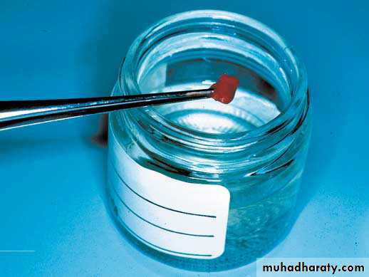
5.HANDLING OF SPECIMEN
6.SUTURING OF THE RESULTING WOUND
If a lesion is large or has different characteristics in various locations more than one area may need to be sampled
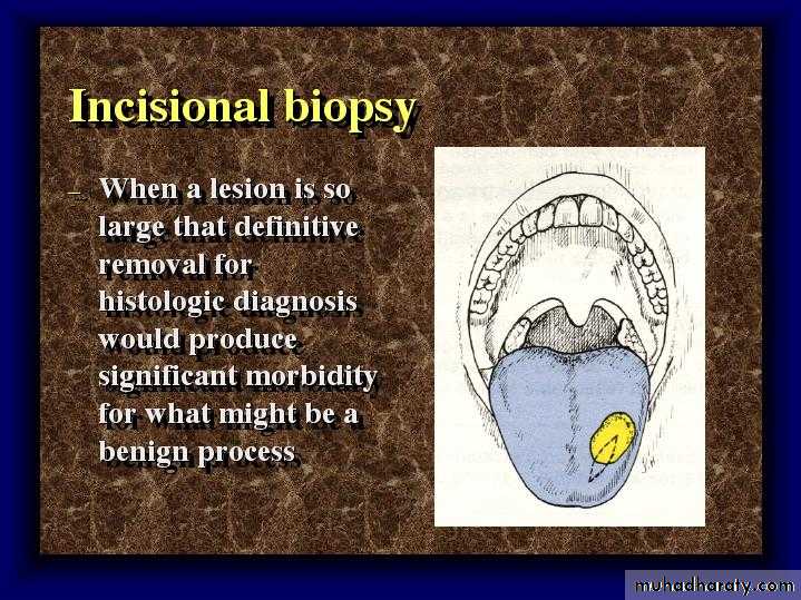
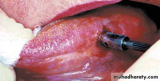
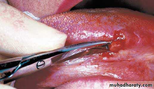
INCISIONAL BIOPSY
Incision should extend from the ulceration out onto clinically normal tissue
Grasp area to be removed with forceps and make an elliptical incision from the centre out onto clinically normal tissue: wound after removal of incised tissue: suturing completedIncisional Biopsy
Indications:Size limitations
Hazardous location of the lesion
Great suspicion of malignancy
Technique:
Representative areas are biopsied in a wedge fashion.
Margins should extend into normal tissue on the deep surface.
Necrotic tissue should be avoided.
A narrow deep specimen is better than a broad shallow one.
DISADVANTAGES:
1. Crush, splits and haemorrhage are the artefacts most frequently found in incisional oral biopsies.2. Theoretical seeding of cancer cells into the adjoining tissues.
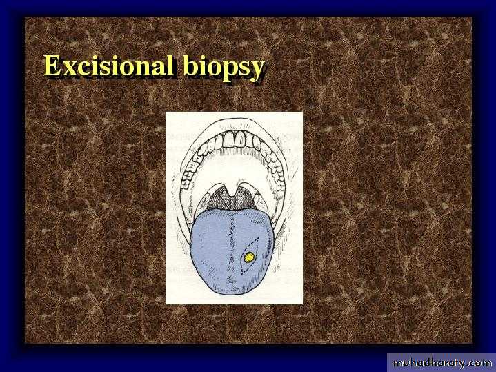
Excisional Biopsy
•• The entire lesion with 2 to 3mm of normal appearing tissue surrounding the lesion is excised if benign.
Excisional Biopsy
An excisional biposy implies the complete removal of the lesion.Indications:
Should be employed with small lesions. Less than 1cm
The lesion on clinical exam appears benign.
When complete excision with a margin of normal tissue is possible without mutilation.
Excisional Biopsy
Technique:
The entire lesion with 2 to 3mm of normal appearing tissue surrounding the lesion is excised if benign.
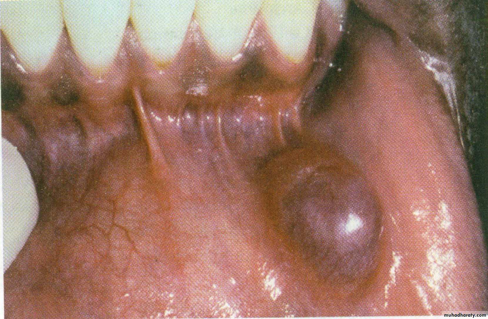
FOR MUCOCELE LESIONS – CAREFUL EXCISIONAL BIOPSY
Punch Biopsy
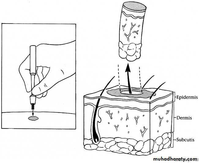
Punch biopsy
Advantages :
Ease of technique
Sutures may not be required if small diameter punch
May produce a more satisfactory specimen in bound down tissues (e.g. hard palate)
Drawbacks:
May not be adequate for biopsy of deeper pathologyMay be difficult to biopsy freely movable tissues (e.g. soft palate, floor of mouth)
Technique of punch biopsy
biopsy punches should range in size from 2-10 mm in diameterthe smaller diameters should be avoided due to the risk of over-manipulating and crushing the tissue .
The technique is easily performed with a low incidence of postsurgical morbidity.
Suturing in regards to a punch biopsy procedure is usually not required as the surgical wounds heal by secondary intention.
CORE BIOPSY
Fine needle biopsy has been established as a safe procedure and is routinely performed under local anaesthesia. Many pathologists believe that for histologic study, core tissue is more useful than cytologic material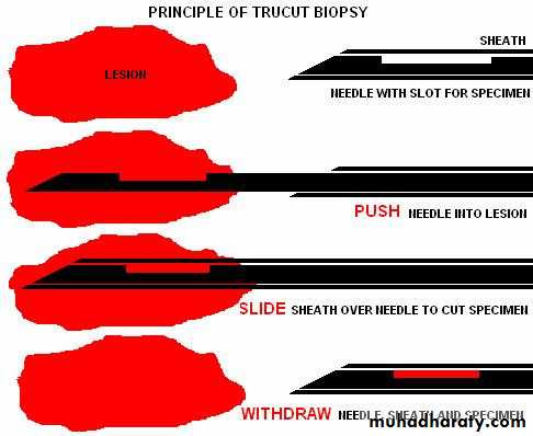
Fine needle aspiration cytology
It is the “Technique of aspiration of cells/ fluid/ tissue fragments using a fine needle for examination under a microscope”ADVANTAGES
1. The technique is relatively painless, produces speedy results.2. It is an inexpensive technique.
3. It requires little equipment.
4. The technique can be done as an out patient or a bed side procedure.
5. There is no problem with wound healing.
6. The technique is readily repeatable
INDICATIONS
1. Non palpable lesions, or area difficult to biopsy but can be localized by CT, MRI, Ultrasound.2. To rule out vascular lesions prior to open surgery.
3. In cases where Biopsy is contraindicated on medical background.
4. Used as a diagnostic screening test at community level for head and neck masses.
5. Indicated for known tumors to assess effect of treatment.
6- To determine the presents of fluid within a lesion and to a certain the type of fluid within a lesion.
7-When exploration of an intraosseous lesion is indicated
Aspiration
• Procedures:
• An 18-gauge needle is connected to a 5 or 10 ml syringe and is inserted into the center of the mass via a small hole in the lesion.
• The tip of the needle may need to be positioned in multiple directions to locate a potential fluid center.
• The material withdrawn during aspiration biopsy can be submitted for pathologic examination and/or culturing.
•
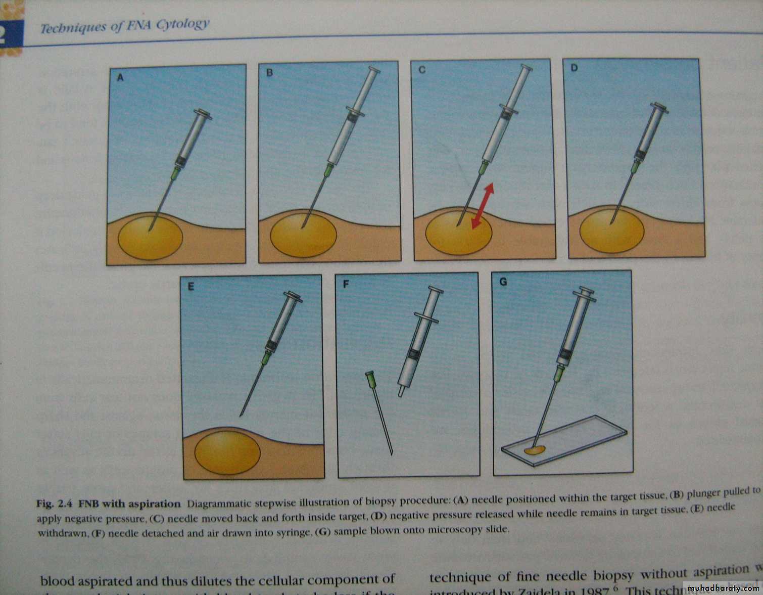
FINE NEEDLE WITH ASPIRATION
The inability to withdraw fluid or air indicates that the lesion is probably solid.A radiolucent lesion in the jaw that yields straw-colored fluid on aspiration is most likely a cystic lesion.
If purulent exudate (pus) is withdrawn, then an inflammatory or infectious process should be considered..
The aspiration of blood might indicate a vascular malformation within the bone.
Any intrabony radiolucent lesion should be aspirated before surgical intervention to rule out a vascular lesion.If the lesion is determined to be vascular in nature, the flow rate (high versus low) should be determined because uncontrollable hemorrhage can occur if incised
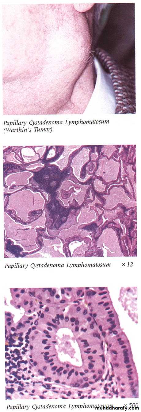
FOR MAJOR SALIVARY GLAND/LYMPH GLAND LESIONS FNAC MAY BE USEFUL
Brush BiopsyDiagnosis of oral epithelial dysplasia has traditionally been based upon histopathological evaluation of a full thickness biopsy specimen from lesional tissue.
It has recently been proposed that cytological examination of “brush biopsy” samples is a non-invasive method of determining the presence of cellular atypia, and hence the likelihood of oral epithelial dysplasia.
Exfoliative Cytology
It is a quick and simple procedure, is an important alternative to biopsy in certain situations. In exfoliative cytology, cells shed from body surfaces, such as the inside of the mouth, are collected and examined. This technique is useful only for the examination of surface cells and often requires additional cytological analysis to confirm the results.The Advantages and Disadvantage of oral cytological procedures include:
AdvantagesCytology may be helpful when large areas of mucosal change are noted, or in areas with difficult surgical access
Disadvantages
Not very reliable with many false positives.
Expertise in oral cytology is not widely available
For red & white lesions include both red & white area
For Vesiculobullous lesions
Fluid is more representative. Intact vesicle or bulla should be biopsied.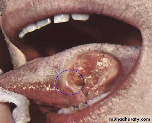
ULCERS
Include margin, deep part of ulcer and site of maximal clinical activity.AVOID Superficial ulcers & necrotic tissue
INJECTION
Intraosseous and Hard Tissue Biopsy
Intraosseous lesions are most often the result of problems associated with the dentition
indications
Any intraosseous lesion that fails to respond to routine treatment of the dentition.Any intraosseous lesion that appears unrelated to the dentition.
Principles of surgery
Mucperiosteal flaps should be designed to allow adequate access for incisional/excisional biopsy.Incisions should be over sound bone
Cortical perforation must be considered when designing flaps
Flaps should be full thickness
Major neurovascular structures should be avoided
Principles of surgery
Osseous windows should be submitted with the specimenOsseous preformations can be enlarged to gain access
Avoid roots and neurovascular structures
The tissue consistency and nature of the lesion will determine the ease of removal
Principles of surgery
Incisional biopsies only require removal of a section of tissueSoft tissue overlying the lesion should be reapproximated following thorough irrigation of the operative site.
The specimen should be handled as previously described
BIOPSY DATA SHEET
PATIENT DATA
HISTORY
CLINICAL DESCRIPTION
NATURE OF BIOPSY
RADIOGRAPHS & PHOTOGRAPHS
DISCRIPTION OF BIOPSY SPECIMEN
BIOPSY REPORT
IT SHOULD INCLUDE DIAGNOSIS AS WELL AS A COMPLETE MICROSCOPIC DESCRIPTIONIt is not easy to procure a good biopsy specimen, nor is it very difficult, but the procedure must be carefully planned and carried out.