
1
Course: Immunology
Lecturer: Dr. Weam Saad
Lecture: Lymphatic System
Lymphatic System
Tissues and Organs Involved in the Immune System Response
Lymphoid Tissues and Organs
The immune system is organized on several special tissues, called
lymphoid or immune tissues. Some lymphoid tissues have a remarkable degree
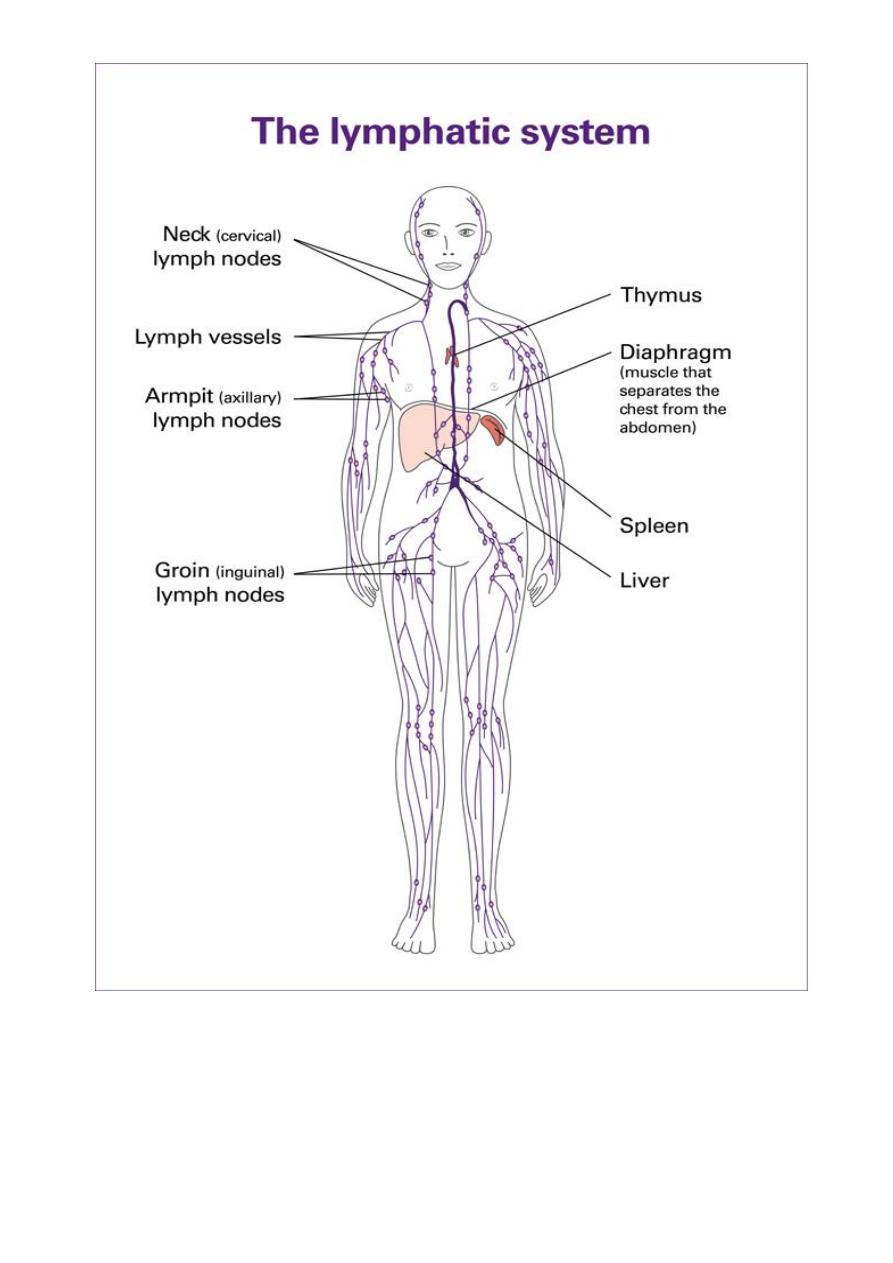
of organization called as lymphoid organs. The lymph nodes are the most
important and are located in groups along major blood vessels and loose
connective tissues. Other mammalian lymphoid organs are the thymus and the
spleen (white pulp). Lymphoid tissues include the gut-associated lymphoid
tissues (GALT)—tonsils, Peyer's patches, and appendix—as well as aggregates
of lymphoid tissue in the submucosal spaces of the respiratory and genitourinary
tracts.
Lymphoid tissues can be subdivided into primary and secondary as
follow:
A. Primary Lymphoid Tissues:
Lymphoid tissues have the ability to produce cells of the lymphocytic
lineage, which is the characteristic of primary lymphoid tissues.
1. Bone Marrow
It is a flexible tissue in the interior of bones. It produces RBCs by the
cores of bone marrow in the heads of long bones in a process known as
hematopoiesis. On average, bone marrow constitutes 4% of the total body mass
of humans; in an adult weighing 65 kilograms. The hematopoietic component
of bone marrow produces approximately 500 billion blood cells per day. Bone
marrow is a key component of the lymphatic system, producing the lymphocytes
and other cells of the immune system via stem cells,
Physiological role. Moreover production of blood cells, the red bone marrow
is a key element of the lymphatic system, it generates lymphocytes from
2
immature hematopoietic progenitor cells. The bone marrow and thymus
constitute the primary lymphoid tissues involved in the production and early
selection of lymphocytes. Furthermore, bone marrow performs a valve-like
function to prevent the backflow of lymphatic fluid in the lymphatic system.
Other important role is maturation and differentiation of B-lymphocytes.
2. Lymph Nodes.
The lymph nodes are many and distributed all over the body. They
measure 1 to 25 mm in diameter and play a very important role in the initiation
of the immune response.
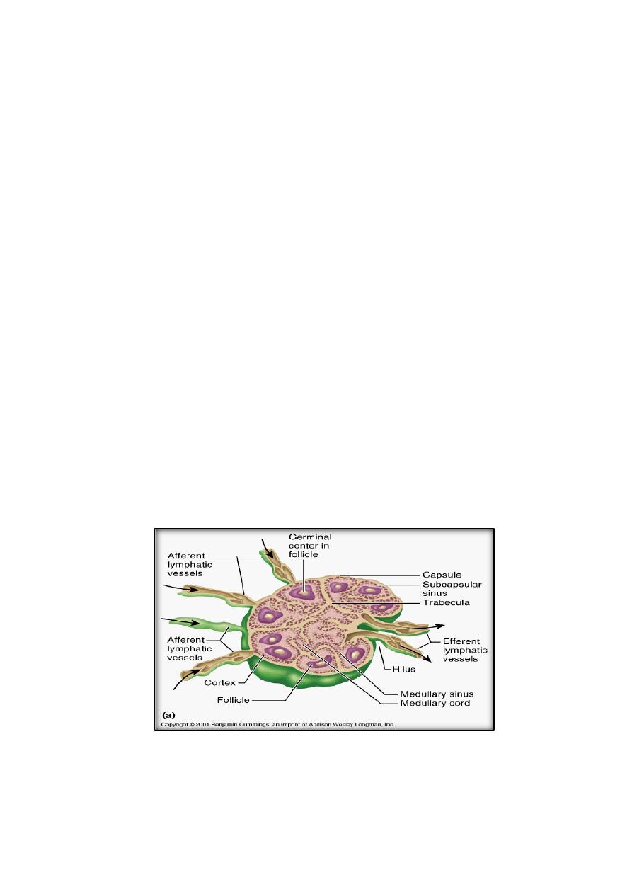
Anatomical organization. The lymph nodes are circulated by a
connective tissue capsule and open into the subcapsular sinus. The lymph node
also receives blood from the systemic circulation through the hilar arteriole.
Two main regions can be distinguished in a lymph node: the cortex and the
medulla. The cortex and the deep cortex (also known as paracortical area)
contain many lymphocytes, the cortex also containing packed lymphocytes and
termed follicles or nodules.
Physiological role. The lymph nodes can be compared to a network of
filtration and communication stations where antigens are trapped and messages
are transmitted between the different cells involved in the immune response.
Also have an important role during secondary immune responses.
(Lymph Nodes Anatomy)
Langerhans cells of the dermis. Those cells express MHC-II molecules,
and therefore function as APC, they migrate from the dermis to the paracortical

3
areas, where they interact with the T lymphocytes and helper T lymphocytes to
specifically recognize those MHC-associated peptides in APC which is essential
for proper initiation of the immune response.
3. Thymus
The thymus is a primary lymphoid organ in mammals. It is believed to
play a key role in determining the differentiation of T lymphocytes.
Anatomical organization. The thymus is located in the superior
mediastinum, anterior to the great vessels. It has a connective tissue capsule,
which divide the organ into lobules. Each lobule has a cortex and medulla, and
coated with epithelial cells.
a. Cortex.
Lymphocyte aggregates, composed mainly of immunologically immature T-
lymphocytes, are located in the cortex where these cells proliferation occurs. A
small number of macrophages and plasma cells are also present. In addition, the
cortex contains epithelial cells.
b. Medulla.
Not as densely as the cortex, the medulla contains predominantly mature T-
lymphocytes, and has a larger epithelial cell-to-lymphocyte ratio than the cortex
and the squamous epithelial cells known as Hassall's corpuscles.
Physiological roles:
a. T-lymphocyte differentiation. The thymus is believed to be the organ
where T-lymphocytes differentiate during embryonic life. The Thymic
cortex is an area of cell proliferation and death (only 1% of the cells
generated in the thymus, mature and migrate to the peripheral tissues).
The mechanism of the T-lymphocyte differentiation is believed to involve
the interaction of T-lymphocyte precursors with thymic epithelial cells.
b. Hormone synthesis. The Thymic epithelial cells produce hormonal
factors (e.g., thymosin and thymopoietin), which may play an important
role in the differentiation of T-lymphocytes.

4
B. Secondary Lymphoid Tissues
1.
Spleen: Anatomical organization. Surrounded by a connective tissue
capsule, have the white and the red pulp.
Physiological role. The spleen is the lymphoid organ associated with
filtering and clearing of particulate matter, infectious organisms, and aged cells
of peripheral blood. Also help the macrophages to process the antigen.
2.
Mucosa-Associated Lymphoid Tissues (MALT)
Include the lymphoid tissues of the intestinal tract, genitourinary tract,
tracheobronchial tree, and mammary glands. All of the mucosa-associated
lymphoid tissues are un-encapsulated and contain both T and B lymphocytes.
3.
Gut-Associated Lymphoid Tissue (GALT)
It is found along the digestive tract. Three major sites are the tonsils, the
Peyer's patches (located on the submucosa of the small intestine) and the
appendix. Tonsils, located in the oropharynx, contain mostly B-lymphocytes
and are the sites of antigenic stimulation. While, Peyer's patches, are lymphoid
structures found in the submucosal space of the small intestine.
Physiological roles:
a. The follicles of the intestinal Peyer's patches are rich with B-cells,
which differentiate into IgA-producing plasma cells.
b. T-lymphocytes are also present in the intestinal mucosa, the most
common are memory helper T-cells. This population is involved in the
induction of humoral immune responses (HMI).
5