
Endocrinological Emergencies
Mosul Medical College
Department of Medicine
Presented by:
Dr. Salam Fareed
8/21/2016

Diabetic ketoacidosis

8/21/2016
INTRODUCTION
Diabetic ketoacidosis (DKA) is an acute, major, life-
threatening complication of diabetes that mainly
occurs in patients with type 1 diabetes, but it is not
uncommon in some patients with type 2 diabetes-
20%.
This condition is a complex disordered metabolic
state characterized by:
hyperglycemia: blood glucose level > 200 mg/ dl
Ketoacidosis: ketonuria > ++ on standard urine
sample.
Metabolic acidosis: PH < 7.3, s. bicarbonate < 15

8/21/2016
Pathophysiology
DKA typically occurs in the setting of
hyperglycemia with relative or absolute insulin
deficiency and an increase in counterregulatory
hormones.
Sufficient amounts of insulin are not present
to suppress lipolysis and oxidation of free fatty
acids, which results in ketone body production
and subsequent metabolic acidosis.
DKA occurs more frequently with type 1
diabetes, although 10% to 30% of cases occur in
patients with type 2 diabetes.

8/21/2016
Predisposing Factors
Several risk factors can precipitate the development
of extreme hyperglycemia:
infection(UTI).
intentional or inadvertent insulin therapy omission.
myocardial infarction.
Stress.
trauma.
confounding medications, such as glucocorticoids or
atypical antipsychotic agents.

8/21/2016
Clinical Presentation
The most common early symptoms of DKA are the
insidious increase in polydipsia and polyuria. The following
are other signs and symptoms of DKA:
-may be the 1
st
presentation
Malaise, generalized weakness, and fatigability
Nausea and vomiting; may be associated with diffuse
abdominal pain, decreased appetite, and anorexia
Rapid weight loss in patients newly diagnosed with type 1
diabetes
History of failure to comply with insulin therapy or missed
insulin injections due to vomiting or psychological reasons
or history of mechanical failure of insulin infusion pump
Decreased perspiration
Altered consciousness (eg, mild disorientation, confusion)

8/21/2016
Signs and symptoms of DKA associated with
possible intercurrent infection are as follows
:
Fever
Coughing
Chills
Chest pain
Dyspnea
Arthralgia
Urinary symptoms
Hormones
affect insulin
level
Cortisone
Nor
adrenaline
Gulcagon

8/21/2016
On examination
Ill appearance
Dry skin
Labored respiration
Dry mucous membranes
Decreased skin turgor
Decreased reflexes
Characteristic acetone (ketotic) breath odor
Tachycardia
Hypotension
Tachypnea

8/21/2016
Investigations:
•
Serum glucose levels
•
Serum electrolyte levels
•
Amylase and lipase levels
•
Urine dipstick
•
Ketone levels
•
ABG measurements
•
CBC count
•
BUN and creatinine levels
•
C-RP
•
Urine and blood cultures if intercurrent infection is suspected
•
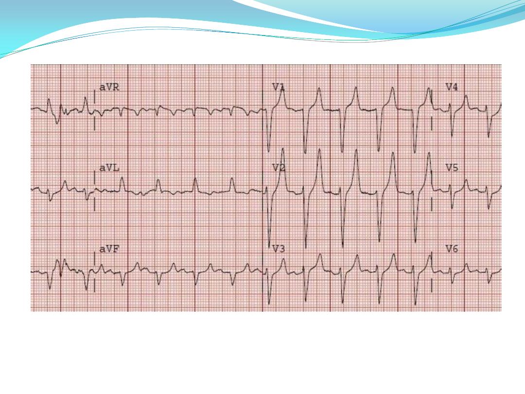
ECG(hyper-k+=peaked T wave+no ST segment+widening of QRS)
•
Chest radiography: to rule out pulmonary infection
•
Head CT scanning: to detect early cerebral edema.
normal urea ( < 40 mg/dl)
Normal creatinine( <1.2 mg/dl)
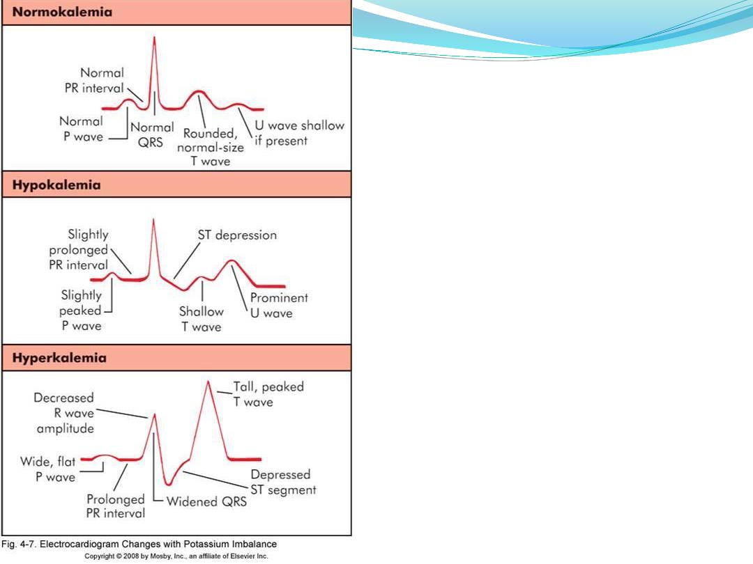
hyper-k+=
peaked T wave+no ST segment+widening of
QRS

8/21/2016
Management:
Managing diabetic ketoacidosis (DKA) in an
intensive care unit during the first 24-48 hours
always is advisable.
Plan for therapy:
When treating patients with DKA, the following
points must be considered and closely monitored:
Correction of fluid loss with intravenous fluids
Correction of hyperglycemia with insulin
Correction of electrolyte disturbances, particularly
potassium loss
Correction of acid-base balance
Treatment of concurrent infection, if present
بDKA يركسلا لزنت ام
بسرعة
ﻻ
ن
اذا نزل بسرعة يعمل
Cerebral odema

8/21/2016
Laboratory studies for diabetic ketoacidosis (DKA)
should be scheduled as follows:
Blood tests for glucose every 1-2 h until patient
is stable, then every 4-6 h
Serum electrolyte determinations every 1-2 h
until patient is stable, then every 4-6 h
Initial blood urea
Initial arterial blood gas (ABG) measurements,
followed with bicarbonate as necessary

Example how to arrange a chart to follow a
DKA patient
S. k
RBS
Output
Input
fluid
Insulin
BP
Time
6.2
410
Nil
2 L/NS
20 units
IM
80/50
3:00 PM
4:00 PM
8/21/2016

S. k
RBS
Output
Input
fluid
Insulin
BP
Time
6.2
620
Nil
1L/NS
20 units
IM or
5u. As
bolus&
5u./hr
100/50
10:00 aM
550
o.5L/hr
N.S
5u./hr
110/6o
11:00 aM
4
490
100cc
0.5l/hr+20
mEq K+
5
110/60
12:00
450
400
0.5l
N.S+20K+
6
120/80
1:00pm
4.5
350
600
=
6
120/80
2:00pm
250
0.5
G.W/3hr
+20mEq k
5
=
3:00pm-
6pm

8/21/2016
Insulin Therapy:
Using soluble (Short acting) insulin administered
either:
I.V infusion(prefered method):
o
Bolus: 0.1 unit/ kg. I.V direct
o
then maintain contiueous iv infusion of 0.1 unit/ kg./
hr. using syringe pump.
I.M:
o
Bolus: 10-20 units
o
Followed by 5 units hourly.

8/21/2016
Target blood sugar:
Falling 55-110 mg/ dl per hr.
(3-6 mmol/l)
Rapid decline → cerebral edema
Failure to reach the target → require
reassessment of insulin therapy.
Shift to subcutaneous insulin regimen
when the patient vomiting stopped and
become biochemically stable.

8/21/2016
Fluid Replacement:
Average of 6 litres fluid deficit exist
3 L are extracellular replaced by 0.9% isotonic saline.
3 L are intracellular replaced by dextrose
Timing and amount as following:
1
st
hr: using normal (isotonic) saline
systolic BP > 90 mmHg → 1 L
systolic BP < 90 mmHg → 2 L
Set 2 wide bore IV line initially

8/21/2016
Then as :
1 L OVER 2 hrs
1 L OVER 2 hrs
1 L EVERY 6 hrs
Shift to 10% dextrose fluid whenever blood sugar level
become < 250 mg/dl (14mmol/l).
Note: be cautious with elderly, pregnant, those with
heart or renal failure.

8/21/2016
Potassium Replacememt
According to serum potassium level as:
> 5.5 mmol/l → non to be given
3.5 – 5.5 (mmol/l) → 40 meq/l
be cautious in replacing K usually hyperkalemia
occurs initially due to prerenal failure secondary to
dehydration for that reason K is not recommended to
be given in the first hour of therapy.

8/21/2016
Other
Acidosis: is usually corrected with the time by
adequate fluid and insulin replacement. Bicarbonate
therapy is not recommended as it can induce cerebral
edema
Infection: should be treated by antibiotcs
accordingly
Brain edema: is the leading cause of death in DKA, it
can exist in spite of metabolic stablisation. It should be
treated by mannitol solution 20%
(7 ml/ kg.)

8/21/2016
Case Scenario
A 20-year-old woman is evaluated in the
emergency department for polyuria,
polydipsia, polyphagia, and an
unintentional 5.4-kg (11.9-lb) weight loss
over the past month. She has had increasing
lethargy over the last 24 hours. Her medical
history and family history are unremarkable.
She takes no medications.

8/21/2016
On physical examination,
temperature is 37.5 °C , blood pressure is
98/52 mm Hg, pulse rate is 120/min, and
respiration rate is 30/min. BMI is 17.
She is lethargic with dry mucous
membranes, tachypnea, and tachycardia.
Chest auscultation is clear. Abdominal
examination shows diffuse mild tenderness
and normal bowel sounds. There is no
rebound tenderness or guarding with
palpation.

8/21/2016
Laboratory studies
:
Hemoglobin= 17 g/dL (170 g/L)
Leukocyte count= 14,200/µL (14.2 × 10
9
/L)
Blood gases, arterial::
pH= 7.25
PCO
2
= 21 mm Hg
Creatinine= 1.3 mg/dL
Electrolytes
Sodium= 130 mEq/L
Potassium= 3.0 mEq/L
Chloride= 99 mEq/L
Bicarbonate= 9 mEq/L
Glucose= 620 mg/dL (34.4 mmol/L)

8/21/2016
An electrocardiogram shows sinus tachycardia 120/min.
Chest radiograph is normal.
What is the most appropriate management?
Means blood glucose level < 63 mg/ dl (3.5 mmol/l)
which is a common complication in diabetes [ those
on insulin therapy or on oral insulin secretagoues
especially sulphonylurea as Glibenclimide].
Risk factors:
1- strict glycemic control
2- extreme of age( elderly & young)
3- renal impairment
4- impaired awareness of hypoglycemia
5- long duration of DM
6- pevious history of hypoglycemia
7-ESRF (bz of insulin retention)

Causes:
1- missed or inadequate meal
2- error in therapy or poorly designed regimen
3- exercise
4- alcohol
5- lipohypertrophy at site of insulin injection.
6- factitious
7- breastfeeding

Clinical presentation:
I.
Autonomic symptoms: sweating, hunger, anxiety,
trembling.
II.
Neuroglycopenic symptoms: confusion, inability to
concentrate, drowsiness, incoordination, slurring of
speech, coma.
III.
Non specific: nausea, headache
Nocturnal hypoglycemia is common and usually not
awake the person, described as poor sleep, morning
headache, vivid dreams. The partner may notice
sweating, twitching and seizure. It can be fatal in
rare cases ( dead-in-bed syndrome)

Spontaneous hypoglycemia
means hypoglycemia that exist in non diabetic person
(uncommon condition).
---------------------------------------
Any Hypoglycemia can be Confirmed by whipple s
criteria:
symptoms of hypoglycemia
low blood glucose
symptoms resolved by correction of blood sugar.
Assessed by doing serum insulin & C-peptide level.

Management of Hypoglycemia:
Mild cases: oral 10-15 gm. Glucose followed by snack
of complex carbohydrate content.
Severe cases:
1- I.V hypertonic glucose 50% (30 ml) or 70 ml 20 %
2- I.M glucagon 1 mg
3- if the patient is conscious; 25 gm of oral refined sugar.

if the patient fail to respond
so we should exclude:
1- cerebral edema
2- alcohol intoxicaion
3- post ictal state
4- cerebral hemorrhage

Adrenal insufficiency
Secondary (↓ACTH)
• Withdrawal of suppressive glucocorticoid therapy
• Hypothalamic or pituitary disease
Primary (↑ACTH)
•
Intra-adrenal haemorrhage
•
(Waterhouse–
•
Friedrichsen syndrome
•
following meningococcal
•
septicaemia)
•
Amyloidosis
•
Haemochromatosis
•
Corticosteroid biosynthetic enzyme
defects
•
Congenital adrenal hyperplasias
•
Drugs
(Metyrapone, ketoconazole,
etomidate)
Addison’s disease
Common causes
• Autoimmune
Sporadic
Polyglandular syndromes
(p. 795)
• Tuberculosis
• HIV/AIDS
• Metastatic carcinoma
• Bilateral adrenalectomy
Rare causes
• Lymphoma

Clinical presentation of adrenal crisis:
presence of intercurrent infection, or surgery.
severe hypotension
Na ↓, K ↑, Ca ↑
hypoglycemia
muscle cramp
nausea, vomiting , diarrhea
unexplained fever

Investigations:
random plasma cortisol
short synacthin test
Management:
volume replacement by isotonic saline to correct
hyponatremia, and hyperkalemia
hydrocortisone 100 mg I.V, then 50-100 mg every 6
hrs. till the patient tolerate oral steroid
correct hypoglycemia
Correct any underlying cause.

Thyrotoxic crisis:
is a rare life threatening condition, usually result from
infection of a previously unrecognised or inadeqately
treated hyperthyroid state.
Presentation:
Fever
Tachycardia or atrial fibrillation
Agitation or confusion
Acute heart failure

Management:
1- rehydration
2- propranolol 80 mg * 4 orally OR 1-5 mg * 4 I.V
3- sodium ipodate
4- Lugol s solution ( k iodide)
5- dexamethasone 2 mg * 4
6- carbimazole 40-60 mg/ day

Myxoedema coma:
is a rare and fatal condition of old age. Usually patient
present with disturb level of consciousness with severe
hypothermia ( as low as 25 C), and convulsions.
Treatment:
1- I.V triiodothyronine ( T 3)… later by levothyroxine 50
Mg
2- rewarming
3- hydrocortisone

Sheehan syndrome(Hypopituitarism)
one of the commonest causes of panhypopituitarism,
which occur as a result of postpartum hemorrhage.
During pregnancy the pituitary gland enlarges, so
when bleeding exist the hypophyseal vessele constrict
(vasospasm) causing necrosis of the pituitary gland.

Clinical presentation:
1- first symptom is inability of breast feeding
2- adrenal insufficiency
3- hypothyroidism
4- amenorrhea
5- diabetes insipidus
6- pallor ?

Management:
1- Cortisol replacement: H.C 15- 20 mg / day
2- Thyroxine 50-150 Mg / day
3- Sex hormone replacement for those below 50 years