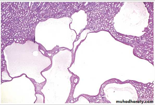
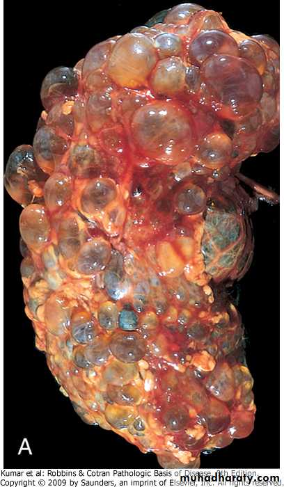
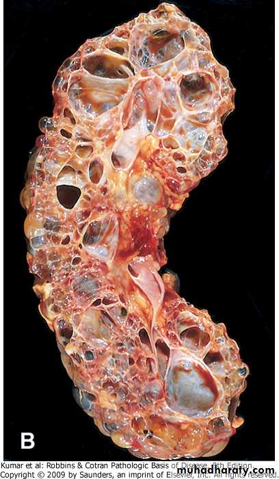
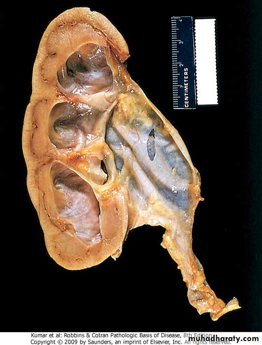
These occur as single or multiple, usually cortical.
The size range from 1-10cm or more.They are translucent and filled with clear fluid.
They are lined by a single layer of cuboidal or flattened epithelium.
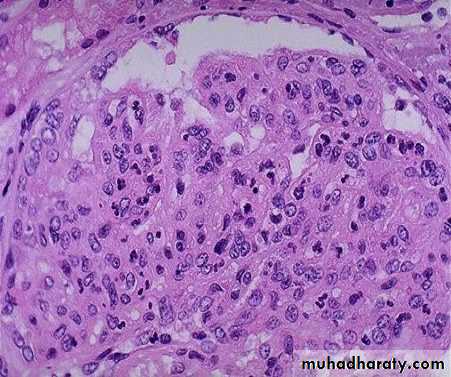
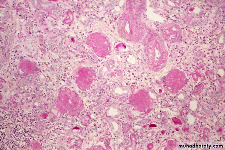
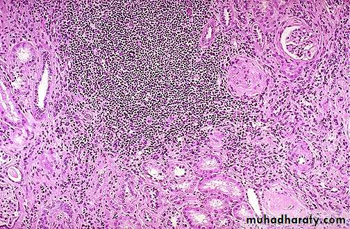
Post-streptococcal glomerulonephritis The hypercellularity of post-streptococcal glomerulonephritis is due to increased numbers of epithelial, endothelial, and mesangial cells as well as neutrophils in and around the capillary loops. This disease may follow several weeks after infection with certain strains of group A beta hemolytic streptococci. Patients typically have an elevated anti-streptolysin O (ASO) titer.
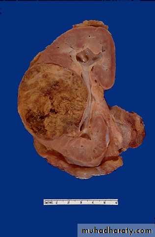
Chronic glomerulonephritis: there is presence of small, shrunken kidneys,diffuse granular surface and thin cortex .
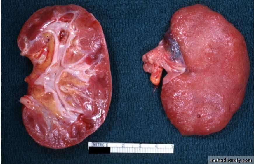
Chronic GN: Defined by the presence of > 2/3 sclerotic glomeruli. Microscopically, there is hyalinization of glomeruli, interstitial fibrosis, atrophy of tubules, and a lymphocytic infiltrate.
Grossly:
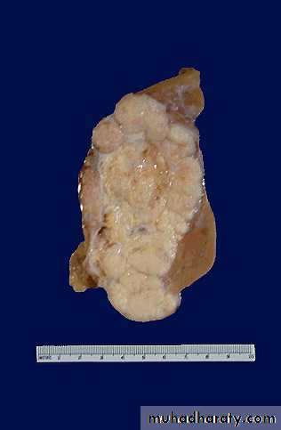
Tumors are solitary, well circumscribed, rounded and of soft in consistency.The size is variable, with a median weight of 550gm.
The cut section is predominantly solid and pale gray or tan and often exhibits areas of cystic change, necrosis, and hemorrhage.
Multicentric foci are found in 7% of cases.
Nephroblastome (Wilm's Tumour)Morphologic features
•
Renal cell carcinoma
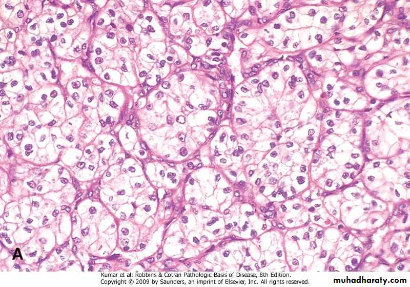
Renal cell carcinoma /Microscopically, tubular and glandular growth of tumor cells with large nuclei, and cytoplasm ranging from granular to clear.
Chronic pyelonephritis microscopical features
Microscopically, tubules are atrophic and dilated in others. There are varying degrees of chronic interstitial inflammation and fibrosis in the cortex and medulla.
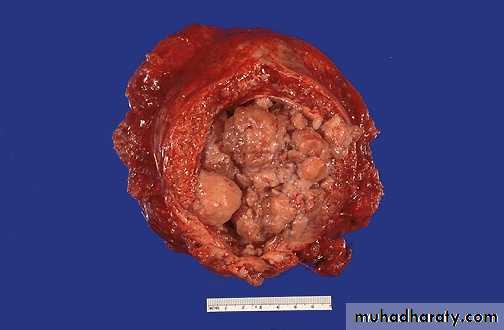
Xanthogranulomapyelonephritis
Grossly, the lesion produces large, yellowish/orange mass that may be confused with renal cell carcinoma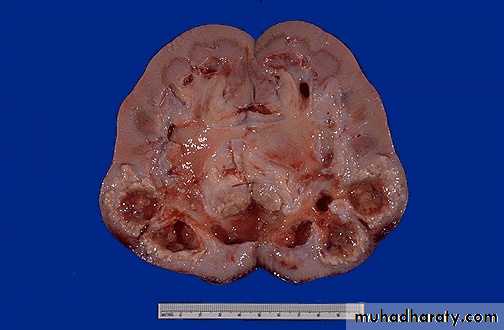
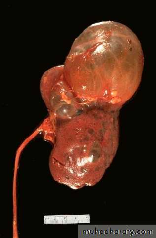
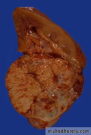
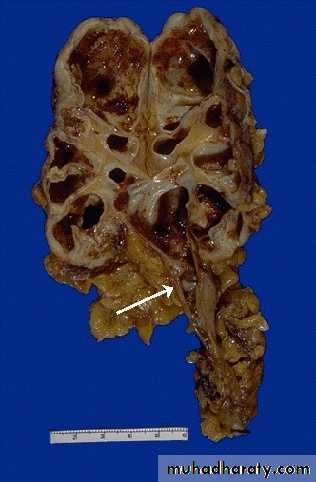
Hydronephrosis: dilatation of renal pelvis and calyces associated with progressive atrophy of the renal parenchyma due to obstruction of urine out flow.
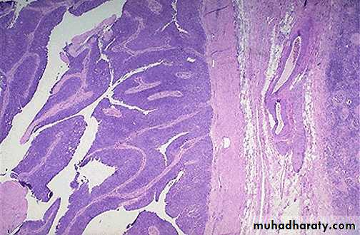
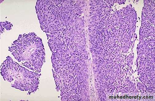
Transitional cell carcinoma TCC The gross patterns vary from purely papillary to nodular or flat. The tumors may also be invasive or non. Papillary lesions range in size between 1-5cm. They may be multicentric.
A transitional cell carcinoma of the urothelium is shown on the left at low power to reveal the frond-like papillary projections of the tumor. on the right higher magnification showinh multilayering of transitional cells with a core of blood vesses