SURGERY OF THE ANAL CANAL
الاستاذالمساعد الدكتور محمدصالح عبداللهرئيس فرع الجراحة
جراح استشاري
Embryology and Anatomy
The anal canal consist of two part of different embryological origin1-proximal anal canal which developed from the hind gut which receive it`s blood supply from the inferior mesenteric artery and nerve supply by autonomic nerve system it is insensitive
2- distal anal canal which developed from ectoderm (cloaca) which receive it`s blood supply from internal pudendal artery and nerve supply by pudendal nerve and it is sensitive
The cloaca
- it is ectodermal invagination- it is common camper to which hind gut and allantois open (urogenital system)
- it is divided by the urorectal septum into the urogenital sinus and the rectum (anal canal).
- the proxmal anal canal unite with distal anal canal to form normal anal canal , and these union at dendate line
- any abnormality in these union and cloaca formation result in congenital abnormality
Anal canal
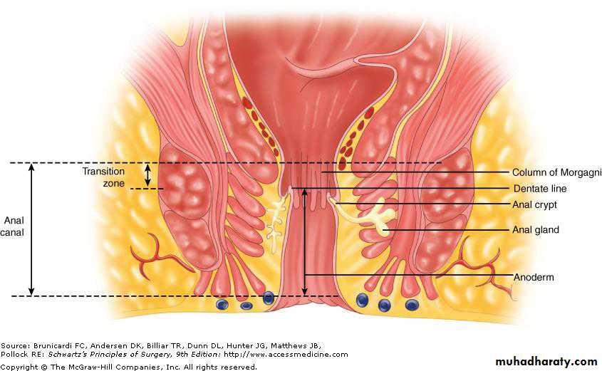
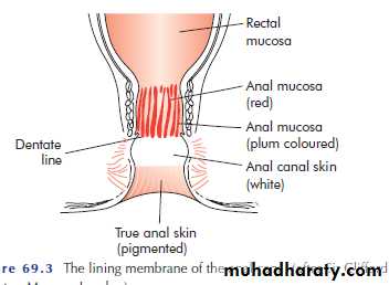
- The surgical anal canal measures 2 to 4 cmIt begins at the anorectal junction and terminates at the anal verge.
The dentate line marks the transition point between columnar rectal mucosa and squamous anoderm.
the anal transition zone The 1 to 2 cm of mucosa just proximal to the dentate line shares histological characteristics of columnar, cuboidal, and squamous epithelium
The dentate line is surrounded by longitudinal mucosal folds, known as the columns of Morgagni,
- there are small pockets between the lower extremmities of the column which is called crypt of Morgagni
- The anal gland situated at inersphinecter space open to the crypt of Morgagni
Infection of the anal gland cause perianal abscessesanal sphincter
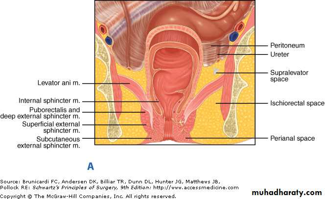
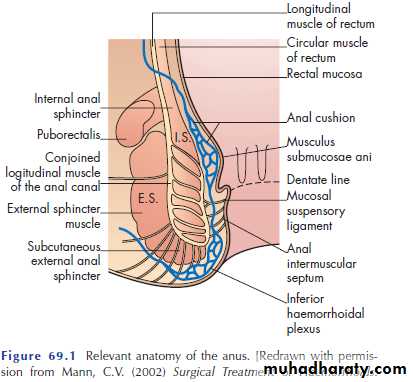
-the anal canal surrounded by two layer of muscle1- internal anal sphincter
the inner smooth-muscle layer of the lower rectum coalesces to form the internal anal sphincter, which surround upper two third of the anal canal
2- The external sphinctor is composed of striated (voluntary) muscle surround the internal sphincter but extends further distally ,it consist of three part? some consider it only one part
1- the subcutaneous
2- superficial
3- deep external sphincter
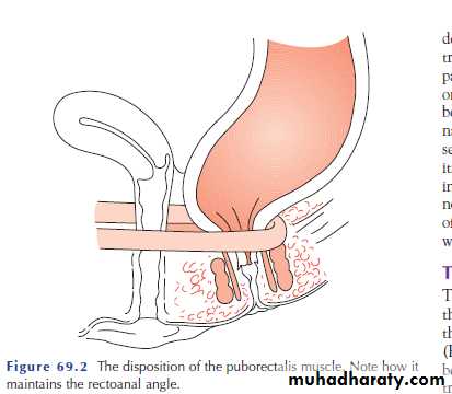
The deep external anal sphincter is an extension of the puborectalis muscle or join it
Puborectalis is part of levator ani muscle arises from pubic bone as loop around the rectum and sling it, the fibers from one side pass to other side
-the external smooth muscle fibers of the rectum intermingled with fibers from pubrecralis, its fibres fan out through the lower part of external sphincter to insert to true anal skin
Blood supply
1-The superior hamorrhoidal artery , which is more important due to it`s relation to the haemorrhoid it arises from the terminal branch of the inferior mesenteric artery and supplies the upper anal canalit divided to
- single left branch
- two right branch
2-The middle hamorrhoidal artery arises from the internal iliac artery
3-The inferior hamorrhoidal artery arises from the internal pudendal artery, which is a branch of the internal iliac artery.
A rich network of collaterals between these arteries
venous drainage
The venous drainage is parallel to the corresponding artery.1-The superior haemorrhoidal vein drains into the portal system via the inferior mesenteric vein.
2-The middle haemorrhoidal vein drains into the internal iliac vein.
3-The inferior haemorrhoidal vein drains into the internal pudendal vein, and subsequently into the internal iliac vein.
A submucosal plexus deep to Morgagni columns forms the hemorrhoidal plexus and drains into all three veins
2/12/2018
15
2/12/2018
16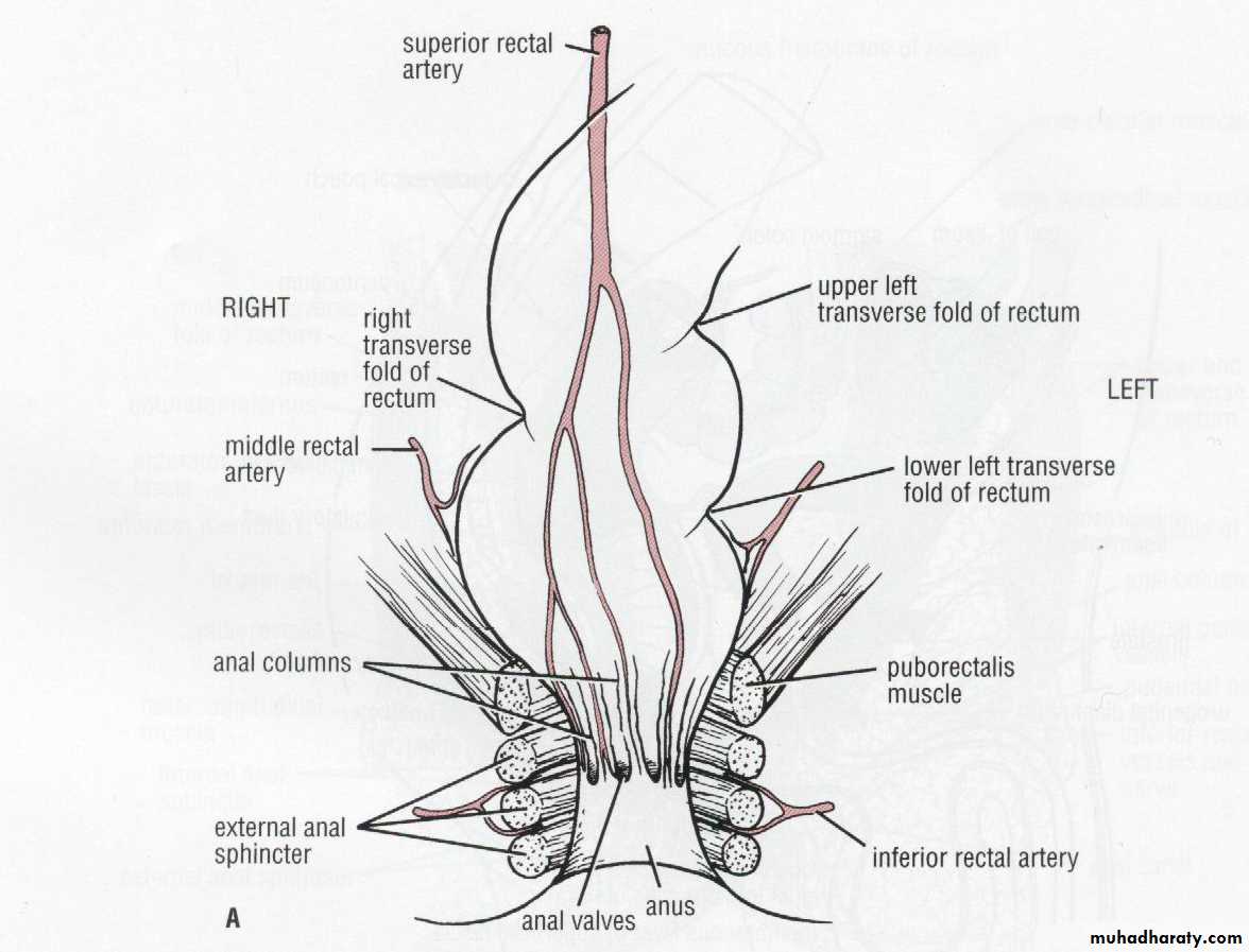
2/12/2018
172/12/2018
182/12/2018
19Lymphatic Drainag
The anal canal.
- Proximal to the dentate line, lymph drains into both the inferior mesenteric lymph nodes and the internal iliac lymph nodes
- Distal to the dentate line drains into the inguinal lymph nodes, but can also drain into the inferior mesenteric lymph nodes and internal iliac lymph nodes.
2/12/2018
21Nerve Supply
- Parasympathetic nerve fibers are known as the nervi erigentes , it originate from S2-S4- Sympathetic nerve fibers are derived from L1-L3 and join the preaortic plexus. The preaortic nerve fibers then extend below the aorta to form the hypo gastric plexus
- hypo gastric plexus joins the parasympathetic fibers(nervi erigentes) to form the pelvic plexus. Which supply the upper part of anal canal
,
-The internal anal sphincter is innervated by sympathetic and parasympathetic nerve fibers; both types of fibers inhibit sphincter contraction
-The external anal sphincter and puborectalis muscles are innervated by the inferior rectal branch of the internal pudendal nerve
Sensory innervations to the anal canal is provided by the inferior rectal branch of the pudendal nerve
the anal canal below the dentate line is sensitive.
. While the rectum is relatively insensitive
physiology
defecation
Distention of the rectum causes a reflex relaxation of the internal anal sphincter (the rectoanal inhibitory reflex) that allows the contents to make contact with the anal canal.
This "sampling reflex" allows the sensory epithelium to distinguish solid stool from liquid stool and gas
Defecation proceeds by coordination of
1-increasing intra-abdominal pressure via the Valsalva maneuver
2- increased rectal contraction
3- relaxation of the puborectalis muscle4- and opening of the anal canal
The internal sphincter is responsible for most of the resting, involuntary sphincter tone (resting pressure).The external sphincter is responsible for most of the voluntary sphincter tone (squeeze pressure)
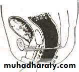
• Imperforated Anus
• - One infant in 4500 birth is born with imperforate anus or imperfect fusion of the post-allantoic gut with the proctodaeum
• -it is divided to two groups according to the termination of the bowel above or below the pelvic floor
• A- Low variety:
• bowel terminate below the pelvic floor
• 1-The covered anus, the anal canal covered by bar of skin
• 2- Ectopic anus, anus situated anteriorly at perineum in the male or vulva in the female
• (3) Stenosed anus, small, minute opening
• (4) Membranous stenosis anus at normal position but covered by thin membrane
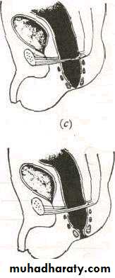
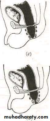
B - High variety :-
the anal canal end above the pelvic floor1- Anorectal agenesis associated usually with fistula, the fistulous tract connect to bladder in male and to posterior fornix of vagina in female
2- Rectal atresia anal canal is normal but end blindly at the level of pelvic floor and rectum end blindly above the pelvic floor
3- Cloaca only in female, bowel, urinary and genital tract open to a common wide cavity
2/12/2018
29The covered anus

2/12/2018
30• Ectopic anus
2/12/2018
31• Stenosed anus
2/12/2018
32• Membranous stenosis
2/12/2018
33•
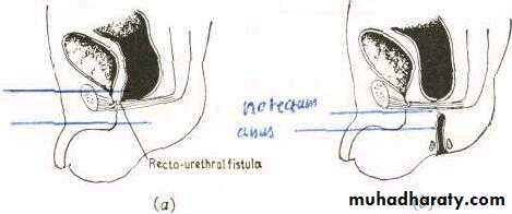
Anorectal agenesis
2/12/2018
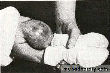
34Sacro-coccygeal teratoma
The tumor, which arises between the sacrum and the rectum, is firmly attached to the coccyx .
- Teratoma are tumors composed of tissue from all three embryonic germ layers
• - Although rare, is among the most common of the large size tumors seen during the first three months of life
• - Females are more often affected than males
• - tumur might be huge at birth
• - Is prone to become malignant
• - MRI for diagnosis
• .
Treatment:
- surgical excision soon after birth- delay is liable to result in
1- fatal ulceration
2-infection
3- rectal or urinary obstruction
4- malignant change
2/12/2018
36
Sacro-coccygeal teratoma
Post- anal dermoid
- cystic swelling in front of lower sacrum and coccyx- It is simple form of teratoma
- it is symptomless until adult life when prone to infection
- May be large size cause difficulty in defecation
Treatments is surgical excision
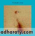
Pilonidal sinuspilonidal sinus or pilonidal cyst, pilonidal abscess if there is infection
is a cyst or abscess near or on the natal cleft of the buttocks that often contains hair and skin debris
Pilonidal means "nest of hair", and is derived from the Latin words for hair ("pilus") and nest ("nidus").
- it is common condition of the skin overlying the skin of sacrum
- It affect male more than female
- usually it affect young, male, with dense, dark , strong hair
- young adult ages of 15 and 24.
Rarely after 35years
Aetiology
1-Congenital theory
-Congenital dermal inclusion of the skin overlying the lower sacral region
- after puberty ,this dermal inclusion get infection and presented as infected pilonidal sinus
2-Acquired theory
that hair from head, back, gravitate to the skin over sacrum and coccyx ,and the hair are drawn through the skin to accumulate in the subcutaneous tissue with debris and organism
The following factor confirm this theory
1- it also occur at other area like umblicus, and at web of finger of barber2-there is only dead hair and there is no hair follicle
3- tip of hair is directed inward
4- it usually occurs at hairy man
5-liability to recurrence after complete surgical excision
Pathlogy
- There is the cavity in subcutaneous tissue, overlying the sacrum and it open to the skin by one, or more opening at midline- a cavity, lined by granulation tissue and their tracts partially epithelized
- contain loose hair
- intermittently, it infected by aerobe and anaerobe bacteria and forming abscess
clinical feaures
- asymptomatic- local discomfort, discharge
- loose hair pouting out of them
- pain ,due to infection and abscess formation
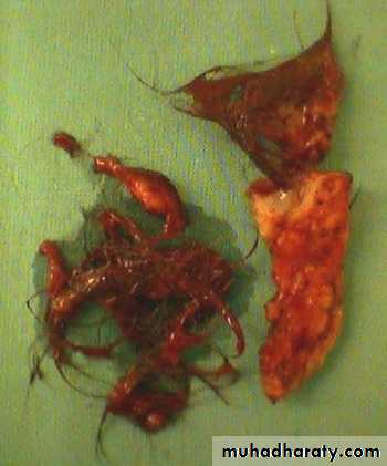
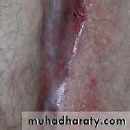
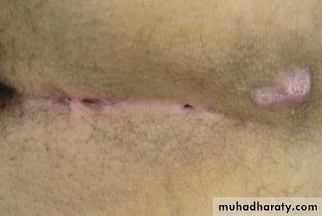
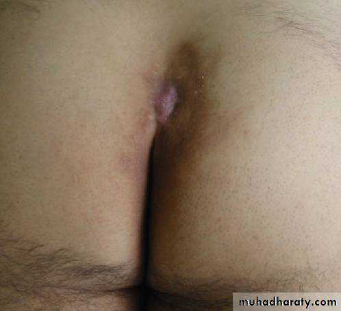
Pilonidal sinus
2/12/2018
44PILONIDAL' SINUS
2/12/2018
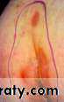
46Infected pilonidal sinus
Diferential dignosis
1- perianal abscess2- anal fistula
3- dermoid cyst
4 – sacrococcygeal teratoma
Treatment
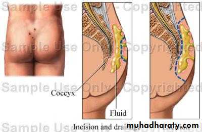
A-Pilonidal abscess
1-incision and drainage,
- incision
- clean the abscess
- remove loose hair
- left the wound open and daily dressing , it will heal by granulation tissue
2-antibiotic
3- analgesic
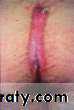
B-Pilonidal sinus
- localized excision of the cavity with it`s side tracts and and usually we determine the extension of the tracts by injection of methylene blue to the cavity- the wound either left open to heal by granulation tissue which takes time
- or is closed by sutures to hasten healing
- There is high percentage of recurrence
2/12/2018
50PILONIDAL' SINUSTreatment

2/12/2018
51PILONIDAL' SINUSTreatment
2/12/2018
52hair removed from pilonidal sinus