Lec.4 Oral Histology
د
.
سحر غانم القزاز
AMELOGENSIS
Mean the process of production & development
(mineralization) of enamel, and begins when the
crown is forming during the bell stage of tooth
development.
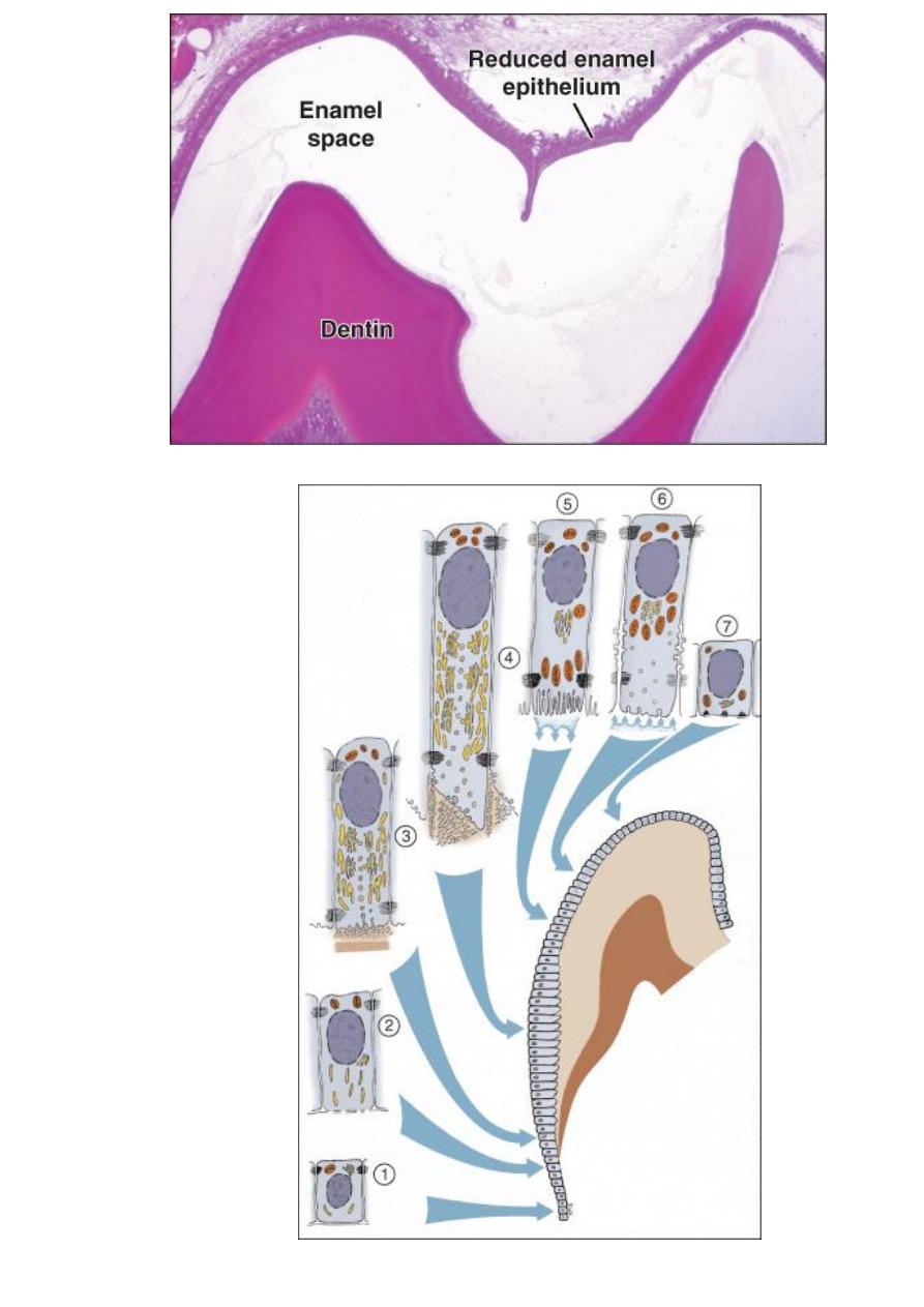
A- Life cycle of the ameloblast: The life span of the
cells of the inner enamel epithelium can be divided
into six stages.
1-morphogenic stage: the inner enamel epithelium
interacts with the adjacent mesenchymal cells of
dental papillae, determining the shape of the
dentinioenamel junction & the crown.
During this morphogenic stage the cells are short
columnar, with large oval nuclei.
Terminal bars appear represent points of close
contact between cells. The inner enamel epithelium
is separated from the C.T of dental papillae by basal
lamina.
2-organizing stage: the inner enamel epithelium cells
become longer & come into close contact with C.T.
cells of the pulp which differentiate into odontoblasts
.Reverse functional polarity of cells takes place by the

migration of the centrioles and Golgi complex from
proximal ends of the cell into the distal ends.
The 1st appearance of dentin is a critical phase in the
life cycle of the inner enamel epithelium as it’s in
contact with the C.T. of dental papillae; it receives
nutrient material from the blood vessels of this
tissue. When dentin forms, it cuts off the ameloblasts
from their original source of nourishment, then they
are supplied by the capillaries that surround &
penetrate the outer enamel epithelium.
3-formative stage: the ameloblasts enter their
formative stage after the 1st layer of dentin has been
formed. During formation of the enamel matrix the
ameloblasts retain the same length & arrangement.
The earliest change is the development of cell
process on the ameloblast surface, which penetrate
the predentin & known as Tome’s processes.
(conical projection of the ameloblast surface.
4-maturative stage: enamel maturation
(full mineralization) occurs after most of the
thickness of the enamel matrix has been formed in
the occlusal or incisal area.
During enamel maturation the ameloblasts are
slightly reduced in length & closely attached to

enamel matrix & display microvilli at their distal ends
& cytoplasmic vacuoles.
These 2 structures indicate an absorptive function of
the ameloblasts
5- Protective stage: when the enamel has completely
developed & has fully calcified, the ameloblasts can
no differentiated
.( from the ameloblast cells and cells of the stratum
intermedium & stellate reticulum and outer enamel
epithelium which fuse together to form the reduced
enamel epithelium).
The function of reduced enamel epithelium is to
protect the mature enamel by separating it from the
C.T, until the tooth erupts.
6- Desmolytic stage: the reduced enamel epithelium
proliferates & elaborate enzymes that atrophied &
destroyed the C.T, fibers by desmolysis separating it
from the oral epithelium so that fusion of the two
epithelium can occur then the tooth erupted into the
oral cavity, in this time the reduced enamel
epithelium contribute to form the junctional
epithelium.

B-formation of the enamel matrix
Ameloblasts begin enamel deposition after a few
amount of dentin have been deposited at the
dentinoenamel junction.
The ameloblasts maintains cell-to-cell attachments at
both the proximal & distal ends of the cell.
Short conical processes (Tome’s processes) develop
at the apical end of the ameloblasts during the
formative or secretory stage.
Junctional complexes called the terminal bar appear
at the junction of the cell bodies & Tome’s processes
& maintain contact between adjacent cells.
As the ameloblast influenced by dentin, the matrix is
synthesized & deposited first along the dentin &
establishes the dentinoenamel junction.
As the enamel matrix develops, it forms in
continuous rods from the dentinoenamel junction to
the surface of the enamel.
With synthesis of enamel, substances needed for
enamel production arrive via the blood vessels &
pass through the stellate reticulum to the stratum
intermedium & ameloblasts. In this manner the
protein amelogenin is produced.

C- Mineralization & maturation of enamel matrix
As amelogenin is deposited & enamel matrix formed,
the matrix begins to mineralize.
As soon as the small crystals of mineral are
deposited, they begin to grow in length & diameter.
The initial deposition of mineral amounts to
approximately 25% of the total enamel. The other
71% of mineral in enamel is a result of growth of the
crystals (4% of enamel is water & organic materials).
Matrix formation & mineralization continue
peripherally to the tips of the cusps & then laterally
on the sides of the crown, finally the cervical region
mineralized
The ameloblast shorten & contact the stratum
intermedium & stellate reticulum & outer enamel
epithelium to form the reduced enamel epithelium.
The reduced enamel epithelium remains on the
enamel surface until the tooth erupts into the oral
cavity.