Acute Post streptococcal
Glomerulonephritis:
Etiology
Pathology
Clinical manifestation
Prevention
Treatment
Prognosis
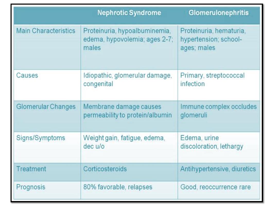
Acute poststreptococcal glomerulonephritis
(APSGN) is a classic example of the acute
nephritic syndrome characterized by the sudden
onset of:
Gross hematuria
Edema
Hypertension
Renal insufficiency.
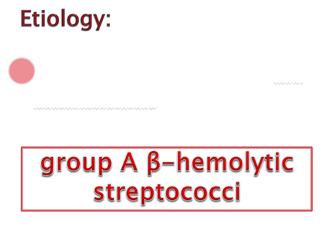
APSGN follows infection of the
throat or skin by certain
“nephritogenic” strains of

Glomeruli appear enlarged and
relatively bloodless and show diffuse
mesangial cell proliferation, with an
increase in mesangial matrix.
Polymorphonuclear leukocyte
infiltration in glomeruli is common.

PSGN is most common in children ages 5-12
yr. and uncommon before the age of 3 yr.
The typical patient develops an acute nephritic
syndrome
1-2 wk. after an antecedent streptococcal
pharyngitis or
3-6 wk. after a streptococcal pyoderma.
CLINICAL MANIFESTATIONS:

The onset is usually abrupt.
• dark color urine
• mild periorbital edema
• and decreased urine output
The earliest symptoms are:
nonspecific symptoms such as malaise, lethargy,
abdominal or flank pain.
The severity of kidney involvement varies from
asymptomatic microscopic hematuria with normal renal
function to gross hematuria with acute renal failure

Patients are at risk for developing encephalopathy
and/or heart failure secondary to hypertension or
hypervolemia.
Hypertensive encephalopathy must be considered
in patients with blurred vision, severe headaches,
altered mental status, or new seizures.
Respiratory distress, orthopnea, and cough may
be symptoms of pulmonary edema and heart
failure.

The acute phase generally resolves within 6-8
wks.
Although urinary protein excretion and
hypertension usually normalize by 4-6 wk.
after onset,
persistent microscopic hematuria can persist
for 1-2 yr. after the initial presentation.
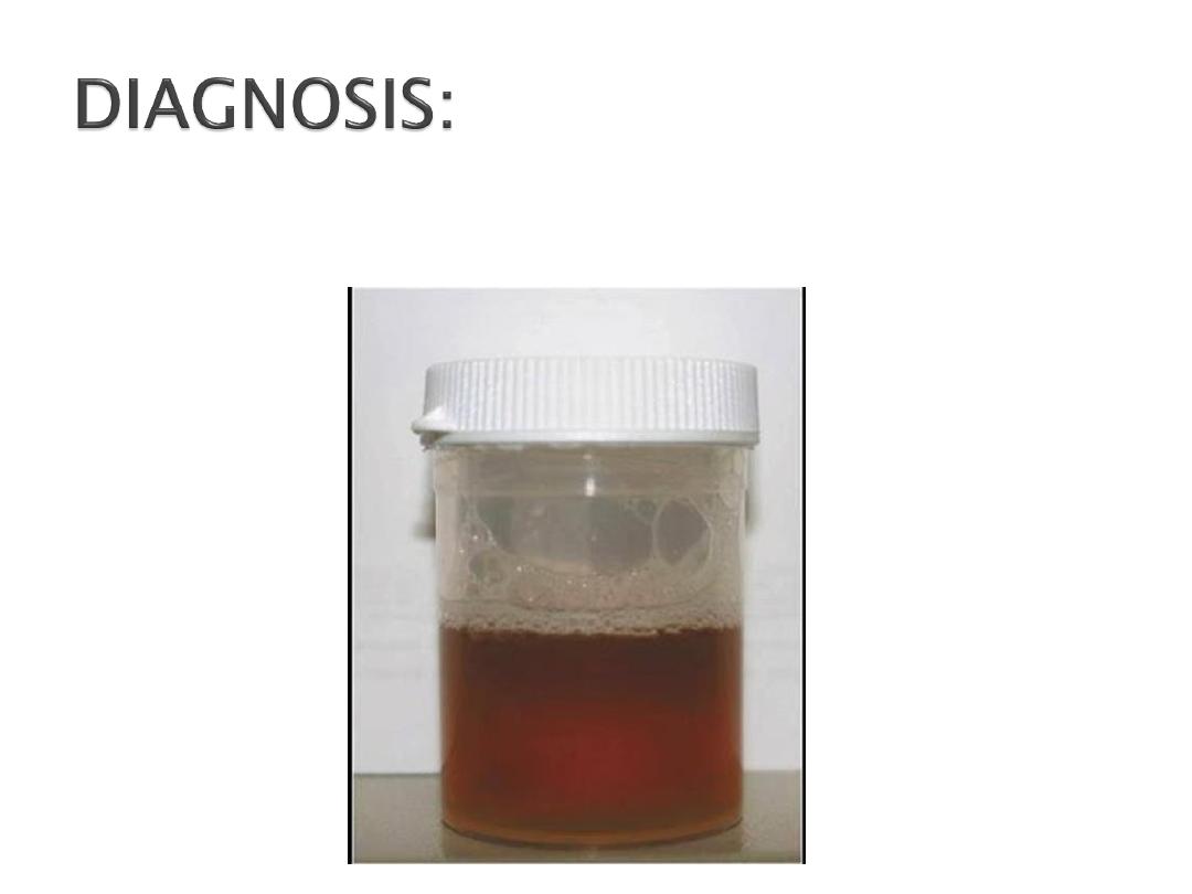
Urine color is usually reddish brown

Urinalysis demonstrates :
• Red blood cells
• Red blood cell casts
• Proteinuria "less than 1 gm/24”
• Polymorphonuclear leukocytes
• Granular cast.
Urine volume is reduced during the first 3-
5 days occasionally the patient is anuric

A mild normochromic anemia may be present
from hemodilution and low-grade hemolysis
Elevated blood urea and serum creatinine
levels , hyperkalemia and metabolic acidosis
The serum C3 level is significantly reduced in
>90% of patients.
C4 normal

Anti dnaease B level is the best single antibody titer
to document cutaneous streptococcal infection.
The ASOT titer is elevated after a pharyngeal
infection but rarely after skin infections.
Confirmation of the diagnosis requires evidence of a
streptococcal infection.

Magnetic resonance imaging (MRI)of
the brain is indicated in patients
with severe neurologic symptoms
Chest x-ray is indicated in those
with signs of heart failure or
respiratory distress
Early systemic antibiotic
therapy for streptococcal
throat and skin infections will
reduce but not eliminate the
risk of GN

Any child with PSGN should be hospitalized.
Systemic antibiotic therapy with 10 days course of
penicillin is recommended to limit the spread of the
nephritogenic organisms.
The major life threating complications during initial
1-2 weeks are acute renal insufficiency and acute
hypertension.
Fluid restriction
to 400
ml/m2/24 hr.
which is the
insensible loss +
UOP
If still anuria with
evidence of volume
overload, restrict
fluids further
.
If still anuria, do
renal dialysis.
Management of
other sequelae of
ARF: hyperkalemia,
hypertension and
metabolic acidosis

serum K level > 6 mEq/L can lead to cardiac arrhythmia, cardiac arrest,
and death.
The earliest electrocardiographic change seen in patients with
hyperkalemia is the appearance of peaked T waves.
This may be followed by widening of the QRS intervals
ST segment depression,
ventricular arrhythmias, and cardiac arrest
Hyperkalemia:
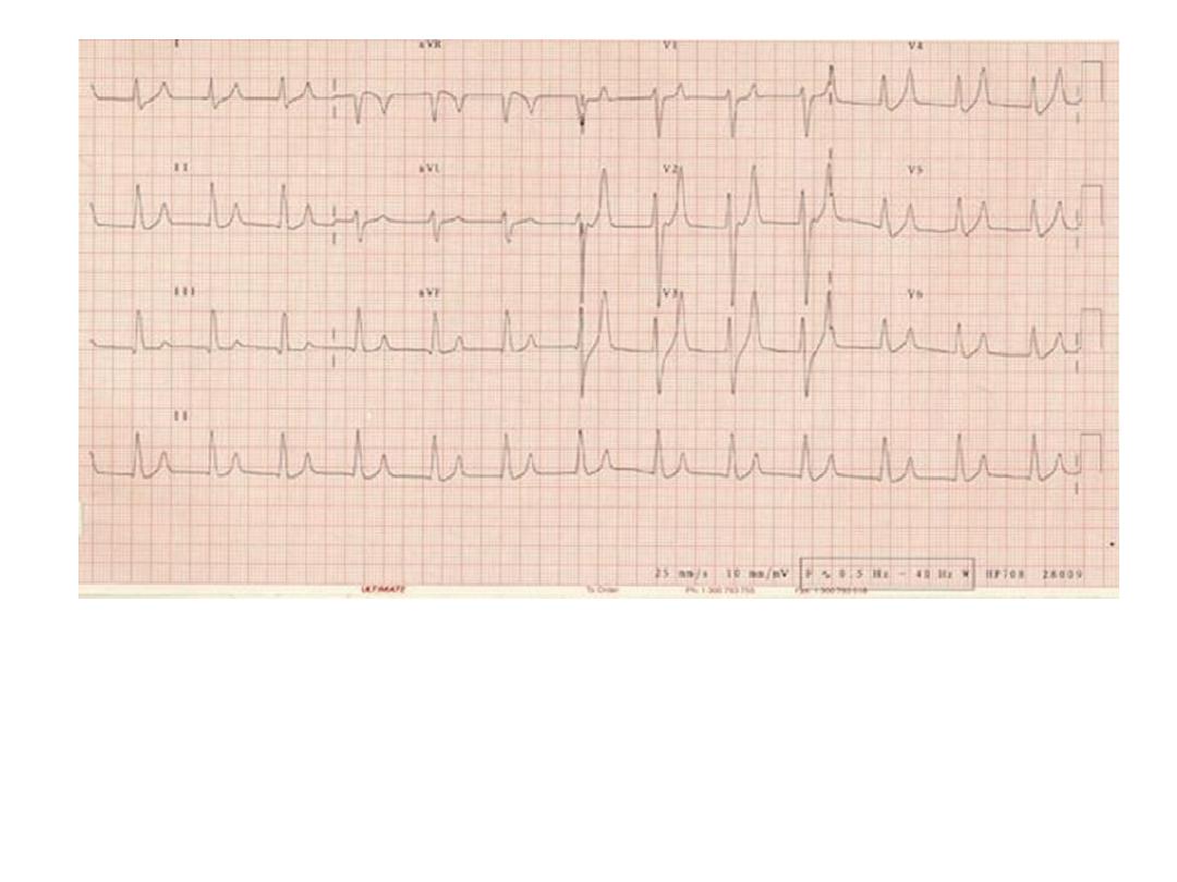
Early ECG changes showing Peaked T waves

S. K+ value rises to >6.0 mEq/L:
• Exogenous sources of K should be
eliminated.
• Kayexalate, 1 g/kg, should be given orally or
by retention enema,
a single dose of 1 g/kg can
decrease the s.k+ level by 1 mEq/L.

More severe elevations in s.K+ (>7 mEq/L) require emergency
measures in addition to Kayexalate ,the following agents should be
administered
:
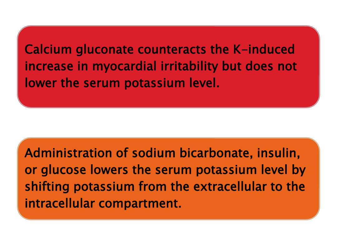
Calcium gluconate 10% solution, 1.0 mL/kg IV, over 3-5 min
Sodium bicarbonate, 1-2 mEq/kg IV, over 5-10 min
Regular insulin, 0.1 units/kg, with glucose 50% solution, 1 mL/kg,
over 1 hr.
A similar effect has been reported with the acute administration of
β-adrenergic agonists
persistent hyperkalemia should be managed by dialysis.
Bp should be checked at interval of 4-6 hrs,
with evidence of hypertensive encephalopathy
or signs of pulmonary edema or diastolic
Bp>95mmmercury treatment is indicated
.
Salt and water restriction is critical, and
diuretic administration may be useful
Isradipine may be administered for relatively
rapid reduction in blood pressure.

Longer-acting agents such as calcium
channel blockers (amlodipine) or β blockers
(propranolol, labetalol) may be helpful in
maintaining control of blood pressure.
Children with severe symptomatic HT should
be treated with continuous infusions of
nicardipine , sodium nitroprusside , labetalol
, or esmolol and converted to intermittently
dosed antihypertensive when more stable
Complete recovery occurs in
>95% of children. Recurrences
are extremely rare. Mortality in
the acute stage can be avoided
by appropriate management of
ARF, HF and HT