The shoulder joint (glenohumeral joint) is a ball and socket joint between the scapula and the humerus. It is the major joint connecting the upper limb to the trunk.
It is one of the most mobile joints in the human body, at the cost of joint stability.
Structures of the Shoulder Joint
Articulating Surfaces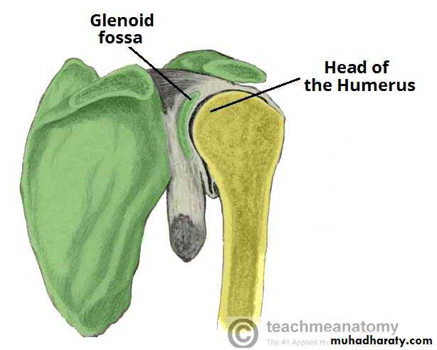
Fig 1.0 – The articulating surfaces of the shoulder joint.
The shoulder joint is formed by the articulation of the head of the humerus with the glenoid cavity (or fossa) of the scapula. This gives rise to the alternate name for the shoulder joint – the glenohumeral joint.Both the articulating surfaces are covered with hyaline cartilage – which is typical for a synovial type joint.
The head of the humerus is much larger than the glenoid fossa, giving the joint inherent instability. To reduce the disproportion in surfaces, the glenoid fossa is deepened by a fibrocartilage rim, called the glenoid labrum.
Joint Capsule and Bursae
The joint capsule is a fibrous sheath which encloses the structures of the joint. It extends from the anatomical neck of the humerus to the border of the glenoid fossa. The joint capsule is lax, permitting greater mobility (particularly abduction).
The synovial membrane lines the inner surface of the joint capsule, and produces synovial fluid to reduce friction between the articular surfaces.
To reduce friction in the shoulder joint, several synovial bursae are present. A bursa is a synovial fluid filled sac, which acts as a cushion between tendons and other joint structures. The bursae that are important clinically are:
Subacromial – Located inferiorly to the deltoid and acromion, and superiorly to the supraspinatus tendon and the joint capsule. It supports the deltoid and supraspinatus muscles. Inflammation of this bursa is the cause of several shoulder problems.
Subscapular – Located between the subscapularis tendon and the scapula. It reduces wear and tear on the tendon during movement at the shoulder joint.
There are other minor bursae present between the tendons of the muscles around the joint, but this is beyond the scope of this article.
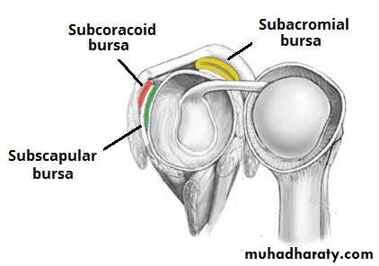
Fig 1.1 – The major bursae of the shoulder joint.
LigamentsIn the shoulder joint, the ligaments play a key role in stabilising the bony structures. The majority of the ligaments are thickenings of the joint capsule:
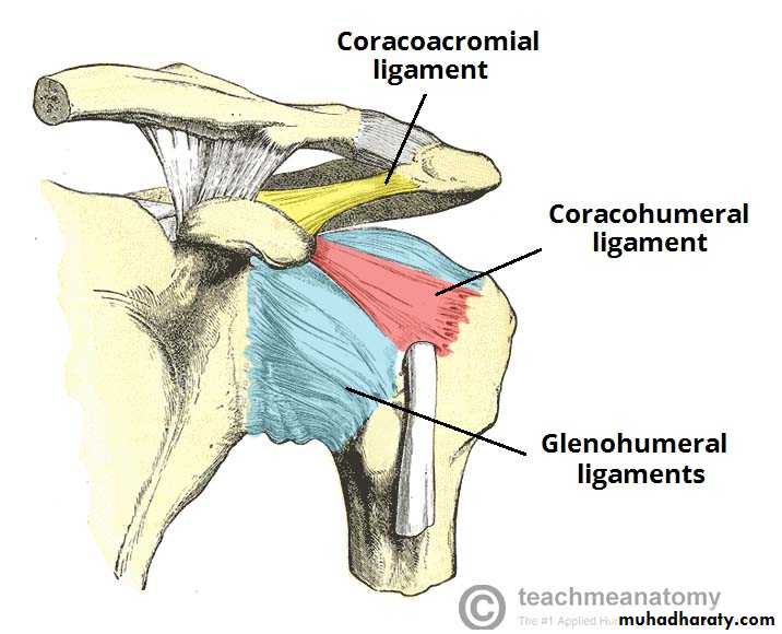
Fig 1.2 – The ligaments of the shoulder joint. The transverse humeral ligament is not shown on this diagram.
Glenohumeral ligaments (superior, middle and inferior) – Consists of three bands, which runs with the joint capsule from the glenoid fossa to the anatomical neck of the humerus. They act to stabilise the anterior aspect of the joint.
Coracohumeral ligament – Attaches the base of the coracoid process to the greater tubercle of the humerus. It supports the superior part of the joint capsule.
Transverse humeral ligament – Spans the distance between the two tubercles of the humerus. It holds the tendon of the long head of the biceps in the intertubercular groove.
The other major ligament is the coracoacromial ligament. Unlike the others, it is not a thickening of the joint capsule. It runs between the acromion and coracoid process of the scapula, forming the coraco-acromial arch. This structure overlies the shoulder joint, preventing superior displacement of the humeral head.
Neurovascular Supply
Arterial supply to the glenohumeral joint is via the anterior and posterior circumflex humeral arteries, and the suprascapular artery. Branches from these arteries form an anastomotic network around the joint.
The joint is supplied by the axillary, suprascapular and lateral pectoral nerves. These nerves are derived from roots C5 and C6 of the brachial plexus..
Movements
As a ball and socket synovial joint, there is a wide range of movement permitted:Extension (upper limb backwards in sagittal plane)Produced by the posterior deltoid, latissimus dorsi and teres major.
Flexion (upper limb forwards in sagittal plane)Produced by the biceps brachii (both heads), pectoralis major, anterior deltoid and coracobrachialis.
Abduction (upper limb away from midline in coronal plane) abduction is produced by the supraspinatus. The middle fibres of the deltoid , trapezius and serratus anterior.
Adduction (upper limb towards midline in coronal plane)Produced by contraction of pectoralis major, latissimus dorsi and teres major.
Medial Rotation (rotation towards the midline, so that the thumb is pointing medially)Produced by contraction of subscapularis, pectoralis major, latissimus dorsi, teres major and anterior deltoid.
Lateral Rotation (rotation away from the midline, so that the thumb is pointing laterally)Produced by contraction of the infraspinatus and teres minor.
Mobility and Stability
The shoulder joint is one of the most mobile in the body, at the expense of stability. Here, we shall consider the factors the permit movement, and those that contribute towards joint structure.
Factors that contribute to mobility:
Type of joint – It is a ball and socket joint.
Bony surfaces – Shallow glenoid cavity and large humeral head –Laxity of the joint capsule.
Factors that contribute to stability:
Rotator cuff muscles – These muscles surround the shoulder joint, attaching to the tubercles of the humerus, whilst also fusing with the joint capsule. The resting tone of these muscles act to ‘pull’ the humeral head into the glenoid cavity.
Glenoid labrum: This is a fibrocartilaginous ridge surrounding the glenoid cavity. It deepens the cavity, reducing the risk of dislocation.
Ligaments – The ligaments act to reinforce the joint capsule, and forms the coraco-acromial arch.
By TeachMeSeries Ltd (2017)
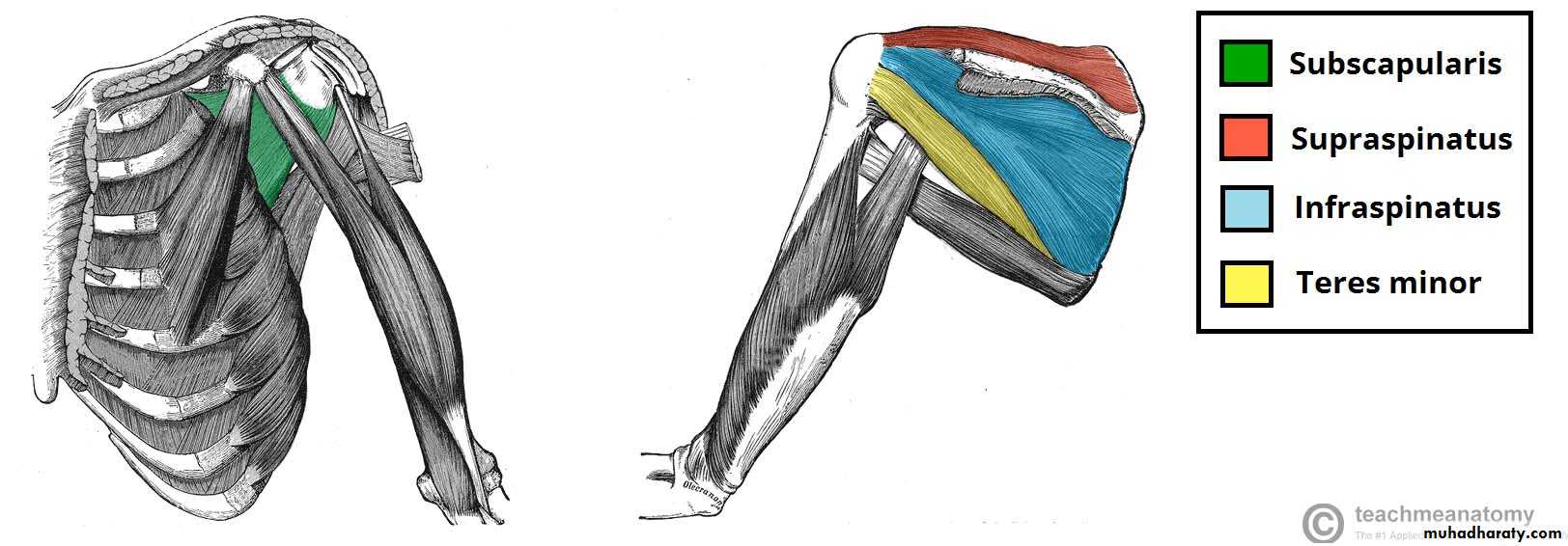
Fig 1.3 – The rotator cuff muscles, which act to stabilise the shoulder joint.
An anterior dislocation is usually caused by excessive extension and lateral rotation of the humerus. The humeral head is forced anteriorly and inferiorly – into the weakest part of the joint capsule. Tearing of the joint capsule is associated with an increased risk of future dislocations.The axillary nerve runs in close proximity to the shoulder joint, and can be damaged in the dislocation. Injury to the axillary nerve causes paralysis of the deltoid, and loss of sensation over regimental badge area. A dislocation can also stretch the radial nerve, as it is tightly bound in the radial groove.
By MB [CC-BY-SA-2.5], via Wikimedia Commons
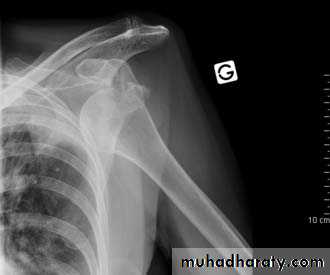
Fig 1.4 – Anterior dislocation of the shoulder joint.
Rotator Cuff TendonitisThe rotator cuff muscles have a very important role in stabilising the glenohumeral joint. They are often under heavy strain, and therefore injuries of these muscles are relatively common.
Tendonitis refers to inflammation of the muscle tendons – usually due to overuse. Over time, this causes degenerative changes in the subacromial bursa, and the supraspinatus tendon. This increases friction between the structures of the joint.
The characteristic sign of rotator cuff tendonitis is the ‘painful arc’ – pain in the middle of abduction, where the affected area comes into contact with the acromion.